Abstract
Purpose of the Study:
The aim of our study was to compare the technetium-99m (Tc-99m)-ethylenedicysteine (EC) renography calculation of differential renal function (DRF) with this measurement using Tc-99m-dimercaptosuccinic acid (DMSA) scintigraphy.
Materials and Methods:
Patients referred to our department were included in our study, and both DMSA and EC scans were performed for each patient according to the standard imaging protocols. A checklist was filled for each patient. Statistical analysis was performed using correlation and regression methods.
Results:
Forty-two patients (mean age: 3.6 ± 3.4 years), including 32 boys and 10 girls, participated in our study. The results of EC scintigraphy were significantly correlated with the values of DMSA scintigraphy (P < 0.001). Performing linear regression, EC renography significantly (P < 0.001) predicted the DRF as it was calculated by DMSA scintigraphy (R2 = 0.92, P < 0.001). This test was significant in both male and female subgroups (P < 0.001).
Conclusions:
Overall, our study findings were similar to the reported results in the other reviewed studies, showing that Tc-99m-EC can be considered as an alternative for DMSA scintigraphy, providing interchangeable results.
Keywords: Differential renal function, technetium-99m-dimercaptosuccinic acid, technetium-99m-ethylenedicysteine
Introduction
Obstructive uropathy is a condition in which urine flow is obstructed on the way reaching the bladder.[1] This obstruction, with any degree of existence and its causes including mechanical or nonmechanical etiologies, can result in renal hydronephrosis. There are different available methods to detect and evaluate this condition in children.
Since the 1970s, diuresis renography has been shown as a potent tool in the evaluation of hydronephrosis alongside other modalities in pediatrics, illustrating the drainage of the hydronephrotic kidney, as well as calculating differential renal function (DRF).[2] To achieve this goal, there are various available radiotracers with various characteristics. These differences made centers to seek the agent of choice to reach maximum imaging yield in their setting.
After preparation of L, L-ethylenedicysteine (EC) complex with technetium-99m (Tc-99m) in the 1990s, introduced as an alternative to orthoiodohippurate, several studies were performed to understand the advantages of using Tc-99m-EC in renal nuclear imaging. As the initial studies showed, Tc-99m-EC is a high-quality agent in renal imaging compared to other agents and, regarding its clearance route, yields a close approximation of the effective renal plasma flow and renal function compared with the gold standard.[3]
In current centers' setting worldwide, Tc-99m-mercaptoacetyltriglycine (MAG3) and Tc-99m-dimercaptosuccinic acid (DMSA) are the common agents used to perform radionuclide renal scintigraphy.[4] Tc-99m-DMSA yields the highest sensitivity in detecting renal parenchymal abnormalities, and there is a broad consensus that it is the most appropriate tracer for cortical scintigraphy.[5] However, the features of tubular fixation and strong plasma protein binding of this radiotracer result in high radiation dose in comparison with other tracers (e.g., DMSA whole-body dose of 1.60 mGy/MBq against 0.25 mGy/MBq in MAG3 scintigraphy).[6] Using Tc-99m-EC, both advantages of high-quality imaging and low radiation dose can come together.
In this study, the estimation of DRF utilizing Tc-99m-EC was compared with Tc-99m-DMSA in children with obstructive and nonobstructive hydronephrosis to investigate the accuracy of Tc-99m-EC.
Materials and Methods
This study included 42 children with obstructive and nonobstructive hydronephrosis who entered the Pediatric Nephrology Department of Ali Asghar Children's Hospital of Tehran, Iran, in a 1-year period. Patients with single kidney, horseshoe kidneys, duplication in collecting systems, and renal agenesis were excluded from the study. A checklist containing age, gender, height, weight, anteroposterior pelvic diameter at the time of diagnosis, renal parenchymal thickness, presence of mechanical obstruction, posterior urethral valve, presence of reflux/pyelonephritis, and received medications was filled for each patient. Hydronephrosis grading was performed using the renal pelvis anteroposterior diameter as three different grades including mild (<7 mm), moderate (7–10 mm), and severe (>10 mm).
Both Tc-99m-DMSA and Tc-99m-EC scans were performed within 2 weeks using a single-headed ADAC gamma-camera equipped with a (Low Energy All Purpose) LEAP collimator. Before diuretic renography, the patients were advised to drink water (10 ml/kg body weight) to be well hydrated. Standard imaging was performed in the posterior view after administration of 3.7 MBq (0.1 mCi)/kg body weight of Tc-99m-EC. F + 20 protocol was adopted and 1 mg/kg of furosemide was intravenously injected 20 min after the radiopharmaceutical region of interest (ROI) for kidneys was drawn on the 2–3 min frame of the study by an experienced technologist encompassing the entire kidney and the background ROI was drawn automatically. This measurement was repeated two times, and the average of measures was registered. Two hours after intravenous injection of the Tc-99m-DMSA (55.5–74 MBq; 1.5–2 mCi), imaging with a gamma-camera was performed in four standard views (anterior, posterior, right posterior oblique, and left posterior oblique). Predefined 700,000 counts were taken for each view. Whole-kidney ROIs were drawn by a skilled technologist, and crescent background ROIs were drawn inferolateral to each kidney. DRF was calculated based on the geometric mean method in two consecutive times, and the average figure was taken into account. In the analysis phase, the DRF calculated by Tc-99m-EC was compared to the measurement by Tc-99m-DMSA. Statistical analysis was performed by an experienced researcher using SPSS so ftware (IBM SPSS Statistics for Windows, Version 25.0. Armonk, NY : IBM Corp). Spearman correlation analysis and linear regression were used, and P < 0.05 was considered significant.
Results
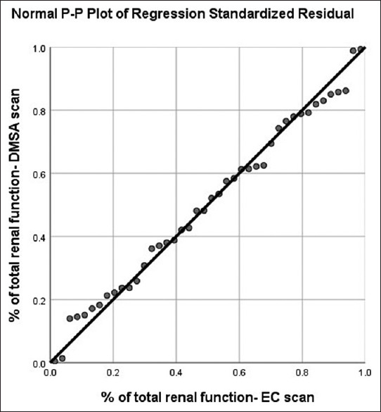
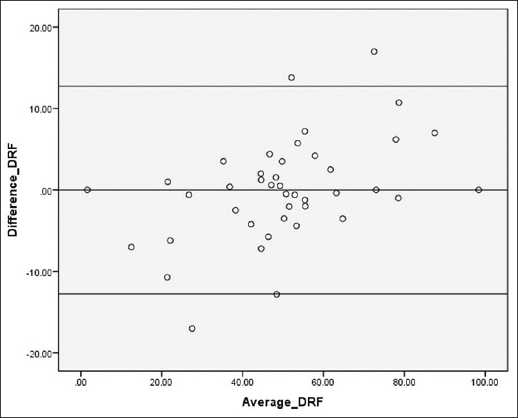
Forty-two participants with a mean age of 3.6 ± 3.4 years (ranging from 1 month old to 12 years old) including 32 boys (76.2%), with a mean age of 4.1 ± 3.6 years, and 10 girls (23.8%), with a mean age of 2 ± 2.2 years, participated in our study. DRF was calculated for each child using Tc-99m-EC and Tc-99m-DMSA. The details are shown in Table 1. Twenty-four patients (57%) had one-sided hydronephrosis, whereas 18 (43%) ones had bilateral hydronephrosis. Figure 1 shows the right kidney renal function calculation using DMSA scintigraphy against what we observed in our study after performing EC scintigraphy in each case. Figure 2 is the Bland–Altman plot of this comparison. Using Spearman (nonparametric) correlation, there was a significant correlation between EC scintigraphy and DMSA scintigraphy measurements in terms of DRF (rs = 0.92, P < 0.001). This correlation was also significant in each gender, showing a ratio of 0.83 (P < 0.001) and 0.98 (P < 0.001) in males and females, respectively. Performing linear regression, EC scintigraphy significantly (P < 0.001) predicts the DRF as it was calculated by DMSA scintigraphy (R2 = 0.92, P < 0.001). This test was significant in both male and female subgroups (P < 0.001). Regarding the effect of one-sided and bilateral hydronephrosis on EC scintigraphy measurements, the Spearman's ratio and linear regression remained significant in predicting the renal function when DMSA scintigraphy sets as the standard test (P < 0.001 for both analyses).
Table 1.
Frequency table of ethylenedicysteine scintigraphy and dimercaptosuccinic acid scintigraphy showing number of patients, mean, standard deviation, and range of measurements
| n | Mean±SD (%) | Range (%) | |
|---|---|---|---|
| Right kidney function (Tc-99m-EC) | 42 | 50±21.2 | 96.4 |
| Right kidney function (Tc-99m) | 42 | 50±18.2 | 96.4 |
| Right kidney function (Tc-99m-EC) DMSA - male | 32 | 50±18.7 | 96.4 |
| Right kidney function (Tc-99m-DMSA) - male | 32 | 50±16.9 | 96.4 |
| Right kidney function (Tc-99m-EC) -female | 10 | 50±29.2 | 85 |
| Right kidney function (Tc-99m-EC) - female | 10 | 50±22.9 | 67.7 |
EC: Ethylenedicysteine, DMSA: Dimercaptosuccinic acid, SD: Standard deviation
Figure 1.

Plot of measured renal function of the right kidney of each patient with ethylenedicysteine scintigraphy versus dimercaptosuccinic acid scintigraphy in children (n = 42)
Figure 2.

Comparisons of differential renal function calculated by technetium-99m-ethylenedicysteine and technetium-99mdimercaptosuccinic acid, using the Bland–Altman plot (n = 42)
Discussion
Evaluation method of renal function is an important part of studying hydronephrosis in children. Nuclear renography is an adjunct test in this field to calculate DRF and the obstruction severity.[7] It is also an indispensable technique for following up patients and tracking their kidney function.[8] Tc-99m-DMSA, a tubular agent, provides a high-quality illustration of the renal parenchyma and cortical scars. However, there are drawbacks in its administration such as high radiation dose and long biological half-life which limit its usage. To search for alternative agents with high-quality imaging properties and low radiation dose, several studies have been performed. Tc-99m-EC is one of the reliable radiotracers with acceptable count statistics and excellent image quality,[9] which can be comparable with Tc-99m-DMSA in terms of calculating DRF. Buyukdereli and Guney[10] studied 124 kidneys of 64 patients, who had various renal disorders, with a mean age of 5 years. The results showed a highly positive correlation between DRF measured by EC renography and DMSA scintigraphy (R = 0.91, P = 0.001). Kibar et al.,[6] by studying 29 children, reported that the sensitivity and specificity of the EC renography in detecting renal parenchymal defects, comparing with DMSA scintigraphy, to be 92.6% and 100%, respectively. They found a close correlation of measured DRF between these two different methods. Mohammadi-Fallah et al.[11] reported specificity and positive predictive value of 100% for EC scintigraphy in their study and concluded that EC scan is a reliable alternative of DMSA scintigraphy. Domingues et al.[12] analyzed 111 patients and found that the DRF measured by EC scintigraphy was close to values obtained by Tc-99m-DMSA with no significant difference. Raja et al.[13] showed that scintigraphy findings using EC were significantly correlated with DMSA imaging findings and the DRF measured by these methods could be reported interchangeably. Atasever et al.,[14] by studying patients with acute pyelonephritis, reached the conclusion that EC scintigraphy measurements were reliable according to DMSA renography as a gold standard. Results of the mentioned studies were similar to our study findings, showing that Tc-99m-EC can be considered as an alternative for DMSA scintigraphy, providing lower radiation dose and interchangeable results. On the other hand, although Yapar et al.[15] found a high correlation between EC images and DMSA study in terms of DRF, they reported that DRF measured by EC scintigraphy was underestimated in their study.
Conclusion
All in all, after studying 42 patients with hydronephrosis and reviewing articles in this field, we concluded that DMSA scintigraphy, as a gold standard and with its advantages, is a method which can be replaced by EC scan because of its significantly close approximation in measuring DRF. However, our study had limitations which can be resolved in future studies. The main weakness of our study was the population size which should be larger in order to perform further analyses (e.g., interpreting results in each grade of hydronephrosis). In addition, the number of detected renal defects could be included to calculate the performance statistics of EC scintigraphy.
Financial support and sponsorship
Nil.
Conflicts of interest
There are no conflicts of interest.
References
- 1.Jones RA, Easley K, Little SB, Scherz H, Kirsch AJ, Grattan-Smith JD. Dynamic contrast-enhanced MR urography in the evaluation of pediatric hydronephrosis: Part 1, functional assessment. AJR Am J Roentgenol. 2005;185:1598–607. doi: 10.2214/AJR.04.1540. [DOI] [PubMed] [Google Scholar]
- 2.Bayne CE, Majd M, Rushton HG. Diuresis renography in the evaluation and management of pediatric hydronephrosis: What have we learned? J Pediatr Urol. 2019;15:128–37. doi: 10.1016/j.jpurol.2019.01.011. [DOI] [PubMed] [Google Scholar]
- 3.Moran JK. Technetium-99m-EC and other potential new agents in renal nuclear medicine. Semin Nucl Med. 1999;29:91–101. doi: 10.1016/s0001-2998(99)80002-6. [DOI] [PubMed] [Google Scholar]
- 4.Piepsz A, Gordon I, Hahn K. Paediatric nuclear medicine. Eur J Nucl Med. 1991;18:41–66. doi: 10.1007/BF00177684. [DOI] [PubMed] [Google Scholar]
- 5.Dick PT, Feldman W. Routine diagnostic imaging for childhood urinary tract infections: A systematic overview. J Pediatr. 1996;128:15–22. doi: 10.1016/s0022-3476(96)70422-5. [DOI] [PubMed] [Google Scholar]
- 6.Kibar M, Yapar Z, Noyan A, Anarat A. Technetium-99m-N, N-ethylenedicysteine and Tc-99m DMSA scintigraphy in the evaluation of renal parenchymal abnormalities in children. Ann Nucl Med. 2003;17:219–25. doi: 10.1007/BF02990025. [DOI] [PubMed] [Google Scholar]
- 7.Nguyen HT, Herndon CD, Cooper C, Gatti J, Kirsch A, Kokorowski P, et al. The Society for Fetal Urology consensus statement on the evaluation and management of antenatal hydronephrosis. J Pediatr Urol. 2010;6:212–31. doi: 10.1016/j.jpurol.2010.02.205. [DOI] [PubMed] [Google Scholar]
- 8.Mendichovszky I, Solar BT, Smeulders N, Easty M, Biassoni L. Nuclear medicine in pediatric nephro-urology: An overview. Semin Nucl Med. 2017;47:204–28. doi: 10.1053/j.semnuclmed.2016.12.002. [DOI] [PubMed] [Google Scholar]
- 9.Piepsz A, Blaufox MD, Gordon I, Granerus G, Majd M, O'Reilly P, et al. Consensus on renal cortical scintigraphy in children with urinary tract infection. Scientific Committee of Radionuclides in Nephrourology. Semin Nucl Med. 1999;29:160–74. doi: 10.1016/s0001-2998(99)80006-3. [DOI] [PubMed] [Google Scholar]
- 10.Buyukdereli G, Guney IB. Role of technetium-99m N, N-ethylenedicysteine renal scintigraphy in the evaluation of differential renal function and cortical defects. Clin Nucl Med. 2006;31:134–8. doi: 10.1097/01.rlu.0000200460.41091.13. [DOI] [PubMed] [Google Scholar]
- 11.Mohammadi-Fallah M, Alizadeh M, Ahmadi Lavin T. Comparison of DMSA scan 99 m and EC scan 99 m in diagnosis of cortical defect and differential renal function. Glob J Health Sci. 2014;6:38–43. doi: 10.5539/gjhs.v6n7p38. [DOI] [PMC free article] [PubMed] [Google Scholar]
- 12.Domingues FC, Fujikawa GY, Decker H, Alonso G, Pereira JC, Duarte PS. Comparison of relative renal function measured with either 99mTc-DTPA or 99mTc-EC dynamic scintigraphies with that measured with 99mTc-DMSA static scintigraphy. Int Braz J Urol. 2006;32:405–9. doi: 10.1590/s1677-55382006000400004. [DOI] [PubMed] [Google Scholar]
- 13.Raja S, Pareek V, Singh B, Sharma S, Rao KL, Mittal BR. Comparison of 99mTc-ethylene dicysteine and 99mTc-dimercaptosuccinic acid scintigraphy for the Evaluation of Cortical Scarring and Differential Renal Function in Children with recurrent urinary tract infection. J Postgrad Med Edu Res. 2012;46:183–6. [Google Scholar]
- 14.Atasever T, Ozkaya O, Abamor E, Söylemezoğlu O, Buyan N, Unlü M. 99mTc ethylene dicysteine scintigraphy for diagnosing cortical defects in acute pyelonephritis: A comparative study with 99mTc dimercaptosuccinic acid. Nucl Med Commun. 2004;25:967–70. doi: 10.1097/00006231-200409000-00016. [DOI] [PubMed] [Google Scholar]
- 15.Yapar AF, Aydin M, Reyhan M, Yapar Z, Yologlu NA. Assessment of the optimal time interval and background region of interest in the measurement of differential renal function in Tc-99m-EC renography. Ann Nucl Med. 2004;18:419–25. doi: 10.1007/BF02984485. [DOI] [PubMed] [Google Scholar]


