Abstract
Context
Arch height is one important aspect of foot posture. An estimated 20% of the population has pes planus and 20% has pes cavus. These abnormal foot postures can alter lower extremity kinematics and plantar loading and contribute to injury risk. Ankle bracing is commonly used in sport to prevent these injuries, but no researchers have examined the effects of ankle bracing on plantar loading.
Objective
To evaluate the effects of ankle braces on plantar loading during athletic tasks.
Design
Cross-sectional study.
Setting
Laboratory.
Patients or Other Participants
A total of 36 participants (11 men, 25 women; age = 23.1 ± 2.5 years, height = 1.72 ± 0.09 m, mass = 66.3 ± 14.7 kg) were recruited for this study.
Intervention(s)
Participants completed walking, running, and cutting tasks in 3 bracing conditions: no brace, lace-up ankle-support brace, and semirigid brace.
Main Outcome Measure(s)
We analyzed the plantar-loading variables of contact area, maximum force, and force-time integral for 2 midfoot and 3 forefoot regions and assessed the displacement of the center of pressure. A 3 × 3 mixed-model repeated-measures analysis of variance was used to determine the effects of brace and foot type (α = .05).
Results
Foot type affected force measures in the middle (P range = .003–.047) and the medial side of the foot (P range = .004–.04) in all tasks. Brace type affected contact area in the medial midfoot during walking (P = .005) and cutting (P = .01) tasks, maximum force in the medial and lateral midfoot during all tasks (P < .001), and force-time integral in the medial midfoot during all tasks (P < .001). Portions of the center-of-pressure displacement were affected by brace wear in both the medial-lateral and anterior-posterior directions (P range = .001–.049).
Conclusions
Ankle braces can be worn to redistribute plantar loading. Additional research should be done to evaluate their effectiveness in injury prevention.
Keywords: foot type, center of pressure, ankle brace
Key Points
Foot type affected force measures of the middle and the medial side of the foot during athletic activities.
Ankle braces affected the contact area, maximum force, and force-time integral in the medial midfoot region of the foot during athletic tasks.
Portions of center-of-pressure displacement were affected by brace wear in both the medial-lateral and anterior-posterior directions.
Foot posture can alter movement and result in lower extremity injuries. One aspect of foot posture or foot type is arch height, which is extremely important because the medial longitudinal arch is responsible for absorbing most of the impact on the foot during daily activities.1 By analyzing arch height and mobility, clinicians can classify individuals as having pes planus (flat feet or low arches), pes rectus (normal arches), or pes cavus (high arches). Approximately 60% of individuals are classified as having normal arch height, 20% as having pes planus, and 20% as having pes cavus.2 Different foot types are often associated with various rearfoot angles, which are the angles between the calcaneus and the shank.3 More specifically, pes planus is often associated with hindfoot valgus, and pes cavus is often associated with hindfoot varus.4 Although foot posture has many aspects, arch height and rearfoot angle are 2 well-known characteristics that are commonly used to determine foot type in clinical settings.
Different foot types lead to different structural characteristics, and they can also result in different plantar-loading patterns.5–7 For example, the pes planus foot has a more medial center of pressure (COP) and greater contact area, forces, and pressures in the medial midfoot, medial forefoot, and hallux during walking.5,6 This is due to increased midfoot mobility and the hindfoot valgus position of the rearfoot, which allows the foot to collapse medially, resulting in increased plantar loading on the medial aspect of the foot.5,6 The pes cavus foot has a more lateral COP and more pressure in the heel and lateral forefoot during walking.5 This is the result of a more rigid foot and the hindfoot varus position of the rearfoot, which cause increased loading on the lateral aspect of the foot.5 Clinicians should consider these plantar-loading patterns of different foot types when evaluating the function of the foot and potential injury risk.
Injuries have been related to abnormal arch height8 from both abnormal plantar loading5–7,9 and kinematics.10,11 Individuals with pes planus or pes cavus are 2 times more likely to develop stress fractures12 and are at increased risk for ankle sprains.13,14 More specifically, those with pes planus are predisposed to injuries involving the soft tissue, knee, and medial side of the lower extremities.11,15 This could be due to the increased mobility of the foot, as well as increased medial plantar loading.5 Conversely, those with pes cavus are more likely to experience bone, foot, and lateral lower extremity injuries.11,12,15 These injuries occur because of the increased rigidity of the foot, reduced shock attenuation, and increased plantar pressure in the rearfoot and lateral forefoot of the foot compared with pes rectus and pes planus.5 The incidence of injuries due to abnormal foot types highlights the need to identify potential injury-prevention options.
Currently, ankle bracing and ankle taping are commonly used in sport to prevent injury and effectively reduce the occurrence and severity of ankle sprains.16–18 Taping has the advantage of being less bulky and providing a more custom fit18; however, tape loosens during exercise, which may reduce its effectiveness in preventing injury.17–19 Bracing maintains support throughout exercise, can be easily readjusted by the athlete, and is reusable.17,18 Bracing more effectively prevented ankle sprains16,17,20; however, few researchers have evaluated the ability of ankle braces to control the arch. Therefore, the purpose of our study was to identify the effects of different ankle braces on foot posture by analyzing plantar-loading patterns during walking, running, and cutting. We hypothesized that ankle braces would reduce medial plantar loading in individuals with pes planus by shifting pressures laterally and reduce lateral plantar loading in individuals with pes cavus by shifting pressures medially.
METHODS
Participants
Based on a power analysis of previously reported effect sizes involving plantar-loading variables for different foot-posture groups,5,6 we recruited 36 individuals. Healthy young adults aged 18 to 30 years who were recreationally active were recruited from Virginia Tech and the surrounding area. Volunteers were considered recreationally active if they were comfortable completing the athletic tasks in the study. Recruits with a history of lower extremity surgery or injury within the 6 months before the study were excluded. Participants provided written informed consent, and the institutional review board approved the study.
Arch height index was used to classify participants as having pes cavus, pes rectus (normal), or pes planus. The index was obtained by measuring the height from the floor to the dorsum at half the total foot length and dividing that measurement by the length from the posterior aspect of the calcaneus to the first metatarsal head using the foot posture measurement system:
 |
We classified participants with a ratio of ≤0.315 as having pes planus (n = 8), between 0.315 and 0.365 as having normal arches (n = 17), and ≥0.365 as having pes cavus (n = 11) based on previously reported normative values21 and the fact that 60% of the population has normal arches.2
Instrumentation
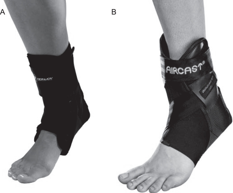
Participants were then fitted for a series of braces before completing the testing session. Brace conditions were no brace, a combination lace-up and figure-of-8 stabilizer brace (ankle-support brace; Performance Anaform Lace-Up Ankle Brace; DonJoy Orthopaedics; Figure 1A), and a brace with semirigid lateral stabilizers and increased midfoot support (semirigid brace; AirCast AirLift PTTD Brace; DonJoy Orthopaedics; Figure 1B). The braces were worn bilaterally, and the order of the brace conditions was randomized.
Figure 1.
A, Ankle-support brace (Performance Anaform Lace-Up Ankle Brace; DonJoy Orthopaedics) and B, semirigid brace (AirCast AirLift PTTD Brace; DonJoy Orthopaedics) worn during the study. DJO® is a registered trademark of DJO, LLC in the US and/or other countries. ©2020 DJO, LLC. Used with permission from DJO, LLC. All rights reserved.
Procedures
Participants were instructed to use a neutral cushioned running shoe (Zoom Pegasus; Nike Inc) during all testing to control for footwear effects.22 For each of the 3 brace conditions, they completed walking, running, and cutting trials (7 of each) in randomized order. The side cut was performed by planting on either foot. The planted foot remained the same for each trial, and this foot was used for analysis across all conditions. Participants completed 3 walking and running practice trials at a self-selected pace, determined by timing gates (TC Timing System; Brower Timing Systems) set 6 m apart, and we calculated the average pace from these trials. Trial completion speed for all subsequent trials was held to within 5% of the average pace in the practice trials. The 3 tasks and 3 brace conditions were combined for a total of 9 conditions. Volitional breaks were provided to prevent fatigue.
The plantar-loading variables of contact area, maximum force, force-time integral (FTI), and COP were evaluated during the dynamic trials using the pedar-X system (Novel Inc) sampling at 50 Hz. Novel Multiproject software was used with an 8-region mask,7,23 and plantar loading was evaluated in the medial and lateral midfoot and the medial, middle, and lateral forefoot during walking, running, and cutting for the foot that participants used to plant during the side cut. Additionally, individuals rated brace comfort on a Likert scale from 1 (least comfortable) to 10 (most comfortable).
Statistical Analysis
We examined the plantar-loading variables for each region of the foot and in each condition, with maximum force normalized to body weight and contact area normalized to the contact area of the entire insole (normalized insole contact area).23 After verifying that all other analysis of variance (ANOVA) assumptions were met, we log transformed the data to produce a normal distribution of variables. A 3 × 3 mixed-model repeated-measures ANOVA, with the α level set at .05, was conducted to assess the effects of brace (within-participant factor) and foot type (between-participants factor) on all variables (SPSS version 26; IBM Corp). Bonferroni-adjusted pairwise comparisons were calculated to determine the simple main effects of brace and foot type on any variables for which a main effect was identified. We evaluated COP displacement for the walking and running trials using the coefficient of multiple correlation (CMC)24 and statistical parametric mapping (SPM)25,26 in MATLAB (The MathWorks) to determine if brace wear had an effect on medial-lateral (ML) or anterior-posterior (AP) COP displacement. The COP curves were separated into AP and ML components for evaluation, as is common for analyses of this type.25,26 Finally, the ordinal data for the brace-comfort ratings were compared using the Wilcoxon signed rank test.
RESULTS
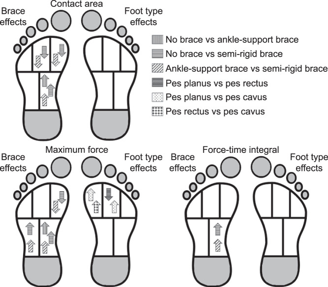
A total of 36 participants (11 men, 25 women; age = 23.1 ± 2.5 years, height = 1.72 ± 0.09 m, mass = 66.3 ± 14.7 kg, arch height index = 0.346 ± 0.035) completed the entire testing protocol. Results from the 3 × 3 mixed-model repeated-measures ANOVA revealed that both brace and foot type had main effects on certain variables. During walking, no foot type-by-brace interaction existed (Figure 2, Table 1). We observed a main effect of brace on contact area in the medial midfoot (P = .005,  = 0.280), medial forefoot (P = .001,
  = 0.338), and middle forefoot (P = .007,
  = 0.264). Brace type also affected maximum force in the medial midfoot (P < .001,
  = 0.710), lateral midfoot (P < .001,
  = 0.561), and medial forefoot (P = .01,
  = 0.247) and FTI in the medial midfoot (P < .001,
  = 0.476). Overall, the semirigid brace condition increased values in the midfoot and decreased them in the forefoot compared with the no-brace or ankle-support–brace condition. We observed a main effect of foot type during walking for maximum force in the medial forefoot (P = .01,
  = 0.229) and middle forefoot (P = .003,
  = 0.302). In general, compared with the pes rectus and pes planus foot, the pes cavus foot had smaller loads in the midfoot and greater loads in the forefoot in both the brace and no-brace conditions.
Figure 2.
Results from a 3 × 3 mixed-model repeated-measures analysis of variance and post hoc testing comparing the effects of brace condition and foot type on the contact area, maximum force, and force-time integral in the 5 regions of interest during the walking task.
Table 1.
Effects of Brace Condition and Foot Type on the Contact Area, Maximum Force, and Force-Time Integral in the 5 Regions of Interest During the Walking Task
| Variable |
Foot Type, Mean ± SD |
Main Effect |
|||||||||||||
| Pes Planus |
Pes Rectus |
Pes Cavus |
Interaction |
Brace |
Foot Type |
||||||||||
| No Brace |
Ankle-Support Brace |
Semirigid Brace |
No Brace |
Ankle-Support Brace |
Semirigid Brace |
No Brace |
Ankle-Support Brace |
Semirigid Brace |
P Value |
 Value Value |
P Value |
 Value Value |
P Value |
 Value Value |
|
| Contact area, normalized insole contact area | |||||||||||||||
| Medial midfoota–c | 0.140 ± 0.042 | 0.144 ± 0.033 | 0.155 ± 0.006 | 0.137 ± 0.024 | 0.142 ± 0.025 | 0.155 ± 0.004 | 0.129 ± 0.021 | 0.138 ± 0.015 | 0.154 ± 0.004 | .93 | 0.013 | .005 | 0.280d | .92 | 0.005 |
| Lateral midfoot | 0.161 ± 0.007 | 0.162 ± 0.005 | 0.162 ± 0.003 | 0.163 ± 0.007 | 0.162 ± 0.008 | 0.161 ± 0.003 | 0.165 ± 0.005 | 0.165 ± 0.005 | 0.160 ± 0.002 | .09 | 0.112 | .07 | 0.152 | .66 | 0.025 |
| Medial forefootb,c | 0.077 ± 0.002 | 0.078 ± 0.002 | 0.072 ± 0.006 | 0.077 ± 0.007 | 0.078 ± 0.004 | 0.073 ± 0.007 | 0.079 ± 0.004 | 0.078 ± 0.004 | 0.076 ± 0.006 | .67 | 0.034 | .001 | 0.338d | .46 | 0.045 |
| Middle forefootb,c | 0.093 ± 0.005 | 0.093 ± 0.006 | 0.092 ± 0.001 | 0.094 ± 0.005 | 0.094 ± 0.005 | 0.092 ± 0.002 | 0.095 ± 0.003 | 0.095 ± 0.004 | 0.092 ± 0.002 | .84 | 0.021 | .007 | 0.264d | .73 | 0.019 |
| Lateral forefoot | 0.085 ± 0.007 | 0.086 ± 0.005 | 0.085 ± 0.002 | 0.087 ± 0.003 | 0.084 ± 0.008 | 0.085 ± 0.001 | 0.087 ± 0.003 | 0.086 ± 0.002 | 0.084 ± 0.002 | .23 | 0.079 | .14 | 0.116 | .84 | 0.010 |
| Maximum force, body weight | |||||||||||||||
| Medial midfoota–c | 0.171 ± 0.065 | 0.173 ± 0.063 | 0.236 ± 0.039 | 0.120 ± 0.036 | 0.132 ± 0.047 | 0.201 ± 0.055 | 0.094 ± 0.024 | 0.107 ± 0.016 | 0.176 ± 0.023 | .72 | 0.031 | <.001 | 0.710d | .09 | 0.134 |
| Lateral midfootb,c | 0.230 ± 0.033 | 0.235 ± 0.036 | 0.269 ± 0.040 | 0.220 ± 0.048 | 0.217 ± 0.032 | 0.261 ± 0.032 | 0.198 ± 0.043 | 0.220 ± 0.052 | 0.240 ± 0.048 | .49 | 0.050 | <.001 | 0.561d | .28 | 0.073 |
| Medial forefootb,c,e,f | 0.164 ± 0.038 | 0.149 ± 0.031 | 0.125 ± 0.038 | 0.176 ± 0.055 | 0.168 ± 0.063 | 0.151 ± 0.064 | 0.234 ± 0.077 | 0.242 ± 0.088 | 0.228 ± 0.083 | .56 | 0.044 | .01 | 0.247d | .01 | 0.229d |
| Middle forefoote,g | 0.206 ± 0.060 | 0.209 ± 0.043 | 0.194 ± 0.044 | 0.262 ± 0.039 | 0.260 ± 0.046 | 0.243 ± 0.049 | 0.276 ± 0.054 | 0.277 ± 0.062 | 0.260 ± 0.057 | .92 | 0.014 | .13 | 0.119 | .003 | 0.302 |
| Lateral forefoot | 0.153 ± 0.050 | 0.164 ± 0.052 | 0.164 ± 0.042 | 0.201 ± 0.051 | 0.203 ± 0.049 | 0.213 ± 0.048 | 0.185 ± 0.045 | 0.193 ± 0.055 | 0.173 ± 0.047 | .30 | 0.070 | .15 | 0.111 | .07 | 0.147d |
| Force-time integral, N·s | |||||||||||||||
| Medial midfootb,c | 130.3 ± 182.9 | 127.5 ± 157.9 | 199.7 ± 261.3 | 67.8 ± 65.1 | 99.2 ± 105.9 | 173.9 ± 147.4 | 81.0 ± 100.1 | 108.3 ± 103.1 | 127.4 ± 134.1 | .75, | 0.028 | <.001 | 0.476d | .94 | 0.004 |
| Lateral midfoot | 187.7 ± 207.9 | 198.5 ± 239.7 | 216.9 ± 267.3 | 168.2 ± 174.5 | 174.2 ± 165.3 | 226.5 ± 181.1 | 132.5 ± 143.3 | 174.3 ± 052.3 | 147.8 ± 146.1 | .61 | 0.040 | .20 | 0.095 | .81 | 0.013 |
| Medial forefoot | 81.3 ± 75.7 | 74.7 ± 83.1 | 48.6 ± 42.0 | 80.5 ± 72.7 | 84.2 ± 80.1 | 76.7 ± 64.0 | 116.9 ± 134.2 | 145.6 ± 145.6 | 110.4 ± 129.1 | .59 | 0.041 | .35 | 0.064 | .60 | 0.031 |
| Middle forefoot | 108.8 ± 101.6 | 105.1 ± 104.4 | 104.9 ± 115.2 | 129.3 ± 114.0 | 140.8 ± 123.0 | 140.4 ± 114.1 | 156.4 ± 185.1 | 198.4 ± 188.5 | 145.1 ± 175.0 | .73 | 0.030 | .62 | 0.030 | .59 | 0.032 |
| Lateral forefoot | 77.3 ± 72.0 | 80.1 ± 84.5 | 72.4 ± 64.6 | 120.2 ± 125.3 | 111.1 ± 104.8 | 128.2 ± 108.3 | 110.5 ± 126.9 | 143.6 ± 132.8 | 99.8 ± 118.8 | .56 | 0.043 | .71 | 0.021 | .51 | 0.040 |
Difference between no-brace and ankle-support–brace conditions.
Difference between no-brace and semirigid-brace conditions.
Difference between ankle-support–brace and semirigid-brace conditions.
Indicates difference (P < .05).
Difference between pes planus and pes cavus.
Difference between pes rectus and pes cavus.
Difference between pes planus and pes rectus.
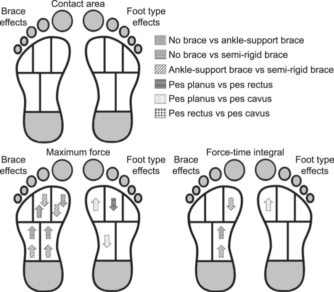
During running, we observed a brace-by-foot type interaction for the maximum force in the medial forefoot (P = .045,  = 0.136; Figure 3, Table 2). A main effect of brace on the maximum force existed in the medial midfoot (P < .001,
  = 0.827), lateral midfoot (P < .001,
  = 0.379), and middle forefoot (P = .001,
  = 0.349). Brace main effects also were present for the FTI in the medial midfoot (P < .001,
  = 0.570) and medial forefoot (P = .006,
  = 0.276). Compared with the no-brace and ankle-support–brace conditions, the semirigid-brace condition usually resulted in greater loads in the midfoot and smaller loads in the forefoot. Maximum force was different in the medial midfoot (P = .04,
  = 0.179) and middle forefoot (P = .01,
  = 0.235) based on foot type. Foot type also affected the FTI in the medial forefoot (P = .03,
  = 0.188) and middle forefoot (P = .047,
  = 0.169). Overall, values were decreased in the midfoot and increased in the forefoot with pes cavus.
Figure 3.
Results from a 3 × 3 mixed-model repeated-measures analysis of variance and post hoc testing comparing the effects of brace condition and foot type on the contact area, maximum force, and force-time integral in the 5 regions of interest during the running task.
Table 2.
Effects of Brace Condition and Foot Type on the Contact Area, Maximum Force, and Force-Time Integral in the 5 Regions of Interest During the Running Task
| Variable |
Foot Type, Mean ± SD |
Main Effect |
|||||||||||||
| Pes Planus |
Pes Rectus |
Pes Cavus |
Interaction |
Brace |
Foot Type |
||||||||||
| No Brace |
Ankle-Support Brace |
Semirigid Brace |
No Brace |
Ankle-Support Brace |
Semirigid Brace |
No Brace |
Ankle-Support Brace |
Semirigid Brace |
P Value |
 Value Value |
P Value |
 Value Value |
P Value |
 Value Value |
|
| Contact area, normalized insole contact area | |||||||||||||||
| Medial midfoot | 0.151 ± 0.013 | 0.154 ± 0.007 | 0.158 ± 0.003 | 0.157 ± 0.004 | 0.159 ± 0.005 | 0.159 ± 0.006 | 0.153 ± 0.009 | 0.154 ± 0.008 | 0.157 ± 0.002 | .45 | 0.054 | .054 | 0.167 | .11 | 0.128 |
| Lateral midfoot | 0.162 ± 0.006 | 0.160 ± 0.004 | 0.162 ± 0.004 | 0.160 ± 0.011 | 0.160 ± 0.007 | 0.162 ± 0.007 | 0.161 ± 0.002 | 0.160 ± 0.003 | 0.160 ± 0.001 | .36 | 0.063 | .32 | 0.069 | .93 | 0.005 |
| Medial forefoot | 0.075 ± 0.006 | 0.077 ± 0.001 | 0.075 ± 0.009 | 0.079 ± 0.003 | 0.079 ± 0.003 | 0.079 ± 0.003 | 0.078 ± 0.001 | 0.078 ± 0.001 | 0.078 ± 0.001 | .64 | 0.037 | .45 | 0.049 | .08 | 0.142 |
| Middle forefoot | 0.091 ± 0.002 | 0.092 ± 0.003 | 0.093 ± 0.003 | 0.092 ± 0.003 | 0.093 ± 0.003 | 0.092 ± 0.004 | 0.092 ± 0.001 | 0.092 ± 0.001 | 0.092 ± 0.001 | .37 | 0.062 | .68 | 0.024 | .84 | 0.011 |
| Lateral forefoot | 0.085 ± 0.003 | 0.084 ± 0.002 | 0.085 ± 0.002 | 0.086 ± 0.003 | 0.084 ± 0.008 | 0.086 ± 0.003 | 0.084 ± 0.002 | 0.084 ± 0.002 | 0.084 ± 0.001 | .82 | 0.022 | .62 | 0.030 | .92 | 0.005 |
| Maximum force, body weight | |||||||||||||||
| Medial midfoota–c | 0.324 ± 0.092 | 0.334 ± 0.093 | 0.400 ± 0.073 | 0.275 ± 0.069 | 0.279 ± 0.064 | 0.359 ± 0.083 | 0.219 ± 0.043 | 0.232 ± 0.048 | 0.327 ± 0.057 | .15 | 0.096 | <.001 | 0.827d | .04 | 0.179d |
| Lateral midfoota,b | 0.390 ± 0.085 | 0.424 ± 0.099 | 0.443 ± 0.075 | 0.364 ± 0.098 | 0.369 ± 0.097 | 0.419 ± 0.087 | 0.319 ± 0.050 | 0.338 ± 0.051 | 0.376 ± 0.055 | .75 | 0.028 | <.001 | 0.379d | .20 | 0.093 |
| Medial forefoota–c | 0.234 ± 0.066 | 0.237 ± 0.046 | 0.183 ± 0.050 | 0.312 ± 0.094 | 0.312 ± 0.105 | 0.277 ± 0.095 | 0.319 ± 0.091 | 0.331 ± 0.091 | 0.304 ± 0.063 | .045 | 0.136d | <.001 | 0.497 | .03 | 0.195 |
| Middle forefootb,e,f | 0.283 ± 0.078 | 0.305 ± 0.065 | 0.265 ± 0.072 | 0.382 ± 0.082 | 0.393 ± 0.079 | 0.376 ± 0.085 | 0.372 ± 0.107 | 0.381 ± 0.100 | 0.351 ± 0.080 | .42 | 0.056 | .001 | 0.349d | .01 | 0.235d |
| Lateral forefoot | 0.214 ± 0.083 | 0.230 ± 0.073 | 0.227 ± 0.070 | 0.246 ± 0.067 | 0.255 ± 0.082 | 0.268 ± 0.067 | 0.243 ± 0.064 | 0.248 ± 0.059 | 0.227 ± 0.054 | .20 | 0.086 | .19 | 0.098 | .48 | 0.043 |
| Force-time integral, N·s | |||||||||||||||
| Medial midfoota,b | 23.9 ± 10.8 | 26.6 ± 12.2 | 35.0 ± 13.7 | 24.6 ± 10.4 | 26.5 ± 14.3 | 43.4 ± 39.9 | 38.1 ± 61.8 | 50.4 ± 66.0 | 63.3 ± 75.5 | .78 | 0.026 | <.001 | 0.570d | .73 | 0.019 |
| Lateral midfoot | 36.2 ± 11.7 | 40.3 ± 14.3 | 39.1 ± 9.9 | 41.3 ± 16.6 | 40.2 ± 19.1 | 51.5 ± 40.4 | 57.5 ± 87.5 | 71.9 ± 88.9 | 70.9 ± 89.9 | .57 | 0.043 | .07 | 0.151 | .82 | 0.012 |
| Medial forefootb,c | 23.0 ± 14.2 | 23.3 ± 11.1 | 17.4 ± 9.3 | 34.8 ± 13.8 | 35.2 ± 16.1 | 32.9 ± 16.5 | 60.7 ± 89.9 | 76.7 ± 109.7 | 55.5 ± 60.9 | .18 | 0.089 | .006 | 0.276d | .03 | 0.188d |
| Middle forefoot | 27.4 ± 16.0 | 30.7 ± 15.9 | 26.6 ± 15.0 | 45.1 ± 17.5 | 46.9 ± 19.8 | 47.6 ± 23.2 | 83.4 ± 150.2 | 106.8 ± 176.0 | 77.5 ± 105.6 | .64 | 0.037 | .07 | 0.151 | .047 | 0.169d |
| Lateral forefoot | 19.2 ± 8.5 | 22.2 ± 11.6 | 19.9 ± 9.0 | 31.1 ± 12.8 | 31.0 ± 15.8 | 34.2 ± 17.7 | 55.8 ± 101.1 | 70.7 ± 111.5 | 52.0 ± 74.1 | .50 | 0.049 | .41 | 0.054 | .18 | 0.099 |
Difference between no brace and semirigid brace conditions.
Difference between ankle-support brace and semirigid brace conditions.
Difference between pes planus and pes cavus.
Indicates difference (P < .05).
Difference between no brace and ankle-support brace conditions.
Difference between pes planus and pes rectus.
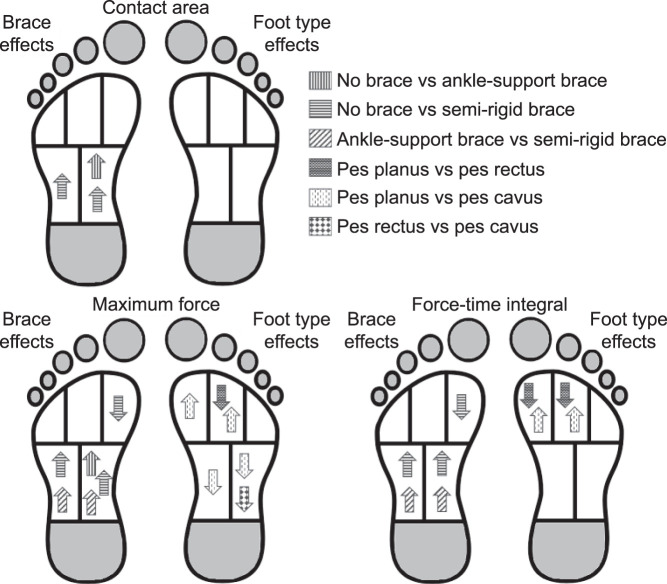
During cutting, we found no interaction between brace and foot type (Figure 4, Table 3). A main effect of brace on contact area was evident in the medial midfoot (P = .01,  = 0.234) and lateral midfoot (P = .006,
  = 0.275) and on maximum force in the medial midfoot (P < .001,
  = 0.785), lateral midfoot (P < .001,
  = 0.546), and medial forefoot (P = .043,
  = 0.178). We noted a main effect of brace on the FTI in the medial midfoot (P < .001,
  = 0.786), lateral midfoot (P < .001,
  = 0.465), and medial forefoot (P = .02,
  = 0.230). On average, the semirigid-brace condition resulted in smaller loads in the forefoot and greater loads in the midfoot compared with the no-brace and ankle-support–brace conditions. We demonstrated a main effect of foot type on maximum force in the medial midfoot (P = .01,
  = 0.232), lateral midfoot (P = .001,
  = 0.341), medial forefoot (P = .004,
  = 0.282), and middle forefoot (P = .02,
  = 0.221) and a main effect of the FTI on the medial forefoot (P = .005,
  = 0.276) and middle forefoot (P = .008,
  = 0.256). A higher arch usually led to decreased loads in the midfoot and increased loads in the forefoot.
Figure 4.
Results from a 3 × 3 mixed-model repeated-measures analysis of variance and post hoc testing comparing the effects of brace condition and foot type on the contact area, maximum force, and force-time integral in the 5 regions of interest during the cutting task.
Table 3.
Effects of Brace Condition and Foot Type on the Contact Area, Maximum Force, and Force-Time Integral in the 5 Regions of Interest During the Cutting Task
| Variable |
Foot Type, Mean ± SD |
Interaction |
Main Effect |
||||||||||||
| Pes Planus |
Pes Rectus |
Pes Cavus |
|||||||||||||
| Brace |
Foot Type |
||||||||||||||
| No Brace |
Ankle-Support Brace |
Semirigid Brace |
No Brace |
Ankle-Support Brace |
Semirigid Brace |
No Brace |
Ankle-Support Brace |
Semirigid Brace |
P Value |
 Value Value |
P Value |
 Value Value |
P Value |
 Value Value |
|
| Contact area, normalized insole contact area | |||||||||||||||
| Medial midfoota,b | 0.153 ± 0.012 | 0.155 ± 0.005 | 0.160 ± 0.005 | 0.154 ± 0.007 | 0.156 ± 0.008 | 0.158 ± 0.003 | 0.154 ± 0.007 | 0.155 ± 0.004 | 0.157 ± 0.003 | .83 | 0.022 | .01 | 0.234c | .98 | 0.001 |
| Lateral midfootb | 0.161 ± 0.005 | 0.160 ± 0.002 | 0.163 ± 0.005 | 0.158 ± 0.005 | 0.156 ± 0.013 | 0.161 ± 0.002 | 0.159 ± 0.002 | 0.158 ± 0.003 | 0.160 ± 0.003 | .92 | 0.014 | .006 | 0.275c | .26 | 0.079 |
| Medial forefoot | 0.076 ± 0.006 | 0.077 ± 0.003 | 0.073 ± 0.009 | 0.077 ± 0.002 | 0.077 ± 0.004 | 0.076 ± 0.004 | 0.077 ± 0.002 | 0.076 ± 0.004 | 0.077 ± 0.002 | .43 | 0.056 | .19 | 0.100 | .43 | 0.050 |
| Middle forefoot | 0.092 ± 0.004 | 0.092 ± 0.002 | 0.093 ± 0.004 | 0.091 ± 0.002 | 0.092 ± 0.003 | 0.091 ± 0.001 | 0.092 ± 0.002 | 0.092 ± 0.003 | 0.092 ± 0.002 | .055 | 0.129 | .28 | 0.077 | .53 | 0.038 |
| Lateral forefoot | 0.085 ± 0.002 | 0.084 ± 0.001 | 0.086 ± 0.003 | 0.083 ± 0.004 | 0.082 ± 0.008 | 0.084 ± 0.003 | 0.084 ± 0.002 | 0.085 ± 0.002 | 0.084 ± 0.002 | .68 | 0.034 | .16 | 0.109 | .29 | 0.072 |
| Maximum force, body weight | |||||||||||||||
| Medial midfoota,b,d,e | 0.366 ± 0.126 | 0.386 ± 0.107 | 0.448 ± 0.088 | 0.289 ± 0.088 | 0.301 ± 0.088 | 0.407 ± 0.097 | 0.224 ± 0.051 | 0.240 ± 0.049 | 0.348 ± 0.050 | .09 | 0.112 | <.001 | 0.785c | .01 | 0.232c |
| Lateral midfootb,d–f | 0.344 ± 0.078 | 0.352 ± 0.077 | 0.407 ± 0.083 | 0.298 ± 0.080 | 0.275 ± 0.086 | 0.344 ± 0.066 | 0.203 ± 0.035 | 0.214 ± 0.052 | 0.268 ± 0.053 | .11 | 0.107 | <.001 | 0.546c | .001 | 0.341c |
| Medial forefootb,e | 0.262 ± 0.061 | 0.243 ± 0.067 | 0.210 ± 0.069 | 0.367 ± 0.126 | 0.364 ± 0.142 | 0.332 ± 0.125 | 0.430 ± 0.141 | 0.431 ± 0.142 | 0.439 ± 0.129 | .21 | 0.084 | .043 | 0.178c | .004 | 0.282c |
| Middle forefoote,g | 0.272 ± 0.045 | 0.265 ± 0.051 | 0.268 ± 0.067 | 0.347 ± 0.082 | 0.363 ± 0.097 | 0.360 ± 0.100 | 0.373 ± 0.098 | 0.366 ± 0.104 | 0.350 ± 0.094 | .54 | 0.045 | .85 | 0.010 | .02 | 0.221c |
| Lateral forefoot | 0.224 ± 0.056 | 0.218 ± 0.054 | 0.219 ± 0.046 | 0.221 ± 0.063 | 0.220 ± 0.065 | 0.226 ± 0.064 | 0.209 ± 0.049 | 0.193 ± 0.057 | 0.189 ± 0.052 | .59 | 0.041 | .19 | 0.100 | .44 | 0.048 |
| Force-time integral, N·s | |||||||||||||||
| Medial midfootb,d | 34.6 ± 17.0 | 34.6 ± 14.7 | 48.7 ± 19.4 | 29.6 ± 12.4 | 32.2 ± 13.2 | 46.0 ± 14.3 | 26.5 ± 14.7 | 26.3 ± 10.8 | 42.8 ± 14.8 | .54 | 0.045 | <.001 | 0.786c | .50 | 0.042 |
| Lateral midfootb,d | 35.7 ± 13.3 | 35.7 ± 12.7 | 41.5 ± 13.8 | 33.3 ± 13.5 | 32.0 ± 15.0 | 37.6 ± 13.7 | 26.1 ± 13.7 | 25.8 ± 9.7 | 31.1 ± 10.7 | .72 | 0.031 | <.001 | 0.465c | .24 | 0.083 |
| Medial forefootb,e,g | 50.9 ± 23.9 | 27.4 ± 15.3 | 23.5 ± 11.2 | 30.5 ± 18.1 | 51.4 ± 24.0 | 46.5 ± 22.1 | 59.4 ± 31.4 | 57.8 ± 27.5 | 58.4 ± 27.4 | .15 | 0.095 | .02 | 0.230c | .005 | 0.276c |
| Middle forefoote,g | 30.6 ± 19.4 | 28.6 ± 18.0 | 31.3 ± 17.7 | 46.1 ± 13.5 | 49.4 ± 16.6 | 49.2 ± 16.6 | 49.7 ± 24.8 | 46.7 ± 21.9 | 47.2 ± 19.5 | .34 | 0.066 | .78 | 0.016 | .008 | 0.256c |
| Lateral forefoot | 23.2 ± 12.7 | 21.4 ± 11.3 | 22.4 ± 10.1 | 30.8 ± 10.5 | 31.1 ± 11.4 | 30.8 ± 11.1 | 26.8 ± 13.2 | 23.8 ± 11.0 | 24.7 ± 11.4 | .58 | 0.042 | .12 | 0.124 | .16 | 0.106 |
Difference between no-brace and ankle-support–brace conditions.
Difference between no-brace and semirigid-brace conditions.
Indicates difference.
Difference between ankle-support–brace and semirigid-brace conditions.
Difference between pes planus and pes cavus.
Difference between pes rectus and pes cavus.
Difference between pes planus and pes rectus.
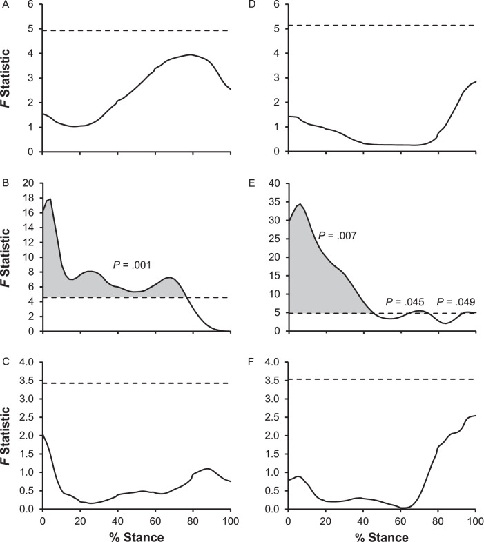
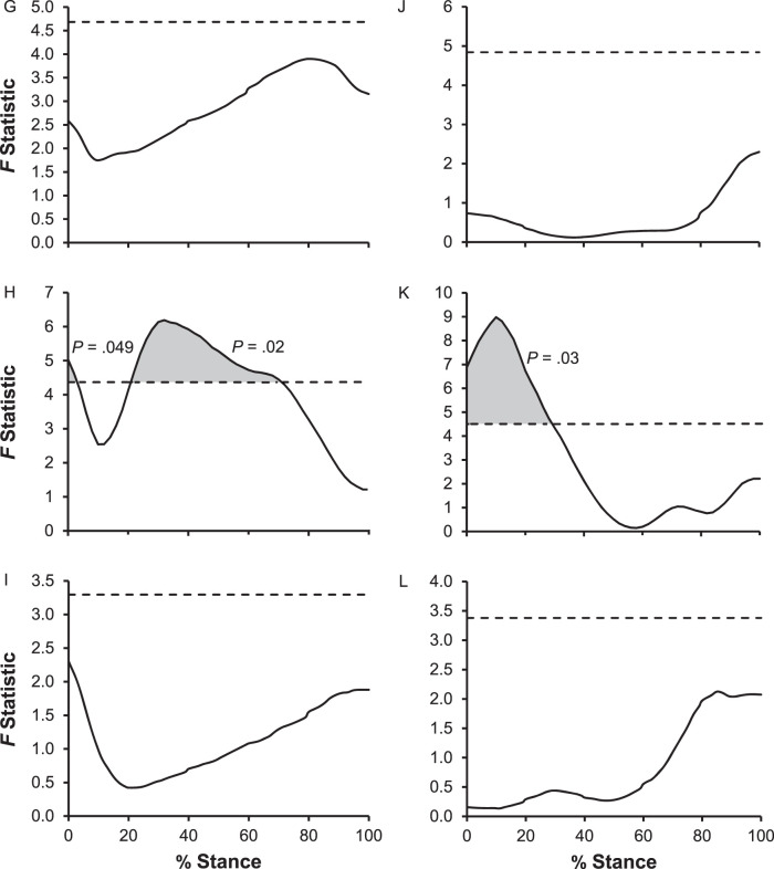
A CMC analysis of the COP displacement curves revealed that the tracings of the COP were highly correlated across brace conditions (Table 4). This was shown in mean CMC values being close to 1 for all foot types in both the walking and running tasks. However, an SPM analysis using a mixed-model repeated-measures ANOVA revealed that brace condition resulted in different COP displacements for certain portions of the COP tracing during both the walking and running tasks (P < .05; Figure 5, Table 5). Specifically, brace condition affected heel strike and midstance in the ML direction and heel strike in the AP direction (P < .05; Figure 5, Table 5). Neither foot type nor the foot type-by-brace interaction had an effect on any portion of the COP curves.
Table 4.
Correlation of Multiple Coefficient Outputs for Comparison of Center-of-Pressure Curves Among Brace Conditions
| Task |
Foot Type |
Center-of-Pressure Curve Direction, Mean ± SDa |
|
| Medial-Lateral |
Anterior-Posterior |
||
| Walking | Pes planus | 0.933 ± 0.071 | 0.991 ± 0.005 |
| Pes rectus | 0.948 ± 0.050 | 0.992 ± 0.009 | |
| Pes cavus | 0.955 ± 0.042 | 0.989 ± 0.007 | |
| Running | Pes planus | 0.958 ± 0.040 | 0.992 ± 0.008 |
| Pes rectus | 0.934 ± 0.074 | 0.987 ± 0.022 | |
| Pes cavus | 0.961 ± 0.042 | 0.979 ± 0.034 | |
Values close to 1 indicate similar curves, and values close to 0 indicate dissimilar curves.
Figure 5.
Graphic representation of statistical parametric mapping analysis of the center of pressure as a percentage of the stance phase, with effects indicated by the shaded regions and associated P values. A, Foot type, B, brace condition, and, C, interaction results for the medial-lateral direction during the walking task. D, Foot type, E, brace condition, and, F, interaction results for the anterior-posterior direction during the walking task. G, Foot type, H, brace condition, and, I, interaction results for the medial-lateral direction during the running task. J, Foot type, K, brace condition, and, L, interaction results for the anterior-posterior direction during the running task. Continued on next page.
Figure 5.
Continued from previous page.
Table 5.
Statistical Parametric Mapping Outputs of a 3 × 3 Repeated-Measures Analysis of Variance Indicating the Regions of Difference in Center-of-Pressure Traces Based on Each Factor
| Task |
Center-of-Pressure Direction |
Factor |
|||
| Foot Type |
Brace Condition |
Interaction |
|||
| Region of Difference, % Stance Range |
P Value |
||||
| Walking | Medial-lateral | NA | 0.0–76.6 | .001 | NA |
| Anterior-posterior | NA | 0.0–45.5 | .007 | NA | |
| NA | 63.9–74.6 | .045 | NA | ||
| NA | 92.9–98.0 | .049 | NA | ||
| Running | Medial-lateral | NA | 0.0–3.0 | .049 | NA |
| NA | 20.9–71.0 | .02 | NA | ||
| Anterior-posterior | NA | 0.0–29.2 | .03 | NA | |
Abbreviation: NA, not applicable.
The Wilcoxon signed rank test between comfort ratings for the ankle-support–brace and semirigid-brace conditions indicated that participants found the ankle-support brace more comfortable than the semirigid brace (P = .03). However, no differences in comfort ratings between the braces based on foot-type classification were seen.
DISCUSSION
The purpose of our study was to determine the effects of 2 ankle braces on plantar loading in different foot types during 3 movement tasks. This was accomplished by analyzing plantar-loading variables in 5 regions of the foot during walking, running, and cutting. We hypothesized that ankle braces would help reduce the irregular plantar loads from abnormal foot postures by shifting plantar-loading patterns medially in individuals with pes cavus and laterally in individuals with pes planus.
We found that foot type indeed affected the plantar-loading variables of maximum force and the FTI, largely independent of brace condition. Specifically, maximum force in the middle forefoot was affected by foot type during walking, running, and cutting; in the medial forefoot during walking and cutting; in the medial midfoot during running and cutting; and in the lateral midfoot during cutting. Post hoc testing revealed that most of these differences were between individuals with pes planus and pes cavus. This finding is in agreement with the results of previous studies, in which researchers5–7 found that maximum force in the medial midfoot was greater for individuals with pes planus and less for those with pes cavus and that individuals with pes planus had reduced forces in the forefoot. We also observed that the FTI was affected by foot type in the medial forefoot and middle forefoot during running and cutting. This outcome agrees with earlier work, in which investigators5,6,9 reported that the FTI was less in the forefoot of individuals with pes planus and more in the forefoot of individuals with pes cavus. However, previous authors5,6,9 also noted that the FTI in the midfoot and lateral forefoot was affected by different foot types, which was not the case in our participants. Furthermore, we did not identify any differences in contact area due to foot type for any region of the foot during any task. This result was not consistent with earlier work in which researchers5–7 demonstrated that individuals with pes planus had increased contact area in the medial midfoot and lateral forefoot and that those with pes cavus also had increased contact area in the lateral forefoot versus those with pes rectus. This lack of agreement could be because the brace conditions resulted in fewer differences among foot types. Despite inconsistent agreement with other studies, we detected differences in force distribution due to foot type during different athletic tasks. Investigators5,8 have shown that this contributes to the increased risk of injury that is related to abnormal foot postures, which highlights the need for a preventive measure.
One intervention that could be used to prevent lower extremity injuries by reducing irregular plantar loads is ankle braces. The risk of injuries could be reduced if ankle braces bring the plantar loading of individuals with pes planus and pes cavus to a more neutral position, where the foot best attenuates force. Therefore, we also examined the effects of an ankle-support brace and a semirigid brace on plantar loading during various athletic tasks. Both the ankle-support and semirigid braces increased the contact area in the midfoot during walking and cutting and decreased the contact area in the medial and middle forefoot during walking, independent of foot type. Ankle braces also increased the maximum force in the medial and lateral midfoot during walking, running, and cutting and decreased the maximum force in the medial forefoot during all tasks. Finally, the FTI in the medial midfoot was increased by brace wear during all 3 tasks and was increased in the lateral midfoot and decreased in the medial forefoot during cutting. Overall, the contact area, maximum force, and FTI were increased in the midfoot and decreased in the forefoot in braced conditions, largely independent of foot type. Post hoc testing revealed that most of these differences occurred in the semirigid-brace condition when compared with both the no-brace and the ankle-support–brace conditions. Whereas no previous authors have assessed plantar-loading differences due to ankle braces, arch taping produced inconsistent results, affecting19 or not affecting27 plantar loading. Although bracing and taping reduced the risk of lower extremity injuries,16–18 ankle bracing was more effective.16,17,20 Despite our results not supporting the original hypothesis of a medial or lateral shift, brace wear did cause a redistribution of plantar loading. Future researchers should focus more specifically on how the altered plantar loads resulting from brace wear affect injury occurrence and severity.
Furthermore, in addition to the regional differences evident in the contact area, maximum force, and FTI, braces modified the COP distribution. The COP was analyzed in the AP and ML directions, as is common in studies of this nature,25,26 because the curves were being compared across multiple conditions. Whereas a CMC analysis showed that the COP distribution was highly correlated across the 3 brace conditions, the SPM analysis identified portions of the COP distribution that were affected by brace wear. In the AP direction, the brace affected the beginning of the COP tracing in both the walking and running tasks, with a larger portion of the curve being altered during walking. Thus, most of the brace effects in the AP direction were at heel strike, indicating that participants contacted the ground with a different heel-strike angle while wearing the brace. In the ML direction, the brace altered the middle portion of the curve during both walking and running, with a larger portion of the curve being affected by brace wear during the former. Hence, most of the ML changes due to brace wear occurred at midstance, which was likely a result of the brace controlling the pronation and supination of the foot during the greatest weight-bearing phase. The ML shifts are important to consider because individuals with pes planus have displayed a more medial COP and those with pes cavus had a more lateral COP than individuals with a normal foot posture.5 Although the SPM analysis did not indicate the direction of the shift for each foot type, the ML changes at midstance caused by the brace combined with the facts that braces are easily adjustable, can be applied without an athletic trainer, and are reusable17,18 supports the need for further research into the possibility of using ankle braces for injury prevention.
Our study had a few limitations. First, we included an unequal number of participants for each foot-type group, with 8 participants classified as having pes planus, 17 as having normal arches, and 11 as having pes cavus. Whereas this was expected because a larger portion of the population has normal arches,2 a more equally distributed sample could have resulted in a more robust analysis. Second, the AirLift PTTD Brace was designed with an inflatable pocket under the arch, which was not inflated during our study. This was done to prevent variations in inflation levels among participants from biasing the results but is not standard for the intended use of the brace. Third, not all participants were comfortable cutting, despite this being an inclusion criterion. Furthermore, the cutting speed for each participant was not standardized among trials. This could have resulted in speed variations that affected the plantar-loading measures. However, we minimized this risk by averaging plantar-loading values across multiple trials. Nevertheless, these aspects should be considered by future investigators.
CONCLUSIONS
Our study is one of the first analyses of the effects of ankle braces, combined with foot type, on plantar-loading variables. We determined that foot type affected plantar forces in the foot independent of brace wear, which agrees with previous work. Additionally, we found that ankle braces, specifically semirigid braces, altered COP and plantar forces in the midfoot and forefoot, independent of foot type. Although we could not draw conclusions about using ankle braces for preventing injuries related to foot type, these results support the use of braces to redistribute plantar loads. Therefore, additional work should be done to determine the effectiveness of ankle braces as an injury-prevention method and further quantify the effects of ankle braces on foot type.
REFERENCES
- 1.Nilsson MK, Friis R, Michaelsen MS, Jakobsen PA, Nielsen RO. Classification of the height and flexibility of the medial longitudinal arch of the foot. J Foot Ankle Res. 2012;5:3. doi: 10.1186/1757-1146-5-3. [DOI] [PMC free article] [PubMed] [Google Scholar]
- 2.Subotnick SI. The biomechanics of running: implications for the prevention of foot injuries. Sports Med. 1985;2(2):144–153. doi: 10.2165/00007256-198502020-00006. [DOI] [PubMed] [Google Scholar]
- 3.Razeghi M, Batt ME. Foot type classification: a critical review of current methods. Gait Posture. 2002;15(3):282–291. doi: 10.1016/S0966-6362(01)00151-5. [DOI] [PubMed] [Google Scholar]
- 4.Franco AH. Pes cavus and pes planus: analyses and treatment. Phys Ther. 1987;67(5):688–694. doi: 10.1093/ptj/67.5.688. [DOI] [PubMed] [Google Scholar]
- 5.Buldt AK, Allan JJ, Landorf KB, Menz HB. The relationship between foot posture and plantar pressure during walking in adults: a systematic review. Gait Posture. 2018;62:56–67. doi: 10.1016/J.GAITPOST.2018.02.026. [DOI] [PubMed] [Google Scholar]
- 6.Buldt AK, Forghany S, Landorf KB, Levinger P, Murley GS, Menz HB. Foot posture is associated with plantar pressure during gait: a comparison of normal, planus and cavus feet. Gait Posture. 2018;62:235–240. doi: 10.1016/J.GAITPOST.2018.03.005. [DOI] [PubMed] [Google Scholar]
- 7.Chuckpaiwong B, Nunley JA, Mall NA, Queen RM. The effect of foot type on in-shoe plantar pressure during walking and running. Gait Posture. 2008;28(3):405–411. doi: 10.1016/j.gaitpost.2008.01.012. [DOI] [PubMed] [Google Scholar]
- 8.Tong JWK, Kong PW. Association between foot type and lower extremity injuries: systematic literature review with meta-analysis. J Orthop Sports Phys Ther. 2013;43(10):700–714. doi: 10.2519/jospt.2013.4225. [DOI] [PubMed] [Google Scholar]
- 9.Sneyers CJ, Lysens R, Feys H, Andries R. Influence of malalignment of feet on the plantar pressure pattern in running. Foot Ankle Int. 1995;16(10):624–632. doi: 10.1177/107110079501601008. [DOI] [PubMed] [Google Scholar]
- 10.Buldt AK, Levinger P, Murley GS, Menz HB, Nester CJ, Landorf KB. Foot posture is associated with kinematics of the foot during gait: a comparison of normal, planus and cavus feet. Gait Posture. 2015;42(1):42–48. doi: 10.1016/J.GAITPOST.2015.03.004. [DOI] [PubMed] [Google Scholar]
- 11.Buldt AK, Murley GS, Butterworth P, Levinger P, Menz HB, Landorf KB. The relationship between foot posture and lower limb kinematics during walking: a systematic review. Gait Posture. 2013;38(3):363–372. doi: 10.1016/J.GAITPOST.2013.01.010. [DOI] [PubMed] [Google Scholar]
- 12.Kaufman KR, Brodine SK, Shaffer RA, Johnson CW, Cullison TR. The effect of foot structure and range of motion on musculoskeletal overuse injuries. Am J Sports Med. 1999;27(5):585–593. doi: 10.1177/03635465990270050701. [DOI] [PubMed] [Google Scholar]
- 13.Morrison KE, Kaminski TW. Foot characteristics in association with inversion ankle injury. J Athl Train. 2007;42(1):135–142. [PMC free article] [PubMed] [Google Scholar]
- 14.Mei-Dan O, Kahn G, Zeev A, et al. The medial longitudinal arch as a possible risk factor for ankle sprains: a prospective study in 83 female infantry recruits. Foot Ankle Int. 2005;26(2):180–183. doi: 10.1177/107110070502600211. [DOI] [PubMed] [Google Scholar]
- 15.Williams DS, McClay IS, Hamill J. Arch structure and injury patterns in runners. Clin Biomech (Bristol Avon) 2001;16(4):341–347. doi: 10.1016/S0268-0033(01)00005-5. [DOI] [PubMed] [Google Scholar]
- 16.Verhagen EA, van Mechelen W, de Vente W. The effect of preventive measures on the incidence of ankle sprains. Clin J Sport Med. 2000;10(4):291–296. doi: 10.1097/00042752-200010000-00012. [DOI] [PubMed] [Google Scholar]
- 17.Dizon JMR, Reyes JJB. A systematic review on the effectiveness of external ankle supports in the prevention of inversion ankle sprains among elite and recreational players. J Sci Med Sport. 2010;13(3):309–317. doi: 10.1016/j.jsams.2009.05.002. [DOI] [PubMed] [Google Scholar]
- 18.Callaghan MJ. Role of ankle taping and bracing in the athlete. Br J Sports Med. 1997;31(2):102–108. doi: 10.1136/bjsm.31.2.102. [DOI] [PMC free article] [PubMed] [Google Scholar]
- 19.Newell T, Simon J, Docherty CL. Arch-taping techniques for altering navicular height and plantar pressures during activity. J Athl Train. 2015;50(8):825–832. doi: 10.4085/1062-6050-50.5.05. [DOI] [PMC free article] [PubMed] [Google Scholar]
- 20.Papadopoulos ES, Nicolopoulos C, Anderson EG, Curran M, Athanasopoulos S. The role of ankle bracing in injury prevention, athletic performance and neuromuscular control: a review of the literature. Foot. 2005;15(1):1–6. doi: 10.1016/J.FOOT.2004.07.002. [DOI] [Google Scholar]
- 21.Butler RJ, Hillstrom H, Song J, Richards CJ, Davis IS. Arch height index measurement system: establishment of reliability and normative values. J Am Podiatr Med Assoc. 2008;98(2):102–106. doi: 10.7547/0980102. [DOI] [PubMed] [Google Scholar]
- 22.Wiegerinck JI, Boyd J, Yoder JC, Abbey AN, Nunley JA, Queen RM. Differences in plantar loading between training shoes and racing flats at a self-selected running speed. Gait Posture. 2009;29(3):514–519. doi: 10.1016/j.gaitpost.2008.12.001. [DOI] [PubMed] [Google Scholar]
- 23.Queen RM, Haynes BB, Hardaker WM, Garrett WE. Forefoot loading during 3 athletic tasks. Am J Sports Med. 2007;35(4):630–636. doi: 10.1177/0363546506295938. [DOI] [PubMed] [Google Scholar]
- 24.Kadaba MP, Ramakrishnan HK, Wootten ME, Gainey J, Gorton G, Cochran GV. Repeatability of kinematic, kinetic, and electromyographic data in normal adult gait. J Orthop Res. 1989;7(6):849–860. doi: 10.1002/jor.1100070611. [DOI] [PubMed] [Google Scholar]
- 25.Vieira MF, de Brito AA, Lehnen GC, Rodrigues FB. Center of pressure and center of mass behavior during gait initiation on inclined surfaces: a statistical parametric mapping analysis. J Biomech. 2017;56:10–18. doi: 10.1016/j.jbiomech.2017.02.018. [DOI] [PubMed] [Google Scholar]
- 26.Pataky TC, Robinson MA, Vanrenterghem J, Savage R, Bates KT, Crompton RH. Vector field statistics for objective center-of-pressure trajectory analysis during gait, with evidence of scalar sensitivity to small coordinate system rotations. Gait Posture. 2014;40(1):255–258. doi: 10.1016/j.gaitpost.2014.01.023. [DOI] [PubMed] [Google Scholar]
- 27.Kim T, Park JC. Short-term effects of sports taping on navicular height, navicular drop and peak plantar pressure in healthy elite athletes: a within-subject comparison. Medicine (Baltimore) 2017;96(46):e8714. doi: 10.1097/MD.0000000000008714. [DOI] [PMC free article] [PubMed] [Google Scholar]


