Abstract
Background
Neurofilament light in serum (sNfL) is a biomarker for axonal damage with elevated levels in many neurological disorders, including neurodegenerative dementias. Since within-group variation of sNfL is large and concentrations increase with aging, sNfL’s clinical use in memory clinic practice remains to be established. The objective of the current study was to evaluate the clinical use of serum neurofilament light (sNfL), a cross-disease biomarker for axonal damage, in a tertiary memory clinic cohort.
Methods
Six neurologists completed questionnaires regarding the usefulness of sNfL (n = 5–42 questionnaires/neurologist). Patients that visited the Alzheimer Center Amsterdam for the first time between May and October 2019 (n = 109) were prospectively included in this single-center implementation study. SNfL levels were analyzed on Simoa and reported together with normal values in relation to age, as part of routine diagnostic work-up and in addition to cerebrospinal fluid (CSF) biomarker analysis.
Results
SNfL was perceived as useful in 53% (n = 58) of the cases. SNfL was more often perceived as useful in patients < 62 years (29/48, 60%, p = 0.05) and males (41/65, 63%, p < 0.01). Availability of CSF biomarker results at time of result discussion had no influence. We observed non-significant trends for increased perceived usefulness of sNfL for patients with the diagnosis subjective cognitive decline (64%), psychiatric disorder (71%), or uncertain diagnosis (67%). SNfL was mostly helpful to neurologists in confirming or excluding neurodegeneration. Whether sNfL was regarded as useful strongly depended on which neurologist filled out the questionnaire (ranging from 0 to 73% of useful cases/neurologist).
Discussion
Regardless of the availability of CSF biomarker results, sNfL was perceived as a useful tool in more than half of the evaluated cases in a tertiary memory clinic practice. Based on our results, we recommend the analysis of the biomarker sNfL to confirm or exclude neurodegeneration in patients below 62 years old and in males.
Supplementary Information
The online version contains supplementary material available at 10.1186/s13195-021-00841-4.
Keywords: Neurofilament light protein, Serum, Biomarkers, Dementia, Alzheimer’s disease, Subjective Cognitive Decline, Clinical implementation, Diagnostic work-up
Background
Neurofilament light (NfL) is a major protein of the axonal cytoskeleton and an accurate blood biomarker for axonal damage across the spectrum of neurological diseases [1]. Increased levels of NfL in cerebrospinal fluid have been reported in many neurodegenerative and neurological diseases with estimated fold-changes of 1–5 for the dementias in comparison to healthy controls [2, 3]. Recent technological advancements allow the ultrasensitive detection of NfL in blood [4], facilitating an exponential increase in studies across the field of neurology that identify blood NfL as powerful biomarker for diagnostic, prognostic, or disease monitoring purposes [5–9]. Also, for neurodegenerative dementias, including Alzheimer’s disease (AD) and frontotemporal dementia (FTD), higher levels of blood NfL are informative biomarkers to separate dementia from controls on a group level [10–18].
The positive correlation of NfL levels with increasing age [2, 16, 19] complicates the interpretation of NfL and has delayed the establishment of cut-off values for clinical practice. Also, NfL levels across the different types of neurodegenerative dementias are largely overlapping [2], questioning the clinical value of NfL on the individual patient level in memory clinic practice. Prospective studies are thus needed to evaluate the usefulness of blood NfL in the diagnostic work-up for dementia, similar to the evaluation studies that were performed for the classical AD biomarkers in cerebrospinal fluid (CSF) [20, 21].
We here aimed to evaluate the clinical use of serum NfL (sNfL) in a prospective study performed in a tertiary memory clinic setting. Patients visiting the memory clinic were sampled for NfL analysis and neurologists per patient indicated how they appreciated the sNfL result. We analyzed the frequency of NfL results perceived as useful and studied whether patient characteristics, i.e., age, sex, and diagnosis, were associated with an increased percentage of perceived usefulness of sNfL.
Methods
Patients
We prospectively and consecutively included patients that had their first visit at the memory clinic of the Alzheimer Center Amsterdam between May and October 2019. The Alzheimer Center Amsterdam is a tertiary center, which means that patients are referred for analysis of their cognitive complaints by their general practitioner or via their local specialist physician, in case of second or third opinions, as is customary in the Netherlands [22]. At their first visit, all patients received a standardized and multidisciplinary work-up, including medical and neurological examination including history taking and cognitive examination by a neurologist, assessment of vital functions, informant based history, neuropsychological investigation, brain magnetic resonance imaging (MRI), electroencephalogram (EEG), standard laboratory work, and lumbar puncture for collection of cerebrospinal fluid (CSF). Clinical diagnosis was made by consensus in a multidisciplinary meeting. Subjects were diagnosed with subjective cognitive decline (SCD; n = 28) when cognitive complaints were present but criteria for mild cognitive impairment (MCI), dementia, or any other neurological or psychiatric disorders were not met and all other examinations were normal [23]. Subjects were diagnosed with MCI (n = 5), of which n = 3 due to AD and n = 2 clinically defined without underlying neurodegenerative disorders, or dementia due to Alzheimer’s disease (AD; n = 29), according to the established National Institute on Aging - Alzheimer’s Association diagnostic guidelines [24, 25]. Subjects were diagnosed with other types of dementia according to clinical guidelines for frontotemporal dementia (FTD; n = 3) [26, 27], dementia with Lewy bodies (DLB; n = 3) [28], primary progressive aphasia (PPA; n = 2) [29], progressive supranuclear palsy (PSP; n = 2) [30, 31], or vascular dementia (VaD; n = 1) [32]. Patients were diagnosed with psychiatric disorders and referred to a psychiatrist for further examination when signs of neurological or neurodegenerative diseases could not be objectified (n = 17). Some patients were diagnosed with other neurological diseases (n = 11), due to drug abuse (n = 2), Parkinson’s disease (n = 1), cerebral amyloid angiopathy (n = 1), temporal epilepsy (n = 1), recent subdural hematoma in combination with corticobasal syndrome (n = 1), suspected hydrocephalus (n = 2) in combination with AD (n = 1) or vascular damage or psychiatric disorders (n = 1), suspected functional disorder after cerebrovascular accident (n = 1), or unknown cause (n = 2). For some patients the diagnosis could not be established after the first visit and was therefore postponed (n = 8), of which n = 6 had a syndrome diagnosis of MCI or dementia, n = 1 had a suspected functional disorder, and for n = 1, a severe language barrier impeded further testing. Two weeks after the first visit, the neurologist discussed the diagnostic outcomes with the patient. All patients included in this study gave written informed consent for the storage of their clinical data for research purposes as part of the Amsterdam Dementia Cohort (ADC) and the ADC was approved by the local ethics committee [22, 33].
Biomarker analysis
Blood and CSF samples were collected during routine diagnostic investigations and collected and processed at room temperature within 2 h (Alzheimer Center and Clinical Chemistry, Amsterdam UMC location VUmc, the Netherlands).
CSF biomarkers amyloid-beta(1-42) (aβ1-42), phosphorylated Tau(P181) (pTau), and total Tau (tTau) were analyzed using the Elecsys assays (Roche Diagnostics GmbH, Mannheim, Germany) as previously described [34, 35]. Cut-offs for biomarker abnormality were determined at < 1000 pg/mL for aβ1-42, > 19 pg/mL for pTau and > 235 pg/mL for tTau [34, 35].
Serum was stored at − 20 °C for 1–7 days until the weekly routine analysis of NfL. Serum was thawed at room temperature and centrifuged for 10 minutes at 10,000g. Serum was diluted 1:4 and analyzed on Simoa HD-1 (Quanterix, Lexington, MA) using the commercially available NF-Light kit (Quanterix, Lexington, MA) according to manufacturer’s instructions. The intra-assay coefficient of variation (CV; mean ± SD) for NfL was 3.5 ± 0.8 %, calculated as the average of the duplo CV of samples per run, averaged over all runs (n = 25). The inter-assay CV percentage for NfL was calculated for three human quality control (QC) serum samples over all the runs (n = 25): 7.9 % for QC high, 10.0% for QC medium, and 7.2% for QC low.
Questionnaires
Two weeks after the examination day, patients returned to the clinic to discuss their diagnostic result with their neurologist. After this visit, neurologists filled out a questionnaire regarding the use of sNfL. The questionnaire inquired after the suspected diagnosis, including the differential diagnosis or other relevant notes, and asked how the neurologist used the NfL result. The answer to the latter question was one of the following options: “No, not used for diagnosis”; “Yes, confirmation of diagnosis”; “Yes, exclude diagnosis, namely…”; “Yes, differentiate between different forms of dementia”; “Yes, to comfort the patient”; “Yes, prognostic”; “Yes, for trial selection”; “Yes, different, namely….” The backside of the form included a graph and table with the sNfL reference range of healthy controls in relation to age (Additional file 1, Figure e-1 and Table e-1).
Background information regarding the years of experience as neurologist, time spent on research, frequency of performing result consultations, and previous knowledge and experience with sNfL were requested from the participating neurologists.
Data analysis
To define the usefulness of sNfL, the answer to the question how the neurologist used the NfL result was dichotimized as “yes” (“Yes, confirmation of diagnosis”; “Yes, exclude diagnosis, namely…”; “Yes, differentiate between different forms of dementia”; “Yes, to comfort the patient”; “Yes, prognostic”; “Yes, for trial selection”; “Yes, different, namely…”; and “no” (“No, not used for diagnosis”). Diagnoses that had less than five patients per group, i.e., FTD (n = 3), DLB (n = 3), PPA (n = 2), PSP (n = 2), and VAD (n = 1), were grouped together as “other dementia (OD)” for further analyses. The variable “doubt regarding diagnosis” was constructed based on whether the neurologist indicated one diagnosis (doubt: “no”) or more than one diagnosis or an additional comment on the questionnaire (doubt: “yes”). To study the reasons underlying the use of NfL, patients with a diagnosis of MCI, AD, or OD were grouped as “neurodegenerative diseases,” patients with SCD or psychiatric disorder as “no neurodegenerative diseases,” and patients with other neurology or a postponed diagnosis as “other.”
Demographics were stratified for neurologist (Additional file 1, Table e-2), and variables were tested group-wise using Fisher exact test for categorical variables, one-way analysis of variance (ANOVA) for normally distributed continuous variables, and Kruskal Wallis test for non-normally distributed continuous variables. Logistic regression analysis was performed to assess the effect of age on the perceived usefulness of sNfL with neurologist as covariate. For the categorical predictors, i.e., age categories or dichotomized, sex, availability of CSF biomarker results, doubt regarding diagnosis or diagnosis, mixed models were applied with neurologist as random intercept. Linear regression analysis was performed to study the effect of age on log-transformed sNfL concentrations. A linear mixed model was applied to assess the differences in log-transformed sNfL concentrations across the diagnostic groups, correcting for age. Post-hoc pairwise comparisons were performed using the “emmeans” package with Tukey p value adjustment for multiple testing. Non-parametric Mann-Whitney U tests were performed to compare sNfL concentrations between CSF amyloid status (CSF aβ1-42), CSF tau status (CSF pTau), and CSF neurodegeneration status (CSF tTau). Statistical analyses were performed in R version 4.0.2 [36], and p values below 0.05 were considered significant.
Results
Questionnaire responses
In total, 109 questionnaires were completed by six neurologists (Table 1). In more than half of the questionnaires (n = 58, 53%), the neurologist perceived the sNfL result as useful. Two neurologists (A and E) together filled out the majority of all questionnaires (n = 82, 75%) and perceived sNfL as useful in 71% of the questionnaires. The other four neurologists each completed 5-10 questionnaires and never perceived sNfL as useful. There was variation across the neurologists’ backgrounds: the number of years working as a neurologist at time of the study ranged from 1 to 30 years; time spent on research varied between 20 and 60%; the frequency of performing result consultations varied from weekly to once every 2 weeks to incidentally. Four neurologists (A, C, D, and E) had prior knowledge on sNfL and two had no or very little, while only one neurologist (C) had used sNfL as biomarker prior to the current study.
Table 1.
Frequencies of questionnaire responses across six neurologists of the Alzheimer Center Amsterdam and details on neurologists’ background. Abbreviation: sNfL, serum Neurofilament Light.
| Total | Neurologist | ||||||
|---|---|---|---|---|---|---|---|
| A | B | C | D | E | F | ||
| Number of completed questionnaires (%) | 109 | 40 (37) | 6 (6) | 6 (6) | 10 (9) | 42 (39) | 5 (5) |
| sNfL useful? | |||||||
| Yes, n (%) | 58 (53) | 29 (73) | 0 (0) | 0 (0) | 0 (0) | 29 (69) | 0 (0) |
| No, n (%) | 51 (47) | 11 (27) | 6 (100) | 6 (100) | 10 (100) | 13 (31) | 5 (100) |
| Years as neurologist | 4 | 3 | 14 | 18 | 30 | 1 | |
| Time spent on research (%) | 40% | 60% | 20% | 25% | 40% | 50% | |
| Frequency of performing results consultations | 1x/week | 1x/2 weeks | 1x/week(on average) | Incidental | 1x/week | 1x/2 weeks | |
| Had prior knowledge of sNfL | Yes | No | Yes | Yes | Yes | Very little | |
| Had prior experience with sNfL as biomarker | No | No | No | Yes | No | No | |
Cohort characteristics
For 109 patients, a questionnaire was completed (Table 2). The average age of the patients was 63 ± 9 years old, 40% was female, and the median mini-mental state examination (MMSE) score was 26. For 69% of the patients, biomarker results for CSF aβ1-42, pTau and tTau were available. For 17% of the patients, neurologists indicated to have a doubt regarding the diagnosis. Patients were mainly diagnosed with AD, SCD, or psychiatric diagnoses. The demographics of this study were comparable to those of the ADC [33], indicating that a representative sample of the tertiary memory clinic population was included in the current study. To examine potential bias in the type of patients seen by the different neurologists, the cohort was additionally stratified by neurologist (see Additional file 1 Table e-2). Other than neurologists A and E seeing all the FTD and PPA patients (n = 5 in total), the distributions of the patient characteristics among neurologists were in proportion to the total cohort and the number of completed questionnaires per neurologist.
Table 2.
Patient demographics of the cohort that was prospectively studied for the neurologist’s perceived usefulness of biomarker sNfL
| Total | |
|---|---|
| n | 109 |
| Age, mean (SD) | 63 (9) |
| Sex = f, n (%) | 44 (40) |
| MMSE, median [IQR] | 26 [23, 28] |
| CSF biomarkers available = Y (%) | 76 (69) |
| Doubt diagnosis = yes (%) | 18 (17) |
| Diagnosis, n (%) | |
| Subjective cognitive decline (SCD) | 28 (26) |
| Mild cognitive impairment (MCI), of which 3 due to AD, 2 clinically defined | 5 (5) |
| Dementia due to AD | 29 (27) |
| Other dementia (OD) | 11 (10) |
| Other neurology (ON) | 11 (10) |
| Psychiatric disorder | 17 (16) |
| Postponed | 8 (7) |
| aβ1-42, pg/mL (median [IQR]) | 889 [559, 1446] |
| pTau, pg/mL (median [IQR]) | 16 [11, 26] |
| tTAU, pg/mL (median [IQR]) | 200 [135, 290] |
| sNfL, pg/mL (median [IQR]) | 14 [10, 19] |
Variables are represented as number (percentage) or as median [interquartile range] concentration in pg/mL
Abbreviations: amyloid-beta1-42: aβ1-42; CSF: cerebrospinal fluid; MMSE: Mini Mental State Examination; pTau: phosphorylated Tau; sNfL: serum Neurofilament Light; tTau: total Tau
Serum NfL is more often perceived as useful in younger patients
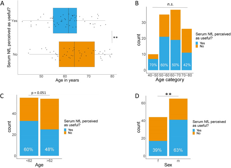
Age was a significant predictor of the perceived usefulness of sNfL with a beta estimate of -0.0975 ± 0.0331 (p < 0.005). Thus, patients in which sNfL was perceived as useful were on group level younger than patients in which sNfL was not perceived as useful (Fig. 1a). Zooming in on age categories, the perceived usefulness of sNfL decreased with every decade of older age: in 70% of subjects aged 40–50 years, sNfL was perceived as useful; in subjects aged 50–60 years, this was 60%; in subjects aged 60–70 years, this was 50%, compared to 42% in those aged 70–80 years, but these differences were not statistically significant (Fig. 1b). When dichotomizing age at the median (62 years), sNfL was perceived as useful in 60% of the youngest half of the cohort and in 48% of the oldest half of the cohort, which was borderline significant (p = 0.051, Fig. 1c).
Fig. 1.
Perceived usefulness of sNfL in relation to age and sex. a Age as continuous variable. b Age as categorical variable. c Age dichotomized at the median. d Sex. Abbreviations: f, female; m, male; sNfL, serum neurofilament light; N.s., not significant, *p value < 0.05; **p value < 0.01, ***p value < 0.001
Serum NfL is more often perceived as useful in male patients
Sex was a significant predictor of the perceived usefulness of sNfL with a beta estimate of 1.58 ± 0.52 (p < 0.005). Patients in which sNfL was perceived as useful were more often male than female (Fig. 1d). After adjusting for age, the effect of sex on the perceived usefulness of sNfL was even stronger, with a beta estimate of 1.98 ± 0.62 (p < 0.005). Age distribution was similar between males and females with 60% males in the youngest group (< 62 years) and 59% males in the oldest group (> 62 year).
Perceived usefulness of sNfL does not depend on CSF biomarker availability
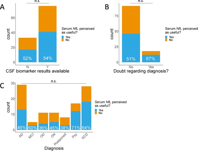
In the 33 (30%) patients of which no CSF biomarker result was available, sNfL was perceived as useful in 52%, which was a similar proportion as in patients with a CSF biomarker result (54%; Fig. 2a).
Fig. 2.
Perceived usefulness of sNfL in relation to availability of CSF biomarker results (a); doubt regarding the diagnosis (b); and diagnosis (c). Abbreviations: AD, Alzheimer’s disease; MCI, mild cognitive impairment; OD, other dementia; ON, other neurology; Psy, psychiatric disorder; N, no; Y, yes; N.s., not significant; *p value < 0.05; **p value < 0.01, ***p value < 0.001
Perceived usefulness of sNfL in relation to doubt regarding the diagnosis
In 18 (17%) patients, the neurologist expressed a doubt regarding the diagnosis. In 67% of these patients, sNfL was perceived as useful, while for the patients without uncertainty regarding their diagnosis, 51% of the sNfL results were perceived as useful (Fig. 2b). This difference was not statistically significant.
Perceived usefulness of serum NfL in relation to diagnosis
Serum NfL was more often perceived as useful in patients with a diagnosis of psychiatric disorder (71%) or SCD (64%) in comparison to a diagnosis of AD (45%); however, these differences did not reach statistical significance, also not when corrected for age and gender (Fig. 2c). The psychiatric disorder and SCD groups had a male predominance, with 12 out of 17 (71%) males in the psychiatric disorder group and 19 out of 28 (68%) in the SCD group, compared to 15 out of 29 (48%) in the AD group, which may underlie the trend in differences in perceived usefulness across diagnoses.
Serum NfL was perceived as useful mainly to confirm or exclude neurodegeneration
Next, we zoomed in on the cases in which sNfL was perceived as useful and listed the reasons of sNfL’s contribution as indicated by the neurologist (Table 3). We grouped the different diagnoses into neurodegenerative diagnosis (n = 20, 34%), no neurodegenerative diagnosis (n = 30, 52%), and other neurology or postponed diagnosis (n = 8, 14%). Reasons for perceived usefulness of sNfL were often quite similar, e.g., “excluding neurodegeneration” and “confirm no neurodegeneration,” or with subtle differences, e.g., “confirm neurodegeneration” versus “support neurodegeneration.” Upon grouping reasons that point in the same direction, the summarized main reasons for perceived usefulness of sNfL were per group: confirming neurodegeneration in cases with neurodegeneration (75%), excluding neurodegeneration in cases without neurodegeneration (87%) and to either confirm or exclude neurodegeneration in 63% of the other neurology or postponed cases. We also observed that in a few patients, sNfL was perceived as useful for follow-up, as indicator of vessel damage, or was explained by a recent heart attack.
Table 3.
Reasons for neurologists on how they used the sNfL result in case they perceived it as useful
| Neurodegenerative diagnosis (n = 20) | No neurodegenerative diagnosis (n = 30) | Other (n = 8) | |||
|---|---|---|---|---|---|
| Confirm AD | N = 10 | Exclude neurodegeneration | N = 8 | Confirm neurological disorder | N = 3 |
| Confirm neurodegeneration, e.g., MCI, FTD, other | N = 5 | Confirm SCD | N = 6 | Exclude AD | N = 1 |
| Support AD | N = 2 | Confirm no neurodegeneration | N = 4 | Exclude neurodegeneration | N = 1 |
| Support neurodegeneration | N = 1 | To reassure patient | N = 4 | For follow-up | N = 1 |
| Exclude FTD, confirm AD | N = 1 | Exclude FTD | N = 2 | Due to recent heart attack | N = 1 |
| For follow-up | N = 1 | Less support for FTD | N = 1 | Other | N = 1 |
| Exclude AD | N = 1 | ||||
| Exclude CTE | N = 1 | ||||
| Support SCD | N = 1 | ||||
| For patient management | N = 1 | ||||
| Indication for vessel damage | N = 1 | ||||
“Neurodegeneration” included subjects diagnosed with MCI, AD, or OD; “no neurodegeneration” included subjects with SCD or psychiatric disorder; “other” included subjects with other neurology or a postponed diagnosis. Abbreviations: AD Alzheimer’s disease, MCI mild cognitive impairment
Serum NfL concentrations increase with age, differ across diagnoses, and are elevated in CSF biomarker positive cases
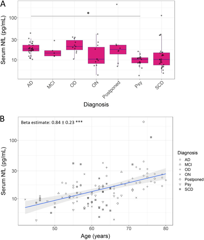
The concentrations of sNfL increased with age in our total cohort (Fig. 3b). The increase of sNfL (± standard error) was on average 0.84 ± 0.23 pg/mL per year (p < 0.001). We also found trends for differences in the average sNfL concentrations across the diagnostic groups after correcting for age (Fig. 3a). The median [interquartile range (IQR)] sNfL concentrations per diagnostic group were 18 [6.2] pg/mL for AD, 15 [4.2] pg/mL for MCI, 21 [12] pg/mL for OD, 10 [12] for ON, 18 [8.2] pg/mL for postponed, 10 [2.9] pg/mL for psychiatric disorder, and 10 [7.2] pg/mL for SCD. The only significant difference in pairwise post-hoc comparisons after correction for multiple testing was reported between the AD and psychiatric disorder groups (p < 0.05). SNfL levels were elevated in positive versus negative CSF biomarker profiles (median [IQR]): 16.2 [8.1] pg/mL versus 11.1 [6.7] pg/mL for CSF amyloid status (p < 0.05); 16.6 [5.1] pg/mL versus 10.5 [7.5] pg/mL for CSF tau status (p < 0.001); and 16.6 [5.1] pg/mL versus 10.5 [7.5] pg/mL for CSF neurodegeneration status (p < 0.001).
Fig. 3.
Concentrations of sNfL across different diagnoses (a) and in relation to age (b). Abbreviations: AD, Alzheimer’s disease; MCI, mild cognitive impairment; OD, other dementia; ON, other neurology; Psy, psychiatric disorder; SCD, subjective cognitive decline. *p value < 0.05; **p value < 0.01, ***p value < 0.001
Discussion
Literature supporting blood NfL as biomarker for neurodegenerative disorders is expanding, but prospective studies to support the clinical use of sNfL for individual patients are lacking. Our study prospectively and systematically monitored the added value of sNfL for individual patients in tertiary memory clinic setting from the neurologist’s perspective. We found that in more than half (53%) of the patients visiting the memory clinic, the treating neurologist evaluated the sNfL result as useful, mainly to detect neurodegeneration. SNfL results were evaluated as part of the diagnostic workup at patients’ first visit. We identified patient characteristics that increased the perceived usefulness of sNfL, such as younger age and male sex, and additionally observed a bias in perceived usefulness across neurologists.
Our main result showed that sNfL was perceived as useful by neurologists mostly in younger patients (< 62 years, 60%) and in male patients (63%). These characteristics were independent predictors of sNfL’s perceived usefulness, meaning that gender and age were not related; males were not younger than females in our cohort. We did not find a significant effect of diagnosis on the perceived usefulness of sNfL, although sNfL was more often perceived as useful in the SCD and psychiatric disorder group (64 and 71%). Interestingly, younger age and male gender were predominant characteristics in the SCD and psychiatric disorder groups, which may have biased the effect of suspected diagnosis on the perceived usefulness of sNfL. The effect of diagnosis, in particular that of SCD or psychiatric disorder, might be underestimated in our study due to the overrepresentation of young male patients in these groups. A typical clinical picture of neurodegeneration versus non-neurodegeneration is that of a young, male patient that enters the memory clinic with a suspected diagnosis of SCD or psychiatric disorder versus neurodegeneration, for example in phenocopy FTD syndrome [37, 38]. Our results support sNfL as helpful biomarker for such clinical questions in particular [12, 17, 18].
We anticipated that sNfL would be more often interpreted as useful in subjects without CSF biomarker results, due to the paucity of add-on objective tests to sustain the diagnosis, but surprisingly, the availability of CSF biomarker results had no influence on the perceived usefulness of sNfL. This indicates that sNfL contributes additional information on top of the information provided by the CSF biomarker results. The CSF biomarkers reflect amyloid plaque pathology (aβ1-42), tau tangle pathology (pTau), and neurodegeneration (tTau), pathologies specific to AD, while sNfL reflects axonal degeneration which occurs in various neurological disorders and is thus also elevated in various disorders. In dementias other than AD, e.g., FTD or PSP, CSF biomarkers are often in the normal range and sNfL will be useful as indicator for an ongoing neurodegenerative process. NfL thus represents an additional pathology, axonal degeneration, that is a useful biomarker of neurodegenerative pathology.
In subjects for which neurologists expressed doubt regarding the diagnosis, sNfL was perceived as useful in 67% of the cases, compared to in 51% of the subjects in which no doubt regarding diagnosis was expressed. This difference was not statistically significant, which could be a power issue, as the first group comprised less than 20% of the total cohort. Replicate studies should clarify whether sNfL has added value as biomarker in memory clinic patients with indecisive diagnoses.
The main use of sNfL reported by neurologists was either to confirm or to exclude neurodegeneration. In 75% of the subjects with an indication for neurodegenerative disease in which sNfL was perceived as useful, the reason was confirming neurodegeneration in general, or specified as AD, FTD, MCI, or other. Other reasons were supporting neurodegeneration, differentiating between AD and FTD, or for follow-up. In the subjects without an indication for neurodegeneration in which sNfL was perceived as useful, the reason was in 87% summarized as excluding neurodegeneration. In the “other“ diagnostic group, in subjects for which sNfL was perceived as useful, in 63%, the reason was to confirm or exclude neurodegeneration, depending on the suspected diagnosis. Interestingly, for two patients, neurologists indicated that the sNfL result was useful because they related it to a recent heart attack or interpreted it as an indication for vessel damage. Since NfL is not specific to neurodegenerative diseases, it is important to consider other underlying diseases as cause of increased NfL levels, especially in the elderly that often suffer from co-pathologies, such as cardiovascular problems [39].
Regarding the measured levels, sNfL levels were lowest in the psychiatric disorder and SCD groups and highest in the AD, OD, and postponed diagnosis groups, with a statistically significant difference after age correction between the psychiatric disorder and AD groups only. These findings are in line with previous literature [2, 10, 15, 16]. We observed two outliers with sNfL values > 100 pg/mL: one subject had a postponed diagnosis with suspicion of vascular dementia; the other had a diagnosis of SCD with underlying cerebral amyloid angiopathy. Literature supports a role for sNfL in cardiovascular diseases [40–42]. Multiple studies including ours showed an increase of NfL in blood with age and an increase in the variation of NfL levels with more outliers in the higher concentration range starting from the age of 60 [19, 43, 44]. These outliers presumably reflect underlying pathologies in the CNS, e.g., neurodegeneration or hemorrhage, in the preclinical stage [19]. Such high variation in normal NfL ranges in older age might explain the added value of sNfL in the younger population in particular that we observed in the current study.
Limitations
This study also faced some limitations. First, there was a strong bias by neurologist in the number of completed questionnaires as well as in the evaluation of sNfL as useful tool. Although we corrected for neurologist in all statistical analyses, personal experiences of neurologists might have affected the outcomes of our study. We closely examined neurologists’ background and the type of patients they have seen in this study and could not identify direct explanations. A combination of factors likely explains the bias, i.e., number of cases evaluated by the neurologist, his/her experience level with sNfL, and the type of patient in front of him/her. Second, our cohort had low power to assess the effects of the diagnostic group on the perceived usefulness of sNfL, for which independent replication studies will be needed. Third, this study was performed at one memory clinic that is specialized in early onset dementia. Results should therefore first be replicated in an independent cohort before these may be generalized to other memory clinic populations.
Conclusions
In conclusion, we here objectified the usefulness of sNfL in a prospective setting by monitoring the neurologists’ opinion on the added value of NfL for the individual patient. Since NfL is not specific for a specific brain disease, its practical use in the memory clinic was not a priori defined despite convincing retrospective cohort studies in literature. Our study revealed that in a memory clinic population, sNfL is deemed useful, especially in younger and in male patients and regardless of the availability of CSF biomarker results. Also, a bias between neurologists based on their background, type of patients seen, and personal experience influenced the perceived usefulness of sNfL. Our results provide first evidence on the clinical context of use of sNfL in memory clinic practice and need replication in other centers to form a basis for future clinical guidelines.
Supplementary Information
Additional file 1. Serum NfL reference values reported to neurologists together with sNfL results (Figure e-1 and Table e-1) and patient demographics stratified per neurologist (Table e-2).
Acknowledgements
The authors thank all neurologists of the Alzheimer Center Amsterdam, Amsterdam University Medical Center, the Netherlands, for their participation in this study. The authors also thank Sonia Rijnsdorp for her assistance with the processing of questionnaire results.
Abbreviations
- aβ1-42
Amyloid-beta(1-42)
- AD
Alzheimer’s disease
- ADC
Amsterdam Dementia Cohort
- CNS
Central nervous system
- CSF
Cerebrospinal fluid
- cv
Coefficient of variation
- DLB
Dementia with Lewy bodies
- EEG
Electroencephalogram
- FTD
Frontotemporal dementia
- IQR
Interquartile range
- MCI
Mild cognitive impairment
- MRI
Magnetic resonance imaging
- PPA
Primary progressive aphasia
- PSP
Progressive supranuclear palsy
- pTau
Phosphorylated Tau(P181)
- Psy
Psychiatric disorder
- QC
Quality control
- SCD
Subjective cognitive decline
- tTau
Total Tau
- sNfL
Serum neurofilament light
- VaD
Vascular dementia
Authors’ contributions
E.A.J.W. was involved in the study design, statistical analysis, and data interpretation and drafted the manuscript. P.S. was involved in the study design, data collection, and critical revision of the manuscript. C.E.T. was involved in the study design, data interpretation, and critical revision of the manuscript. E.G.B.V. was involved in the study design, data collection and data interpretation, and critical revision of the manuscript. All authors read and approved the final manuscript.
Funding
E.A.J.W. received funding from the Selfridges Group Foundation administered by the Weston Brain Institute. This project has received funding from the European Union's Horizon 2020 research and innovation program under grant agreement No. 860303. The funding sources had no role in the study design, execution, or interpretation, nor in the decision to submit the article for publication.
Availability of data and materials
The datasets used and/or analyzed during the current study are available from the corresponding author on reasonable request.
Declarations
Ethics approval and consent to participate
All patients included in this study gave written informed consent for the storage of their clinical data for research purposes as part of the Amsterdam Dementia Cohort (ADC) and the ADC was approved by the local medical ethical committee of the VU University Medical Center.
Consent for publication
Not applicable.
Competing interests
E.A.J.W. and E.G.B.V. report no declarations of interest.
P.S. has received consultancy fees (paid to the institution) from AC Immune, Alkermes, Alnylam, Alzheon, Anavex, Biogen, Brainstorm Cell, Cortexyme, Denali, EIP, ImmunoBrain Checkpoint, GemVax, Genentech, Green Valley, Novartis, Novo Nordisk, PeopleBio, Renew LLC, Roche. He is PI of studies with AC Immune, CogRx, FUJI-film/Toyama, IONIS, UCB, Vivoryon. He is a part-time employee of Life Sciences Partners Amsterdam. He serves on the board of the Brain Research Center.
C.E.T. has a collaboration contract with ADx Neurosciences and Quanterix, performed contract research or received grants from AxonNeurosciences, Biogen, Boehringer, Brainstorm Therapeutics, Celgene, EIP Pharma, Esai, Janssen prevention center, Roche, Toyama, Vivoryon.
Footnotes
Publisher’s Note
Springer Nature remains neutral with regard to jurisdictional claims in published maps and institutional affiliations.
Contributor Information
E. A. J. Willemse, Email: e.willemse@amsterdamumc.nl
P. Scheltens, Email: p.scheltens@amsterdamumc.nl
C. E. Teunissen, Email: c.teunissen@amsterdamumc.nl
E. G. B. Vijverberg, Email: e.vijverberg@amsterdamumc.nl
References
- 1.Khalil M, Teunissen CE, Otto M, Piehl F, Sormani MP, Gattringer T, et al. Neurofilaments as biomarkers in neurological disorders. Nat Rev Neurol. 2018;14(10):577–89. [DOI] [PubMed]
- 2.Bridel C, van Wieringen WN, Zetterberg H, Tijms BM, Teunissen CE, Alvarez-Cermeño JC, et al. Diagnostic value of cerebrospinal fluid neurofilament light protein in neurology. JAMA Neurol. 2019;76(9):1035–48. [DOI] [PMC free article] [PubMed]
- 3.Palermo G, Mazzucchi S, Della Vecchia A, Siciliano G, Bonuccelli U, Azuar C, et al. Different clinical contexts of use of blood neurofilament light chain protein in the spectrum of neurodegenerative diseases. Mol Neurobiol. 2020;57(11):4667–91. [DOI] [PubMed]
- 4.Kuhle J, Barro C, Andreasson U, Derfuss T, Lindberg R, Sandelius Å, Liman V, Norgren N, Blennow K, Zetterberg H. Comparison of three analytical platforms for quantification of the neurofilament light chain in blood samples: ELISA, electrochemiluminescence immunoassay and Simoa. Clin Chem Lab Med. 2016;54(10):1655–1661. doi: 10.1515/cclm-2015-1195. [DOI] [PubMed] [Google Scholar]
- 5.Thouvenot E, Demattei C, Lehmann S, Maceski-Maleska A, Hirtz C, Juntas-Morales R, et al. Serum neurofilament light chain at time of diagnosis is an independent prognostic factor of survival in amyotrophic lateral sclerosis. Eur J Neurol. John Wiley & Sons, Ltd (10.1111). 2020;27(2):251–7. [DOI] [PubMed]
- 6.Ng ASL, Tan YJ, Yong ACW, Saffari SE, Lu Z, Ng EY, et al. Utility of plasma neurofilament light as a diagnostic and prognostic biomarker of the postural instability gait disorder motor subtype in early Parkinson’s disease. Mol Neurodegener. 2020;15:33. doi: 10.1186/s13024-020-00385-5. [DOI] [PMC free article] [PubMed] [Google Scholar]
- 7.Kuhle J, Kropshofer H, Haering DA, Kundu U, Meinert R, Barro C, et al. Blood neurofilament light chain as a biomarker of MS disease activity and treatment response From the Neurologic Clinic and Policlinic. 2019. [DOI] [PMC free article] [PubMed] [Google Scholar]
- 8.Byrne LM, Rodrigues FB, Blennow K, Durr A, Leavitt BR, Roos RAC, Scahill RI, Tabrizi SJ, Zetterberg H, Langbehn D, Wild EJ. Neurofilament light protein in blood as a potential biomarker of neurodegeneration in Huntington’s disease: a retrospective cohort analysis. Lancet Neurol. 2017;16(8):601–609. doi: 10.1016/S1474-4422(17)30124-2. [DOI] [PMC free article] [PubMed] [Google Scholar]
- 9.Kovacs GG, Andreasson U, Liman V, Regelsberger G, Lutz MI, Danics K, Keller E, Zetterberg H, Blennow K. Plasma and cerebrospinal fluid tau and neurofilament concentrations in rapidly progressive neurological syndromes: a neuropathology-based cohort. Eur J Neurol. 2017;24(11):1326–1e77. doi: 10.1111/ene.13389. [DOI] [PubMed] [Google Scholar]
- 10.Mattsson N, Cullen NC, Andreasson U, Zetterberg H, Blennow K. Association between longitudinal plasma neurofilament light and neurodegeneration in patients with Alzheimer disease. JAMA Neurol. 2019;76(7):791–9. [DOI] [PMC free article] [PubMed]
- 11.Wilke C, Preische O, Deuschle C, Roeben B, Apel A, Barro C, Maia L, Maetzler W, Kuhle J, Synofzik M. Neurofilament light chain in FTD is elevated not only in cerebrospinal fluid, but also in serum. J Neurol Neurosurg Psychiatry. 2016;87(11):1270–1272. doi: 10.1136/jnnp-2015-312972. [DOI] [PubMed] [Google Scholar]
- 12.Katisko K, Cajanus A, Jääskeläinen O, Kontkanen A, Hartikainen P, Korhonen VE, Helisalmi S, Haapasalo A, Koivumaa-Honkanen H, Herukka SK, Remes AM, Solje E. Serum neurofilament light chain is a discriminative biomarker between frontotemporal lobar degeneration and primary psychiatric disorders. J Neurol. Springer. 2020;267(1):162–167. doi: 10.1007/s00415-019-09567-8. [DOI] [PMC free article] [PubMed] [Google Scholar]
- 13.Preische O, Schultz SA, Apel A, Kuhle J, Kaeser SA, Barro C, et al. Serum neurofilament dynamics predicts neurodegeneration and clinical progression in presymptomatic Alzheimer’s disease. Nat Med. 2019. [DOI] [PMC free article] [PubMed]
- 14.van der Ende EL, Meeter LH, Poos JM, Panman JL, Jiskoot LC, Dopper EGP, et al. Serum neurofilament light chain in genetic frontotemporal dementia: a longitudinal, multicentre cohort study. Lancet Neurol. Elsevier. 2019;18(12):1103–11. 10.1016/S1474-4422(19)30354-0. [DOI] [PubMed]
- 15.Lewczuk P, Ermann N, Andreasson U, Schultheis C, Podhorna J, Spitzer P, et al. Plasma neurofilament light as a potential biomarker of neurodegeneration in Alzheimer’s disease. Alzheimers Res Ther. 2018;10:71. doi: 10.1186/s13195-018-0404-9. [DOI] [PMC free article] [PubMed] [Google Scholar]
- 16.Mattsson N, Andreasson U, Zetterberg H, Blennow K. Alzheimer’s disease neuroimaging initiative. association of plasma neurofilament light with neurodegeneration in patients with Alzheimer disease. JAMA Neurol. 2017;74(5):557–566. doi: 10.1001/jamaneurol.2016.6117. [DOI] [PMC free article] [PubMed] [Google Scholar]
- 17.Al Shweiki MR, Steinacker P, Oeckl P, Hengerer B, Danek A, Fassbender K, et al. Neurofilament light chain as a blood biomarker to differentiate psychiatric disorders from behavioural variant frontotemporal dementia. J Psychiatr Res. 2019;113:137–140. doi: 10.1016/j.jpsychires.2019.03.019. [DOI] [PubMed] [Google Scholar]
- 18.Ducharme S, Dols A, Laforce R, Devenney E, Kumfor F, van den Stock J, et al. Recommendations to distinguish behavioural variant frontotemporal dementia from psychiatric disorders. Brain. 2020;143(6):1632–50. [DOI] [PMC free article] [PubMed]
- 19.Khalil M, Pirpamer L, Hofer E, Voortman MM, Barro C, Leppert D, et al. Serum neurofilament light levels in normal aging and their association with morphologic brain changes. Nat Commun. 2020;11:812. doi: 10.1038/s41467-020-14612-6. [DOI] [PMC free article] [PubMed] [Google Scholar]
- 20.de Wilde A, Ossenkoppele R, Pelkmans W, Bouwman F, Groot C, van Maurik I, et al. Assessment of the appropriate use criteria for amyloid PET in an unselected memory clinic cohort: The ABIDE project. Alzheimers Dement. 2019;15:1458–1467. doi: 10.1016/j.jalz.2019.07.003. [DOI] [PubMed] [Google Scholar]
- 21.Duits FH, Prins ND, Lemstra AW, Pijnenburg YAL, Bouwman FH, Teunissen CE, et al. Diagnostic impact of CSF biomarkers for Alzheimer’s disease in a tertiary memory clinic. Alzheimers Dement. 2015;11:523–532. doi: 10.1016/j.jalz.2014.05.1753. [DOI] [PubMed] [Google Scholar]
- 22.van der Flier WM, Pijnenburg YAL, Prins N, Lemstra AW, Bouwman FH, Teunissen CE, van Berckel BNM, Stam CJ, Barkhof F, Visser PJ, van Egmond E, Scheltens P. Optimizing patient care and research: the Amsterdam Dementia Cohort. J Alzheimers Dis. 2014;41(1):313–327. doi: 10.3233/JAD-132306. [DOI] [PubMed] [Google Scholar]
- 23.Jessen F, Amariglio RE, van Boxtel M, Breteler M, Ceccaldi M, Chételat G, Dubois B, Dufouil C, Ellis KA, van der Flier WM, Glodzik L, van Harten AC, de Leon MJ, McHugh P, Mielke MM, Molinuevo JL, Mosconi L, Osorio RS, Perrotin A, Petersen RC, Rabin LA, Rami L, Reisberg B, Rentz DM, Sachdev PS, de la Sayette V, Saykin AJ, Scheltens P, Shulman MB, Slavin MJ, Sperling RA, Stewart R, Uspenskaya O, Vellas B, Visser PJ, Wagner M, Subjective Cognitive Decline Initiative (SCD-I) Working Group A conceptual framework for research on subjective cognitive decline in preclinical Alzheimer’s disease. Alzheimers Dement. 2014;10(6):844–852. doi: 10.1016/j.jalz.2014.01.001. [DOI] [PMC free article] [PubMed] [Google Scholar]
- 24.Albert MS, DeKosky ST, Dickson D, Dubois B, Feldman HH, Fox NC, et al. The diagnosis of mild cognitive impairment due to Alzheimer’s disease: recommendations from the National Institute on Aging-Alzheimer’s Association workgroups on diagnostic guidelines for Alzheimer’s disease. Alzheimers Dement. 2011;7(3):270–279. doi: 10.1016/j.jalz.2011.03.008. [DOI] [PMC free article] [PubMed] [Google Scholar]
- 25.McKhann GM, Knopman DS, Chertkow H, Hyman BT, Jack CR, Kawas CH, et al. The diagnosis of dementia due to Alzheimer’s disease: recommendations from the National Institute on Aging-Alzheimer’s Association workgroups on diagnostic guidelines for Alzheimer’s disease. Alzheimers Dement. 2011;7(3):263–269. doi: 10.1016/j.jalz.2011.03.005. [DOI] [PMC free article] [PubMed] [Google Scholar]
- 26.Neary D, Snowden JS, Gustafson L, Passant U, Stuss D, Black S, Freedman M, Kertesz A, Robert PH, Albert M, Boone K, Miller BL, Cummings J, Benson DF. Frontotemporal lobar degeneration: a consensus on clinical diagnostic criteria. Neurology. 1998;51(6):1546–1554. doi: 10.1212/WNL.51.6.1546. [DOI] [PubMed] [Google Scholar]
- 27.Rascovsky K, Hodges JR, Knopman D, Mendez MF, Kramer JH, Neuhaus J, et al. Sensitivity of revised diagnostic criteria for the behavioural variant of frontotemporal dementia. Brain. 2011;134:2456–2477. doi: 10.1093/brain/awr179. [DOI] [PMC free article] [PubMed] [Google Scholar]
- 28.McKeith IG, Dickson DW, Lowe J, Emre M, O’Brien JT, Feldman H, et al. Diagnosis and management of dementia with Lewy bodies: third report of the DLB Consortium. Neurology. 2005;65(12):1863–1872. doi: 10.1212/01.wnl.0000187889.17253.b1. [DOI] [PubMed] [Google Scholar]
- 29.Gorno-Tempini ML, Hillis AE, Weintraub S, Kertesz A, Mendez M, Cappa SF, et al. Classification of primary progressive aphasia and its variants. Neurology. 2011;76:1006–1014. doi: 10.1212/WNL.0b013e31821103e6. [DOI] [PMC free article] [PubMed] [Google Scholar]
- 30.Litvan I, Agid Y, Calne D, Campbell G, Dubois B, Duvoisin RC, Goetz CG, Golbe LI, Grafman J, Growdon JH, Hallett M, Jankovic J, Quinn NP, Tolosa E, Zee DS. Clinical research criteria for the diagnosis of progressive supranuclear palsy (Steele-Richardson-Olszewski syndrome): report of the NINDS-SPSP international workshop. Neurology. 1996;47(1):1–9. doi: 10.1212/WNL.47.1.1. [DOI] [PubMed] [Google Scholar]
- 31.Höglinger GU, Respondek G, Stamelou M, Kurz C, Josephs KA, Lang AE, Mollenhauer B, Müller U, Nilsson C, Whitwell JL, Arzberger T, Englund E, Gelpi E, Giese A, Irwin DJ, Meissner WG, Pantelyat A, Rajput A, van Swieten JC, Troakes C, Antonini A, Bhatia KP, Bordelon Y, Compta Y, Corvol JC, Colosimo C, Dickson DW, Dodel R, Ferguson L, Grossman M, Kassubek J, Krismer F, Levin J, Lorenzl S, Morris HR, Nestor P, Oertel WH, Poewe W, Rabinovici G, Rowe JB, Schellenberg GD, Seppi K, van Eimeren T, Wenning GK, Boxer AL, Golbe LI, Litvan I, for the Movement Disorder Society-endorsed PSP Study Group Clinical diagnosis of progressive supranuclear palsy: the movement disorder society criteria. Mov Disord. 2017;32(6):853–864. doi: 10.1002/mds.26987. [DOI] [PMC free article] [PubMed] [Google Scholar]
- 32.Román GC, Tatemichi TK, Erkinjuntti T, Cummings JL, Masdeu JC, Garcia JH, et al. Vascular dementia: diagnostic criteria for research studies. Report of the NINDS-AIREN International Workshop. Neurology. 1993;43(2):250–260. doi: 10.1212/WNL.43.2.250. [DOI] [PubMed] [Google Scholar]
- 33.van der Flier WM, Scheltens P. Amsterdam dementia cohort: performing research to optimize care. J Alzheimer’s Dis. 2018;62(3):1091–1111. doi: 10.3233/JAD-170850. [DOI] [PMC free article] [PubMed] [Google Scholar]
- 34.Willemse EAJ, van Maurik IS, Tijms BM, Bouwman FH, Franke A, Hubeek I, et al. Diagnostic performance of Elecsys immunoassays for cerebrospinal fluid Alzheimer’s disease biomarkers in a nonacademic, multicenter memory clinic cohort: the ABIDE project. Alzheimers Dement (Amsterdam, Netherlands) 2018;10:563–572. doi: 10.1016/j.dadm.2018.08.006. [DOI] [PMC free article] [PubMed] [Google Scholar]
- 35.Willemse EAJ, Tijms BM, van Berckel BNM, Le Bastard N, van der Flier WM, Scheltens P, et al. Comparing CSF amyloid-beta biomarker ratios for two automated immunoassays, Elecsys and Lumipulse, with amyloid PET status. Alzheimers Dement Diagn Assess Dis Monit. 2021;13(1):1–11. 10.1002/dad2.12182. [DOI] [PMC free article] [PubMed]
- 36.R Core Team. R: a language and environment for statistical computing. Vienna: R Foundation for Statistical Computing; 2020.
- 37.Vijverberg EGB, Gossink F, Krudop W, Sikkes S, Kerssens C, Prins N, et al. The diagnostic challenge of the late-onset frontal lobe syndrome. J Clin Psychiatry. 2017;78:e1197–e1203. doi: 10.4088/JCP.16m11078. [DOI] [PubMed] [Google Scholar]
- 38.Gossink FT, Dols A, Kerssens CJ, Krudop WA, Kerklaan BJ, Scheltens P, et al. Psychiatric diagnoses underlying the phenocopy syndrome of behavioural variant frontotemporal dementia. J Neurol Neurosurg Psychiatry. 2016;87:64–68. doi: 10.1136/jnnp-2016-314597.22. [DOI] [PubMed] [Google Scholar]
- 39.Moseby-Knappe M, Mattsson N, Nielsen N, Zetterberg H, Blennow K, Dankiewicz J, et al. Serum neurofilament light chain for prognosis of outcome after cardiac arrest Supplemental content. 2018. [DOI] [PMC free article] [PubMed] [Google Scholar]
- 40.De Marchis GM, Katan M, Barro C, Fladt J, Traenka C, Seiffge DJ, et al. Serum neurofilament light chain in patients with acute cerebrovascular events. Eur J Neurol. 2018;25(3):562–568. doi: 10.1111/ene.13554. [DOI] [PubMed] [Google Scholar]
- 41.Korley FK, Goldstick J, Mastali M, Van Eyk JE, Barsan W, Meurer WJ, et al. Serum NfL (neurofilament light chain) levels and incident stroke in adults with diabetes mellitus. Stroke. 2019;50:1669–1675. doi: 10.1161/STROKEAHA.119.024941. [DOI] [PMC free article] [PubMed] [Google Scholar]
- 42.Gattringer T, Pinter D, Enzinger C, Seifert-Held T, Kneihsl M, Fandler S, et al. Serum neurofilament light is sensitive to active cerebral small vessel disease. Neurology. 2017;89:2108–2114. doi: 10.1212/WNL.0000000000004645. [DOI] [PMC free article] [PubMed] [Google Scholar]
- 43.de Wolf F, Ghanbari M, Licher S, McRae-McKee K, Gras L, Weverling GJ, et al. Plasma tau, neurofilament light chain and amyloid-β levels and risk of dementia; a population-based cohort study. Brain. 2020;143(4):1220–32. [DOI] [PMC free article] [PubMed]
- 44.Quiroz YT, Zetterberg H, Reiman EM, Chen Y, Su Y, Fox-Fuller JT, Garcia G, Villegas A, Sepulveda-Falla D, Villada M, Arboleda-Velasquez JF, Guzmán-Vélez E, Vila-Castelar C, Gordon BA, Schultz SA, Protas HD, Ghisays V, Giraldo M, Tirado V, Baena A, Munoz C, Rios-Romenets S, Tariot PN, Blennow K, Lopera F. Plasma neurofilament light chain in the presenilin 1 E280A autosomal dominant Alzheimer’s disease kindred: a cross-sectional and longitudinal cohort study. Lancet Neurol. 2020;19(6):513–521. doi: 10.1016/S1474-4422(20)30137-X. [DOI] [PMC free article] [PubMed] [Google Scholar]
Associated Data
This section collects any data citations, data availability statements, or supplementary materials included in this article.
Supplementary Materials
Additional file 1. Serum NfL reference values reported to neurologists together with sNfL results (Figure e-1 and Table e-1) and patient demographics stratified per neurologist (Table e-2).
Data Availability Statement
The datasets used and/or analyzed during the current study are available from the corresponding author on reasonable request.


