Abstract
Coronavirus disease 2019 (COVID-19) is a highly infectious disease caused by the novel “severe acute respiratory syndrome coronavirus-2” (SARS-CoV-2) and is rapidly spreading worldwide. This review is designed to highlight the most common clinical features and computed tomography (CT) signs of patients with COVID-19 and to elaborate the most significant signs indicative of COVID-19 diagnosis. This review involved five original articles with both clinical and radiological features of COVID-19 published during Jan and Mar 2020. In this review, the most frequent symptoms of COVID-19 were fever and cough. Myalgia, fatigue, sore throat, headache, diarrhea, and dyspnea were less common manifestations. Nausea and vomiting were rare. Ground-glass opacity (GGO) was the most common radiological finding on CT, and mixed GGO with consolidation was reported in some cases. In addition, elevated C-reactive protein and lymphopenia are the pertinent laboratory findings of COVID-19. CT is an effective and important imaging tool for both diagnosis and follow-up COVID-19 patients with varied features, duration, and course of the disease. Bilateral GGOs, especially in the periphery of the lungs with or without consolidation, are the hallmark of COVID-19.
Keywords: Clinical manifestations, COVID-19, CT features, fever, ground-glass opacity
Introduction and Background
Coronavirus disease 2019 (COVID-19) is a highly infectious disease that is rapidly spreading worldwide. It first emerged in Wuhan City, China in December 2019 as an outbreak of a new phenotype of pneumonia caused by the novel “severe acute respiratory syndrome' coronavirus-2” (SARS-CoV-2). It is a single-stranded ribonucleic acid (RNA) virus and classified as a member of the Coronaviridae family.[1,2] The virus transmits through air droplets generated during coughing or sneezing by symptomatic or asymptomatic infected individuals and through respiratory fomites.[3,4,5] The incubation period of the virus varies from 2 to 14 days, and the clinical features vary from asymptomatic infection, fever, dry cough, sore throat, headache, diarrhea, vomiting, myalgia, fatigue, shortness of breath, to acute respiratory distress syndrome. It can progress to pneumonia, respiratory failure, acute kidney injury, multiorgan dysfunction, and even death, especially in patients with comorbidities such as diabetes mellitus, hypertension, and heart disease.[3,5] The seventh edition of guidelines for the diagnosis and treatment of COVID-19 by the Chinese National health commission (NHC) divided the patients with COVID-19 into four clinical types: (1) Mild, patients with no or mild symptoms and no pneumonia on medical imaging, (2) Moderate, patients with symptoms and fever with pneumonia on clinical and medical imaging, (3) Severe, patients with respiratory distress (respiration rate >30 cycle/min) or oxygen saturation at a resting stare <93% or lesion progress more than 50% of the lungs within 48 h on medical imaging, or PaO2/FiO2 <300 mmHg, and (4) Critical, patients with respiratory failure or shock or other multiorgan failure and need intensive care.[6] According to the sixth edition of the official “guidelines for the diagnosis and treatment of COVID-19” declared by the NHC of China, computed tomography (CT) is an effective and important screening tool to diagnose, monitor progression, and evaluate treatment effectiveness. Bilateral ground-glass opacities (GGOs) in the peripheral regions of the lungs with or without consolidation are the hallmark of COVID-19. Diverse CT features like crazy-paving pattern, reverse-halo sign, and airway changes and pleural changes can be present.[7] Features of COVID-19 start to appear on CT in 4–7 days from the onset of the initial clinical manifestations and subsequently appear in all stages of the disease.[8] Significant correlations are found between the severity of clinical manifestations and the severity of lung involvement on CT.[9] The American College of Radiology and the European society of radiology (ESR) recommended against the use of chest CT as a widespread screening tool for patients with COVID-19, including patients with no or mild symptoms. The “Real-time reverse transcription polymerase chain reaction” (PT-PCR) is considered as a standard diagnostic test for patients with COVID-19.[10]
The Duch Radiological Society provided COVID-19 Reporting and Data system (CO-RADS) as a categorical CT assessment scheme for the level of suspicion of lung involvement in patients with moderate to severe symptoms of COVID-19 on a scale ranges from 0 to 6 as follows: (1) CO-RADS 1: no suspicion on normal CT, (2) CO-RADS 2: low suspicion when CT abnormalities are consistent with other infections, (3) CO-RADS 3: intermediate suspicion when CT abnormalities are compatible but unsure of COVID-19, (4) CO-RADS 4: high suspicion when CT abnormalities suspicious for COVID-19, (5) CO-RADS 5: very high suspicion when CT abnormalities are typical of COVID-19, (6) CO-RADS 6: proven by PT-PCR positive for COVID-19. If CT is technically insufficient for assigning the score, this is categorized as CO-RADS 0.[11]
This article was designed to summarize and highlight the most common clinical features and radiological signs seen on the CT of patients with COVID-19 and to elaborate the most significant clinical and radiological signs indicative of COVID-19 diagnosis. This review involved five original articles published between January and March 2020. Inclusion criteria involved the original articles interested in both the clinical and radiological features of COVID-19. We excluded review articles, case reports, articles interested only with clinical or radiological manifestations or with other ideas, articles focused only on a specific point, and articles available in other languages. Overall, five articles that met the inclusion criteria involved in this review.[12,13,14,15,16] The diagnosis of patients with COVID-19 was confirmed with PT-PCR in all cases of the involved studies. This article elaborates the most frequent features of COVID-19 for primary care physicians and family care doctors to be alert for detecting the disease as early as possible and to avoid bad complications resulting from delayed diagnosis during the pandemic.
Review
Clinical manifestation
In this review, the most common symptoms of COVID-19 were fever then cough [Table 1].
Table 1.
Common Clinical manifestations of COVID-19
| Author | Han et al., 2020[12] | Zhao et al., 2020[13] | Xu et al., 2020[14] | Xu et al., 2020[15] | Lomoro et al., 2020[16] |
|---|---|---|---|---|---|
| Study date | From Jan 4-Feb 3, 2020 | NA | From Jan-Feb 2020 | From Jan 23-Feb 4, 2020 | From Feb 15-Mar 15, 2020 |
| Patients (n) | 108 | 101 | 50 | 90 | 58 |
| Age (range) (Mean) | 21-90 | 17-75 (44.44) | 3-85 (43.9±16.8) | 18-86 (50) | 18-98 (66.3±16.6) |
| Gender (m) | 38 (35.2) | 56 (55.4) | 29 (58%) | 39 (43) | 36 |
| Fever | 94 (87) | 79 (78.2) | 43 (86) | 70 (78) | 58 (100) |
| Cough | 65 (60) | 63 (62.4) | 20 (40) | 57 (63) | 33 (57) |
| Sore throat | 14 (13) | 12 (11.9) | 4 (8) | 23 (26) | NA |
| Headache | 14 (13) | NA | 5 (10) | 4 (4) | NA |
| Dyspnea | NA | 1 (1) | 4 (8) | 24 (41) | |
| Myalgia | 12 (11) | 0 (0.0) | 8 (16) | 25 (28) | 0 (0.0) |
| Fatigue | 42 (39) | 0 (0.0) | 8 (16) | 19 (21) | 18 (31) |
| Myalgia or fatigue | 0 (0.0) | 17 (16.8) | 0 (0.0) | 0 (0.0) | 0 (0.0) |
| Diarrhea | 15 (14) | 3 (3) | NA | 5 (6) | NA |
| Nausea/vomiting | NA | 2 (2) | NA | 7 (8) | NA |
| ↓ lymphocyte | 65 (60) | NA | 14 (28) | 48 (83) | |
| ↑ CRP | NA | NA | 26 (52) | 38 (42) | 56 (96.5) |
COVID-19=Corona virus disease 2019, CRP=C-reactive protein (normal range=0-10 mg/liter), ↓ =decrease, ↑ =elevated, NA=Not available
Fever was reported as the presenting symptom in 82%, 98%, 98.5%, 86.7%, and 83% and cough was reported in 61%, 76%, 59.4%, 78.3%, and 82% of patients in similar previous studies.[17,18,19,20,21] However, the less common clinical features of COVID-19 were fatigue, myalgia, sore throat, headache, diarrhea, and dyspnea. Many previous studies reported these symptoms less common than fever and cough.[17,18,22] Furthermore, nausea, vomiting, and other gastrointestinal symptoms were relatively rare clinical features of COVID-19. Previous studies reported these as rare manifestations of COVID-19.[23,24]
In this review, elevated C-reactive protein ((↑) CRP) is a laboratory marker of inflammation and decreased lymphocytes (lymphopenia) were the pertinent laboratory findings of COVID-19. These findings were reported in many previous studies.[25,26,27]
Radiological manifestations
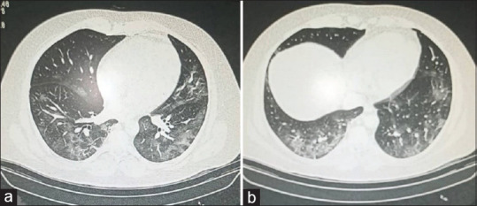
In this review, GGO [Figure 1] was was the most common radiological finding on CT [Table 2].
Figure 1.

Selected computed tomography images of a 37-year-old male patient with COVID-19 on day 3 after symptoms show scattered ground-glass opacities predominantly in the peripheral aspect of both lower lobes
Table 2.
Common Radiological signs of the involved patients (Pattern and distribution)
| Author | Han et al., 2020[12] | Zhao et al., 2020[13] | Xu et al., 2020[14] | Xu et al., 2020[15] | Lomoro et al., 2020[16] |
|---|---|---|---|---|---|
| Patients with CT (n) | 108 | 93 | 41 | 90 | 42 |
| GGO | 65 (60) | 87 (86.1) | 30 (73.17) | 65 (72) | 15 (35.7) |
| Mixed GGO | 44 (41) | 65 (64.4) | 25 (60.97) | NA | 25 (59.5) |
| Consolidation | 6 (5.55) | 44 (43.6) | 15 (36.58) | 12 (13) | 0 (0.0) |
| Crazy-paving | 43 (40) | NA | NA | 11 (12) | 24 (57.1) |
| Pleural effusion | 0 (0.0) | 14 (13.9) | 4 (9.75) | 4 (4) | 3 (7.1) |
| Lymphadenopathy | 0 (0.0) | 1 (1) | 1 (2.43) | 1 (1) | 6 (14.3) |
| Peripheral | 97 (90) | 88 (87.1) | NA | 46 (51) | 27 (64.3) |
| Central | 2 (2) | 1 (1) | NA | NA | 1 (2.4) |
| Mixed | 9 (8) | NA | NA | NA | 12 (28.6) |
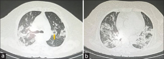
Previous studies reported GGO as the radiological sign in 97.6%, 100%, 91%, and 77% of the cases.[20,27,28,29] Table 2 shows that mixed GGO with consolidation [Figure 2] was the second most common radiological feature, which was reported as 59% and 50% in previous studies.[29,30]
Figure 2.

Selected computed tomography images of a 60-year-old female patient with COVID-19 on day 10 after symptoms show mixed ground-glass and consolidative opacities predominantly in the peripheral zones. Reverse halo sign is also noted (arrow)
Here, the CT findings were peripheral in 67.5% of the cases and reported as 89%, 86%, 75%, and 76% in previous studies.[27,29,30,31]
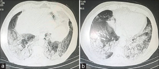
In addition, a crazy-paving pattern [Figure 3] was the third most common radiological feature in this review; however, previous studies reported crazy paving in 36%, 29%, and 19.5% of the patients.[20,28,32]
Figure 3.

Selected computed tomography images of a 60-year-old male patient with COVID-19 on day 14 after symptoms show consolidations predominantly in the peripheral lower lobes, with ground-glass and crazy-paving patterns
Furthermore, here pleural effusion was reported in 6.5% of cases, and it was reported as a rare finding (3%) in previous studies.[27,33] Lymphadenopathy was reported in 2.4% in this review. However, lymphadenopathy was reported as a common finding (58%) in a previous study in Italy,[27] and a rare finding in multiple previous studies.[28,33]
El Homsi et al. reported that CT findings change with stages of COVID-19: (1) Early stage (the first two days), 56% of patients have negative CT; however, GGO was found in 44% and consolidation in the remaining 17%; (2) Intermediate stage (3–5 days), GGO was found in 88% and consolidation in 55%; bilaterally in 76% and peripherally in 64%; (3) Late-phase (6–12 days), GGO was found in 88% and consolidation in 60% of the cases; bilateral in 88% and peripheral in 72%; (4) Absorption stage (more than 14 days), GGO was found in 65% and consolidation in 75%; bilateral in 88% and peripheral in 72%.[10]
Zheng et al. reported that CT features differ at various stages of COVID-19—early stage, progression stage, and absorption stage.[33] Bhandari et al. reported that CT features of COVID-19 vary with the duration and course of the disease. GGO is high in the early stages and low in the late stages.[34] Bernheim et al. reported that lung involvement was bilateral in 28%, 76%, and 88% in the early, intermediate, and late stages, respectively.[35]
Ultimately, CT is crucial and provides an effective diagnosis of COVID-19. In patients with clinical features of COVID-19, low-dose CT can provide a diagnosis with comparable sensitivity to PT-PCR and high specificity to distinguish it from other similar clinical diseases.[36] CT sensitivity for the diagnosis of COVID-19 increases after the fifth day of symptoms.[37] CT is a fast, adequate, and reliable diagnostic modality to refer patients with COVID-19 for hospitalization when the response of PT-PCR is late.[38]
Conclusions
Fever and cough are the most common clinical features of COVID-19. Fatigue, myalgia, sore throat, headache, diarrhea, and dyspnea are less common clinical manifestations. Nausea, vomiting, and other gastrointestinal symptoms are relatively rare. Elevated C-reactive protein and lymphopenia are the pertinent laboratory findings of COVID-19. CT is an effective and important imaging tool for the diagnosis and follow-up of COVID-19 with varied features, duration, and course of the disease. Bilateral GGOs, especially in the peripheral lungs, with or without consolidation, are the hallmark of COVID-19.
Key messages
Fever and cough are the frequent clinical features for diagnosis of COVID-19.
Bilateral GGOs, especially in the peripheral lungs, with or without consolidation, are the hallmark of COVID-19.
Financial support and sponsorship
Nil.
Conflicts of interest
There are no conflicts of interest.
References
- 1.Luo H, Tang QL, Shang YX, Liang SB, Yang M, Robinson N, et al. Can Chinese medicine be used for prevention of corona virus disease 2019 (COVID-19). A review of historical classics, research evidence and current prevention programs? Chin J Integr Med. 2020;26:243–50. doi: 10.1007/s11655-020-3192-6. [DOI] [PMC free article] [PubMed] [Google Scholar]
- 2.Harapan H, Itoh N, Yufika A, Winardi W, Keam S, Te H, et al. Coronavirus disease 2019 (COVID-19): A literature review. J Infect Public Health. 2020;13:667–73. doi: 10.1016/j.jiph.2020.03.019. [DOI] [PMC free article] [PubMed] [Google Scholar]
- 3.Singhal T. A review of coronavirus disease-2019 (COVID-19) Indian J Pediatr. 2020;87:281–6. doi: 10.1007/s12098-020-03263-6. [DOI] [PMC free article] [PubMed] [Google Scholar]
- 4.Rothe C, Schunk M, Sothmann P, Bretzel G, Froeschl G, Wallrauch C, et al. Transmission of 2019-nCoV infection from an asymptomatic contact in Germany. N Engl J Med. 2020;382:970–1. doi: 10.1056/NEJMc2001468. [DOI] [PMC free article] [PubMed] [Google Scholar]
- 5.Jiang F, Deng L, Zhang L, Cai Y, Cheung CW, Xia Z. Review of the clinical characteristics of coronavirus disease 2019 (COVID-19) J Gen Intern Med. 2020;35:1545–9. doi: 10.1007/s11606-020-05762-w. [DOI] [PMC free article] [PubMed] [Google Scholar]
- 6.Li W, Fang Y, Liao J, Yu W, Yao L, Cui H, et al. Clinical and CT features of the COVID-19 infection: Comparison among four different age groups. Eur Geriatr Med. 2020;11:843–50. doi: 10.1007/s41999-020-00356-5. [DOI] [PMC free article] [PubMed] [Google Scholar]
- 7.Ye Z, Zhang Y, Wang Y, Huang Z, Song B. Chest CT manifestations of new coronavirus disease 2019 (COVID-19): A pictorial review. Eur Radiol. 2020;30:4381–9. doi: 10.1007/s00330-020-06801-0. [DOI] [PMC free article] [PubMed] [Google Scholar]
- 8.Zhang Y, Liu Y, Gong H, Wu L. Quantitative lung lesion features and temporal changes on chest CT in patients with common and severe SARS-CoV-2 pneumonia. PLoS One. 2020;15:e0236858. doi: 10.1371/journal.pone.0236858. [DOI] [PMC free article] [PubMed] [Google Scholar]
- 9.Zhou H, Xu K, Shen Y, Fang Q, Chen F, Sheng J, et al. Coronavirus disease 2019 (COVID-19): Chest CT characteristics benefit to early disease recognition and patient classification-a single center experience. Ann Transl Med. 2020;8:679. doi: 10.21037/atm-20-2119a. [DOI] [PMC free article] [PubMed] [Google Scholar]
- 10.El Homsi M, Chung M, Bernheim A, Jacobi A, King MJ, Lewis S, et al. Review of chest CT manifestations of COVID-19 infection. Eur J Radiol Open. 2020;7:100239. doi: 10.1016/j.ejro.2020.100239. [DOI] [PMC free article] [PubMed] [Google Scholar]
- 11.Prokop M, van Everdingen W, van Rees Vellinga T, Quarles van Ufford H, Stöger L, Beenen L, et al. CO-RADS: A categorical CT assessment scheme for patients suspected of having COVID-19-definition and evaluation. Radiology. 2020;296:E97–104. doi: 10.1148/radiol.2020201473. [DOI] [PMC free article] [PubMed] [Google Scholar]
- 12.Han R, Huang L, Jiang H, Dong J, Peng H, Zhang D. Early clinical and CT manifestations of coronavirus disease 2019 (COVID-19) pneumonia. AJR Am J Roentgenol. 2020;215:338–43. doi: 10.2214/AJR.20.22961. [DOI] [PubMed] [Google Scholar]
- 13.Zhao W, Zhong Z, Xie X, Yu Q, Liu J. Relation between chest CT findings and clinical conditions of coronavirus disease (COVID-19) pneumonia: A multicenter study. AJR Am J Roentgenol. 2020;214:1072–7. doi: 10.2214/AJR.20.22976. [DOI] [PubMed] [Google Scholar]
- 14.Xu YH, Dong JH, An WM, Lv XY, Yin XP, Zhang JZ, et al. Clinical and computed tomographic imaging features of novel coronavirus pneumonia caused by SARS-CoV-2. J Infect. 2020;80:394–400. doi: 10.1016/j.jinf.2020.02.017. [DOI] [PMC free article] [PubMed] [Google Scholar]
- 15.Xu X, Yu C, Qu J, Zhang L, Jiang S, Huang D, et al. Imaging and clinical features of patients with 2019 novel coronavirus SARS-CoV-2. Eur J Nucl Med Mol Imaging. 2020;47:1275–80. doi: 10.1007/s00259-020-04735-9. [DOI] [PMC free article] [PubMed] [Google Scholar]
- 16.Lomoro P, Verde F, Zerboni F, Simonetti I, Borghi C, Fachinetti C, et al. COVID-19 pneumonia manifestations at the admission on chest ultrasound, radiographs, and CT: Single-center study and comprehensive radiologic literature review. Eur J Radiol Open. 2020;7:100231. doi: 10.1016/j.ejro.2020.100231. [DOI] [PMC free article] [PubMed] [Google Scholar]
- 17.Borges do Nascimento IJ, Cacic N, Abdulazeem HM, von Groote TC, Jayarajah U, Weerasekara I, et al. Novel coronavirus infection (COVID-19) in humans: A scoping review and meta-analysis. J Clin Med. 2020;9:941. doi: 10.3390/jcm9040941. [DOI] [PMC free article] [PubMed] [Google Scholar]
- 18.Huang C, Wang Y, Li X, Ren L, Zhao J, Hu Y, et al. Clinical features of patients infected with 2019 novel coronavirus in Wuhan, China. Lancet. 2020;395:497–506. doi: 10.1016/S0140-6736(20)30183-5. [DOI] [PMC free article] [PubMed] [Google Scholar]
- 19.Wang D, Hu B, Hu C, Zhu F, Liu X, Zhang J, et al. Clinical characteristics of 138 hospitalized patients with 2019 novel coronavirus-infected pneumonia in Wuhan, China. JAMA. 2020;323:1061–9. doi: 10.1001/jama.2020.1585. [DOI] [PMC free article] [PubMed] [Google Scholar]
- 20.Li K, Wu J, Wu F, Guo D, Chen L, Fang Z, Li C. The clinical and chest CT features associated with severe and critical COVID-19 pneumonia. Invest Radiol. 2020;55:327–31. doi: 10.1097/RLI.0000000000000672. [DOI] [PMC free article] [PubMed] [Google Scholar]
- 21.Chen N, Zhou M, Dong X, Qu J, Gong F, Han Y, et al. Epidemiological and clinical characteristics of 99 cases of 2019 novel coronavirus pneumonia in Wuhan, China: A descriptive study. Lancet. 2020;395:507–13. doi: 10.1016/S0140-6736(20)30211-7. [DOI] [PMC free article] [PubMed] [Google Scholar]
- 22.Liu XH, Lu SH, Chen J, Xia L, Yang ZG, Charles S, et al. Clinical characteristics of foreign-imported COVID-19 cases in Shanghai, China. Emerg Microbes Infect. 2020;9:1230–2. doi: 10.1080/22221751.2020.1766383. [DOI] [PMC free article] [PubMed] [Google Scholar]
- 23.Ai JW, Zi H, Wang Y, Huang Q, Wang N, Li LY, et al. Clinical characteristics of COVID-19 patients with gastrointestinal symptoms: An analysis of seven patients in China. Front Med (Lausanne) 2020;7:308. doi: 10.3389/fmed.2020.00308. [DOI] [PMC free article] [PubMed] [Google Scholar]
- 24.Yang X, Zhao J, Yan Q, Zhang S, Wang Y, Li Y. A case of COVID-19 patient with the diarrhea as initial symptom and literature review. Clin Res Hepatol Gastroenterol. 2020 doi: 10.1016/j.clinre.2020.03.013. doi: 10.1016/j.clinre. 2020.03.013. [DOI] [PMC free article] [PubMed] [Google Scholar]
- 25.Ghayda RA, Lee J, Lee JY, Kim DK, Lee KH, Hong SH, et al. Correlations of clinical and laboratory characteristics of COVID-19: A systematic review and meta-analysis. Int J Environ Res Public Health. 2020;17:5026. doi: 10.3390/ijerph17145026. [DOI] [PMC free article] [PubMed] [Google Scholar]
- 26.Li T, Wang L, Wang H, Gao Y, Hu X, Li X, et al. Characteristics of laboratory indexes in COVID-19 patients with non-severe symptoms in Hefei City, China: Diagnostic value in organ injuries. Eur J Clin Microbiol Infect Dis. 2020;39:2447–55. doi: 10.1007/s10096-020-03967-9. [DOI] [PMC free article] [PubMed] [Google Scholar]
- 27.Caruso D, Zerunian M, Polici M, Pucciarelli F, Polidori T, Rucci C, et al. Chest CT features of COVID-19 in Rome, Italy. Radiology. 2020;296:E79–85. doi: 10.1148/radiol.2020201237. [DOI] [PMC free article] [PubMed] [Google Scholar]
- 28.Wu J, Wu X, Zeng W, Guo D, Fang Z, Chen L, et al. Chest CT findings in patients with coronavirus disease 2019 and its relationship with clinical features. Invest Radiol. 2020;55:257–61. doi: 10.1097/RLI.0000000000000670. [DOI] [PMC free article] [PubMed] [Google Scholar]
- 29.Song F, Shi N, Shan F, Zhang Z, Shen J, Lu H, et al. Emerging 2019 novel coronavirus (2019-nCoV) Pneumonia. Radiology. 2020;295:210–7. doi: 10.1148/radiol.2020200274. [DOI] [PMC free article] [PubMed] [Google Scholar]
- 30.Yoon SH, Lee KH, Kim JY, Lee YK, Ko H, Kim KH, et al. Chest radiographic and CT findings of the 2019 novel coronavirus disease (COVID-19): Analysis of nine patients treated in Korea. Korean J Radiol. 2020;21:494–500. doi: 10.3348/kjr.2020.0132. [DOI] [PMC free article] [PubMed] [Google Scholar]
- 31.Salehi S, Abedi A, Balakrishnan S, Gholamrezanezhad A. Coronavirus disease 2019 (COVID-19): A systematic review of imaging findings in 919 patients. AJR Am J Roentgenol. 2020;215:87–93. doi: 10.2214/AJR.20.23034. [DOI] [PubMed] [Google Scholar]
- 32.Ojha V, Mani A, Pandey NN, Sharma S, Kumar S. CT in coronavirus disease 2019 (COVID-19): A systematic review of chest CT findings in 4410 adult patients. Eur Radiol. 2020;30:6129–38. doi: 10.1007/s00330-020-06975-7. [DOI] [PMC free article] [PubMed] [Google Scholar]
- 33.Zheng Q, Lu Y, Lure F, Jaeger S, Lu P. Clinical and radiological features of novel coronavirus pneumonia. J Xray Sci Technol. 2020;28:391–404. doi: 10.3233/XST-200687. [DOI] [PMC free article] [PubMed] [Google Scholar]
- 34.Bhandari S, Rankawat G, Bagarhatta M, Singh A, Singh A, Gupta V, et al. Clinico-radiological evaluation and correlation of CT chest images with progress of disease in COVID-19 patients. J Assoc Physicians India. 2020;68:34–42. [PubMed] [Google Scholar]
- 35.Bernheim A, Mei X, Huang M, Yang Y, Fayad ZA, Zhang N, et al. Chest CT findings in coronavirus disease-19 (COVID-19): Relationship to duration of infection. Radiology. 2020;295:200463. doi: 10.1148/radiol.2020200463. [DOI] [PMC free article] [PubMed] [Google Scholar]
- 36.Schulze-Hagen M, Hübel C, Meier-Schroers M, Dreher M, Brokmann J, Kopp A, et al. Low-dose chest CT for the diagnosis of COVID-19. Dtsch Arztebl Int. 2020;117:389–95. doi: 10.3238/arztebl.2020.0389. [DOI] [PMC free article] [PubMed] [Google Scholar]
- 37.Guillo E, Bedmar Gomez I, Dangeard S, Bennani S, Saab I, Tordjman M, et al. COVID-19 pneumonia: Diagnostic and prognostic role of CT based on a retrospective analysis of 214 consecutive patients from Paris, France. Eur J Radiol. 2020;131:109209. doi: 10.1016/j.ejrad.2020.109209. [DOI] [PMC free article] [PubMed] [Google Scholar]
- 38.Ducray V, Vlachomitrou AS, Bouscambert-Duchamp M, Si-Mohamed S, Gouttard S, Mansuy A, et al. Chest CT for rapid triage of patients in multiple emergency departments during COVID-19 epidemic: Experience report from a large French university hospital. Eur Radiol. 2020:1–9. doi: 10.1007/s00330-020-07154-4. doi: 10.1007/s00330-020-07154-4. [DOI] [PMC free article] [PubMed] [Google Scholar]


