Abstract
SUMMARY: We describe 2 cases of diffusion-weighted (DW) MR imaging in hypoglycemic coma. One patient, with diffuse cortical lesions, had a poor outcome, but the other, with transient white matter abnormalities, made a complete recovery. The distinctive patterns of DW MR imaging abnormalities in hypoglycemic patients should be recognized and may be a predictor of clinical outcome.
The effects of hypoglycemic coma, especially when severe and prolonged, can be catastrophic.1 Neurologic signs of hypoglycemia are nonspecific and include weakness, confusion, seizures, and coma, and delayed diagnosis can result in a poor outcome or death. On MR imaging, lesions in the cerebral cortex, particularly the temporal lobe and/or hippocampus,2 and the basal ganglia3 have been described. There have only been 2 previous case reports of diffusion-weighted (DW) MR imaging findings in hypoglycemia, with hyperintense signal intensity and reduced apparent diffusion coefficient (ADC) involving the cerebral cortex, hippocampus, and basal ganglia.4
Recently, however, a different pattern of DW MR finding, involving the internal capsules, corona radiate, and frontoparietal cortex, was reported in a patient who later recovered from hypoglycemic coma without neurologic deficit.5 We describe DW MR imaging in 2 patients with different patterns of brain involvement and different clinical outcomes, thus illustrating its potential use for diagnosis and prognosis in hypoglycemic coma.
Case Reports
Case 1
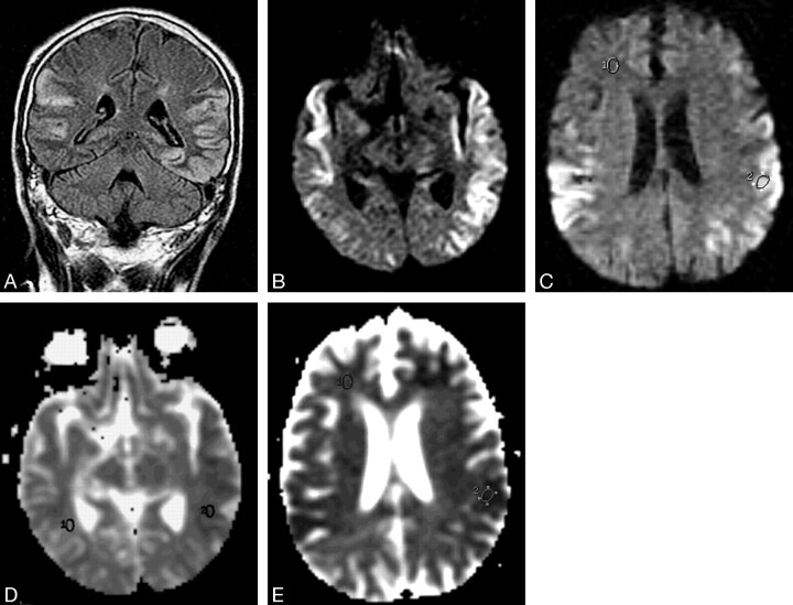
A 65-year-old Chinese man with a history of diabetes mellitus controlled by oral glibenclamide and metformin was admitted to our hospital after a seizure. His family had noticed that he had been drowsy and had become unrousable an hour before the seizure. The patient also had a background of ischemic heart disease, chronic obstructive lung disease, and a cough for the past week. On arrival at the hospital, he was in a coma (Glasgow Coma Scale, 4; eyes 1, verbal 1, motor 2), pupils were nonreactive, but oculocephalic reflex was intact. The patient was tetraplegic, but his vital signs were stable. Immediate bedside blood glucose was 2.1 mmol/L (normal range, 3.1–7.8 mmol/L). Despite emergency infusion of 50% dextrose, and rapid normalization of blood glucose to 12.3 mmol/L, there was no clinical improvement in his level of consciousness. MR imaging of brain performed 5 days after admission showed bilaterally symmetrical hyperintensity involving the temporal, occipital, and part of the frontal lobes on T2-weighted and fluid attenuated inversion recovery (FLAIR) sequences (Fig 1). The affected cortex showed hyperintensity on the corresponding DW MR images (Fig 1B) with decreased ADC. CSF analysis was normal. He developed complications from bronchopneumonia and died on the fifteenth hospital day without regaining consciousness.
Fig 1.
Case 1, a 65-year-old man in a diabetic coma with seizures.
A, Fast spin-echo fluid attenuated inversion recovery (9000 milliseconds/110 milliseconds effective/2200 milliseconds [TR/TE/TI]) MR image shows bilateral hyperintensity of the cortex over the temporal and occipital lobes.
B and C, Diffusion-weighted (10000/105, b value 1000 seconds/mm2) MR images showing corresponding hyperintensity in the cortex.
D and E, ADC maps at the same levels as B and C show decreased ADC in these lesions (618 × 10−3 mm2/s) compared with normal white matter (819 × 10−3 mm2/s).
Case 2
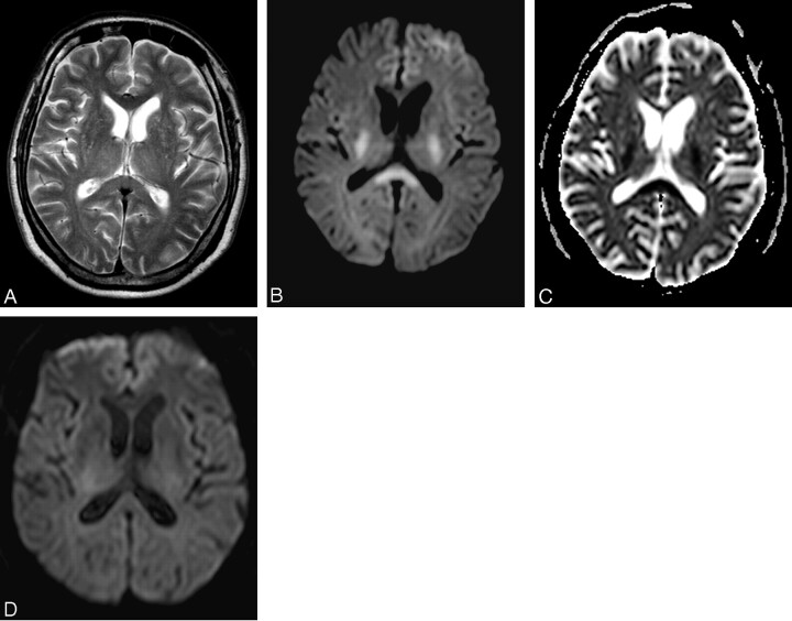
A 69-year-old Chinese man with type 2 diabetes mellitus controlled by oral hypoglycemic agent was seen at the clinic for suspected cardioembolic stoke after several episodes of speech difficulty and generalized weakness. Physical examination showed that he was in atrial fibrillation but had no neurologic deficits. During an echocardiogram, however, the patient suddenly became unresponsive. Urgent MR imaging was performed within 45 minutes for suspected embolic brain stem stroke from atrial fibrillation. Bilaterally symmetrical areas of hyperintensity on DW MR images, with decreased ADC, were seen in the posterior limbs of the internal capsules, corona radiate, and splenium of the corpus callosum (Fig 2B). The T2-weighted MR images and MR angiography were normal. The radiologic impression at the time was acute cerebral infarction. Bedside glucose reading obtained immediately after MR study was 1.9 mmol/L, however, and the patient regained consciousness, recovering rapidly and completely soon after treatment with intravenous dextrose. Follow-up imaging 12 hours later revealed complete resolution of the hyperintense lesions on DW MR, with normalization of the ADC values.
Fig 2.
Case 2, a 69-year-old diabetic man with atrial fibrillation who suddenly became unresponsive.
A, T2-weighted (3000/80 effective) MR image shows subtle increased intensity in the splenium of the corpus callosum (compared with the genu), posterior limbs of the internal capsules, and thalami bilaterally.
B, Corresponding diffusion-weighted (4200/95, b value 1000 seconds/mm2) MR image shows bilaterally symmetrical hyperintensities in the posterior limbs of the internal capsules and the splenium of the corpus callosum.
C, ADC map corresponding to B shows decreased ADC of the splenium of the corpus callosum (485 × 10−3 mm2/s) compared with the genu (890 × 10−3 mm2/s)
D, DW MR image 12 hours later shows complete normalization of previously noted lesions.
Discussion
Hypoglycemia may be caused by overuse of insulin or oral hypoglycemic agents, an undiagnosed insulinoma, or major medical illnesses such as sepsis, renal or hepatic failure, or Addison disease. Glucose is the brain’s main energy substrate, and profound hypoglycemia (with or without complications of ischemia) has been demonstrated to cause neuronal death in pathologic studies.6 There appears to be selective vulnerability of different brain regions to neuronal damage in profound hypoglycemia. Our first patient had widespread signal intensity abnormality in the cerebral cortex of the temporal, occipital, and frontal lobes similar to previous reports.2–4 These lesions were well visualized on late FLAIR and DW MR images, showing reduced ADC.
The mechanism of ADC reduction in hypoglycemia is still unclear, though it may be similar to cerebral infarction and ischemic encephalopathy.7 The usefulness of DW MR imaging in acute cerebral infarction has been well established with intracellular edema and energy failure causing restriction in ADC. Similarly, glucose deprivation may also lead to brain energy failure and membrane ionic pump failure, causing a shift of water into the intracellular space and a drastic reduction of extracellular space volume.8 In an animal study of DW MR imaging in hypoglycemia, Hasegawa et al documented global cortical decline in ADC rapidly spreading to the entire brain. Although the reductions in ADC were transient and were reversed by glucose infusion, neuronal necrosis was observed in the hippocampus and the neocortex in animals with prolonged hypoglycemia.7 Finelli has suggested that DW MR imaging in humans may better define the areas of involvement at an earlier time than conventional MR images and that involvement of the basal ganglia may portend a poor outcome.4 The late MR findings in our first patient, after severe and prolonged hypoglycemia, would be consistent with permanent cytotoxic damage described in these reports.
On the other hand, our second patient did not have gray matter lesions and recovered rapidly after timely treatment. Early DW MR imaging revealed transient ADC decrease with bilaterally symmetrical involvement of the internal capsules, corona radiata, and the splenium of the corpus callosum. This pattern is completely different from our first case and may be the result of a different pathophysiologic process. There has only been one other human case report of reversible DW MR imaging: Aoki et al described bilateral hyperintense lesions within the internal capsule, corona radiate, and frontoparietal cortex in a 73-year-old woman with hypoglycemic coma. After immediate glucose infusion, the patient recovered completely without neurologic deficits, and repeat MR imaging 10 days later showed regression of all lesions.5 Unlike the case described by Aoki et al, our patient did not have any gray matter lesions on DW MR imaging.
The reason the DW changes were limited to the highly directional fibers of the largest white matter tract in our second patient is not known. Experimental evidence in animals suggests that excitatory amino acids released into the extracellular space, and not simply glucose starvation, may be important in the pathogenesis of hypoglycemic neuronal damage.6 Increased sensitivity of DW MR imaging to the bulk movement of water from the extracellular space into the intracellular space early in hypoglycemia may represent changes due to reduction in the extracellular space without cell necrosis. Bulk water movement, which has been suggested as a mechanism for similar reversible DW MR findings limited to the splenium of the corpus callosum in influenza and other childhood infections,9 antiepileptic drug treatment,10 and venous thrombosis (Tang et al, unpublished results) appear to support this hypothesis. The finding in animal studies that ADC depression spreads from the cortex to the subcortical white matter, and not the other way around, however, appears to militate against this theory.7 Furthermore, whether the distribution of DW MR abnormalities is related to the duration of hypoglycemia, whether certain individuals are more resistant to hypoglycemic necrosis, and the time necessary for these lesions to reverse (10 days in the previous report and 12 hours in ours) are unknown. Further studies, perhaps supplemented by diffusion tensor imaging,11 might be useful to elucidate the pathophysiologic cause of these findings.
Our second patient also illustrates the problem of differential diagnosis of DW MR imaging findings in hypoglycemic patients. In both reported cases of reversible DW MR imaging abnormalities, these findings were initially misinterpreted as acute cerebral infarction.5 These examples illustrate a potential pitfall in diabetic patients, who might be at risk for both cerebral infarction and hypoglycemia, especially as DW MR imaging is now a common pulse sequence used to image a wide variety of patients and clinical situations. Moreover, it is important to recognize and consider the possibility of hypoglycemic coma because it can be effectively treated and reversed, leading to complete recovery.
In conclusion, we describe 2 cases of hypoglycemic coma in which transient white matter DW MR abnormalities were associated with complete recovery while widespread cortical lesions were associated with poor outcome and death. The striking difference in the neuroimaging patterns may be helpful in prognosis and differential diagnosis in hypoglycemic coma, and DW MR imaging may be useful in studying the pathophysiology of how low blood glucose affects the brain.
References
- 1.Lockwood AH. Toxic and metabolic encephalopathies. In: Bradley WG, Daroff RB, Fenichal GM, et al, eds. Neurology in clinical practice. 3rd ed. London: Butterworth-Heinemann;2000. :1485
- 2.Boeve BF, Bell DG, Noseworthy JH. Bilateral temporal lobe MRI changes in uncomplicated hypoglycemic coma. Can J Neurol Sci 1995;22:56–58 [DOI] [PubMed] [Google Scholar]
- 3.Fujioka M, Okuchi K, Hiramatsu KI, et al. Specific changes in human brain after hypoglycemic injury. Stroke 1997;28:584–87 [DOI] [PubMed] [Google Scholar]
- 4.Finelli PF. Diffusion-weighted MR in hypoglycemic coma. Neurology 2001;57:933–35 [DOI] [PubMed] [Google Scholar]
- 5.Aoki T, Sato T, Hasegawa K, et al. Reversible hyperintensity lesion on diffusion-weighted MRI in hypoglycemic coma. Neurology 2004;63:392–93 [DOI] [PubMed] [Google Scholar]
- 6.Auer RN. Progress review: hypoglycemic brain damage. Stroke 1986;17:699–708 [DOI] [PubMed] [Google Scholar]
- 7.Hasegawa Y, Formato JE, Latour LL, et al. Severe transient hypoglycemia causes reversible change in the apparent diffusion coefficient of water. Stroke 1996;27:1648–56 [DOI] [PubMed] [Google Scholar]
- 8.Pelligrino D, Almquist LO, Siesjo BK. Effects of insulin-induced hypoglycemia on intracellular pH and impedance in the cerebral cortex of the rat. Brain Res 1981;221:129–47 [DOI] [PubMed] [Google Scholar]
- 9.Takanashi J, Barkovich AJ, Yamaguchi K, et al. Influenza-associated encephalitis/encephalopathy with a reversible lesion in the splenium of the corpus callosum: a case report and literature review. AJNR Am J Neuroradiol 2004;25:798–802 [PMC free article] [PubMed] [Google Scholar]
- 10.Kim SS, Chang KH, Kim ST, et al. Focal lesion in the splenium of the corpus callosum in epileptic patients: antiepileptic drug toxicity? AJNR Am J Neuroradiol 1999;20:125–29 [PubMed] [Google Scholar]
- 11.Lim CC, Yin H, Loh NK, et al. Malformations of cortical development: high-resolution MR and diffusion tensor imaging of fiber tracts at 3T. AJNR Am J Neuroradiol 2005;26:61–64 [PMC free article] [PubMed] [Google Scholar]


