Metachromatic leukodystrophy (MLD) is a set of several disorders caused by deficient lysosomal activity. This deficiency results in accumulation of a metachromatic lipid material, galactosylceramide sulfatide, leading to the breakdown of the myelin sheath in both central and peripheral nervous systems, initially sparing the subcortical “U” fibers.1,2 Several MR imaging features of MLD have been described, but to the best of our knowledge, cranial nerve involvement demonstrated by MR imaging was never mentioned in this setting.
An undernourished 2-year-old girl was hospitalized as a result of uncontrollable vomiting and retarded neuropsychomotor development. Deep tendon reflexes were abolished in the 4 limbs, and the plantar reflex showed bilateral extension response. The routine laboratory tests, cranial nerve examination, and funduscopy were unremarkable. Electroneuromyography showed signs of predominantly sensory peripheral neuropathy in the lower limbs, with a demyelinating pattern that involved all 4 of the limbs, and an ultrastructural examination of the sural nerve was carried out, which showed abnormalities consistent with MLD. The disease was confirmed by a severe decrease of arylsulfatase A activity in leukocytes (1.0 nmol/h/mg of protein; reference range, 5–20 nmol/h/mg).
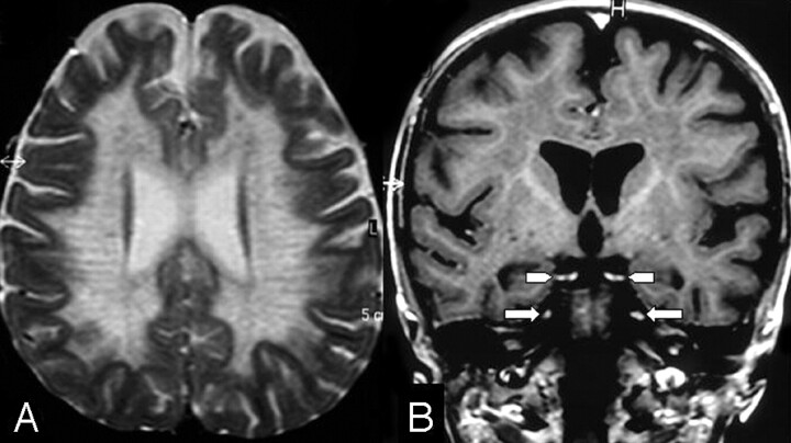
The baseline MR imaging scan showed bilateral and symmetrical involvement of the posterior periventricular white matter and splenium of the corpus callosum, sparing the U fibers. Gadolinium (Gd)-enhanced T1-weighted images showed bilateral and symmetrical abnormal enhancement of several cranial nerves, including the optic, oculomotor, trigeminal, abducens, facial, and vestibulocochlear nerves. The follow-up scan (Fig 1) demonstrated diffuse and symmetrical demyelination in the periventricular white matter and centrum semiovale, with numerous hypointense linear structures in a radiating (“tigroid”) pattern, with involvement of the internal capsules, corticospinal tracts, cerebral peduncles, and cerebellar white matter. There was no evidence of leptomeningeal or parenchymal enhancement. There was no evidence of nerve hypertrophy. There was a considerable worsening of the neurologic deficits characterized by spastic tetraplegia without any contact with the environment.
Fig 1.
A, Axial T2-weighted image shows the white matter involvement with a tigroid pattern.
B, Coronal postcontrast T1-weighted image shows bilateral and symmetrical abnormal Gd enhancement of the oculomotor (arrowheads) and trigeminal nerves (arrows).
Cranial nerve Gd enhancement can be seen in a variety of entities, including neoplastic, infectious, demyelinating, and idiopathic diseases.3–5 Among leukodystrophies, cranial nerve involvement has been documented in infantile Krabbe disease.6,7 Proposed mechanisms for the abnormal nerve root enhancement have included altered vascular permeability with breakdown of the blood-nerve barrier as a result of perivascular inflammation or infiltration8 and enhancement in areas of active myelin breakdown.6
We presume that multiple cranial nerve enhancement in MLD is secondary to the accumulation of the metachromatic lipid material and to the disruption of the myelin sheath, similar to the process that affects peripheral nerves. A future investigation with a larger series is required to evaluate the usefulness of cranial nerve MR imaging analysis in suspected MLD patients.
References
- 1.Cheon JE, Kim IO, Hwang YS, et al. Leukodystrophy in children: a pictorial review of MR imaging features. Radiographics 2002;22:461–76 [DOI] [PubMed] [Google Scholar]
- 2.Faerber EN, Melvin J, Smergel EM. MRI appearances of metachromatic leukodystrophy. Pediatr Radiol 1999;29:669–72 [DOI] [PubMed] [Google Scholar]
- 3.Saremi F, Helmy M, Farzin S, et al. MRI of cranial nerve enhancement. AJR Am J Roentgenol 2005;185:1487–97 [DOI] [PubMed] [Google Scholar]
- 4.da Silva CJ, da Rocha AJ, Mendes MF, et al. Trigeminal involvement in multiple sclerosis: magnetic resonance imaging findings with clinical correlation in a series of patients. Mult Scler 2005;11:282–85 [DOI] [PubMed] [Google Scholar]
- 5.de Fátima Soares M, Braga FT, da Rocha AJ, et al. Optic nerve infiltration by acute lymphoblastic leukemia: MRI contribution. Pediatr Radiol 2005;35:799–802 [DOI] [PubMed] [Google Scholar]
- 6.Bernal OG, Lenn N. Multiple cranial nerve enhancement in early infantile Krabbe's disease. Neurology 2000;54:2348–49 [DOI] [PubMed] [Google Scholar]
- 7.Jones BV, Barron TF, Towfighi J. Optic nerve enlargement in Krabbe's disease. AJNR Am J Neuroradiol 1999;20:1228–31 [PMC free article] [PubMed] [Google Scholar]
- 8.Vasconcellos E, Smith M. MRI nerve root enhancement in Krabbe disease. Pediatr Neurol 1998;19:151–52 [DOI] [PubMed] [Google Scholar]


