Abstract
Viral hepatitis C represents a significant liver pathology worldwide, with a detrimental impact on national health systems. The present study aimed to correlate the levels of serum α-fetoprotein (AFP) with prognostic tools such as Fibroscan®, the presence of mixed cryoglobulinemia, and various demographic and standard biochemical markers, in patients with chronic hepatitis C, unrelated to hepatocellular carcinoma (HCC). A clinical study was designed considering three study groups: Hepatitis C virus (HCV) group including 35 patients with chronic hepatitis C and detectible viral load; sustained viral response (SVR) group including 20 HCV patients without detectable virus load 12 weeks after therapy cessation; a control group represented by 37 healthy volunteers. It was observed that serum AFP was moderately increased in the HCV and SVR groups and was positively correlated with aspartate transaminase (AST), alkaline phosphatase (AP), and γ-glutamyl transferase (GGT). The incidence of mixed cryoglobulinemia was increased in the HCV group, and the degree of fibrosis assessed by Fibroscan® was increased in both the HCV and SVR groups. In conclusion, the data revealed that a moderate increase in AFP levels could be present in patients with HCV even in the absence of HCC, unrelated to viral load or therapy response and that there was a linear positive correlation between serum levels of AFP and the degree of hepatic cytolysis and cholestasis. Additionally, mixed cryoglobulinemia was present in HCV patients with patent viral load, decreasing in those with SVR after therapy cessation unrelated to any renal impairment, while the degree of fibrosis was increased in HCV-infected patients, with no reversibility 12 weeks after successful therapy.
Keywords: hepatitis C virus, α-fetoprotein, mixed cryoglobulinemia, liver fibrosis, sustained viral response
Introduction
Hepatitis C virus (HCV) is a single-stranded RNA virus, a source of debilitating liver disease ranging from hepatitis to cirrhosis and HCC. Approximately 170 million people are infected with HCV worldwide (1).
Among the extrahepatic manifestations of HCV infection, an important role is played by renal involvement, being described as a series of histopathological forms: Proliferative membrane glomerulonephritis, IgA nephropathy, focal segmental glomerulosclerosis, fibrillar glomerulonephritis, tubulointerstitial nephropathy and thrombosis (2,3). The prevalence of HCV infection in patients with chronic kidney disease (CKD) is between 5 and 60% in developed countries, with a certain predominance in hemodialysis patients (3). HCV-positive hemodialysis patients show a decreased survival rate; the risk of death associated with liver disease being aggravated by the increased risk of cardiovascular death attributed to malnutrition and chronic inflammation (4).
HCV induces a low-amplitude T and B lymphocyte-mediated immune response, which leads to long-lasting infection and production of non-specific biochemical markers (3-5). Thus, disease progression is difficult to predict. Efforts should be made to identify patients with HCV and liver inflammation secondary to the viral infection before progression to a more difficult to treat illness such as HCC and cirrhosis (6). The use of routine biochemical markers as the tools for therapeutic response is essential for disease prognosis, similar to biomarker applicability in other pathologies (7,8).
There are different non-specific clinical, biochemical and sonographic tools for the evaluation of the clinical impact of HCV infection: Age, social status, smoking status, platelet count, serum cholesterol and triglycerides, albumin, creatinine, bilirubin, fibrinogen, hemostasis parameters, liver aminotransferases [aspartate transaminase (AST), alanine aminotransferase (ALT)], alkaline phosphatase (AP), γ-glutamyl transferase (GGT), mixed cryoglobulinemia (MC), α-fetoprotein (AFP), and assessment of liver fibrosis by non-invasive methods (sonographic) (9-11).
AFP is a fetal protein synthesized in the same chromosome location as serum albumin. AFP is considered similar to fetal serum albumin, achieving high levels during fetal development and sharing almost the same functions, binding heavy metals, fatty acids and bilirubin. This protein can be found in monomeric, dimeric or trimeric forms. In rodents, it has an important anti-estrogenic property by binding maternal estradiol and preventing virilization of the fetus. The exact role in human adults, however, is not yet elucidated (12). This protein is considered an important serological marker in various hepatic and non-hepatic pathologies such as abnormalities of fetal development (aneuploidies), ataxia-telangiectasia, and various malignancies (yolk sac tumor, HCC, liver metastasis, non-seminomatous germ cell tumors, teratomas, hepatoblastomas, Gruber-Frantz tumor) (13).
Elevated AFP is independently associated with a decreased level of albumin (<3.5 g/dl) (14). Elevated AFP has also been shown to be independently associated with elevated uric acid (10). Hyperuricemia is both a marker of cardiovascular risk and an independent risk factor for the occurrence of high blood pressure and heart failure (15). Arterial stiffness and vascular calcification are major contributors to cardiovascular disease and are independent predictors of cardiovascular mortality in patients with end-stage renal disease (ESRD) (16-18).
Although the role of AFP in HCC is well established, low to moderate plasma levels of AFP in patients with chronic hepatitis C in the absence of HCC have also been described (12). A plasma level higher that 200 ng/ml is considered highly sensitive for HCC in patients with HCV, while levels of 5 to 60 ng/ml are routinely found in patients with HCV, but lacking clinical significance (12).
Correlative biochemical and clinical interactions between AFP and other routinely used markers in HCV infection have been poorly studied. HCV could induce, for example, a greater plasma increase in AFP compared to hepatitis B virus (HBV) (19). The degree of fibrosis and/or inflammation also influences AFP plasma concentration (20).
Research has also revealed correlations between AST, ALT, AST/ALT ratio, bilirubin, platelet count, and the HCV genotype and plasma AFP (20).
AFP could also be used as a marker of antiviral therapy response in HCV infection and could represent a good biochemical tool in quantifying the sustained viral response to therapy (21,22).
Hematologic and metabolic alterations ranging from minor to severe, in accordance with disease progression are generally accepted in the literature. There are, however, contradictory data regarding correlations of AFP levels in HCV patients, without cirrhosis or HCC.
Mixed cryoglobulinemia is another biochemical feature of HVC infection, having a partially elucidated significance (2). According to Schamberg and Lake-Bakaar (2) and Ozkok and Yildiz (23), mixed cryoglobulinemia is related to virus replication, membranoproliferative glomerulonephritis, and therapy response. Cryoglobulinemia is present in a wide range of hematologic, infectious, and rheumatologic diseases. Moreover, mixed cryoglobulinemia is almost consistently present in HCV infections, more so than in HBV infections or other non-infectious diseases (24). The correlative potential of AFP with mixed cryoglobulinemia in patients with HCV infection has not yet been fully explored.
Sonographic imaging of patients with chronic hepatitis is a reliable and non-invasive tool for assessing liver progression to cirrhosis. Using modern interfaces such as Fibroscan®, it is currently able to quantify the degree of fibrosis (25,26). However, the relationship between biochemical and sonographic data in HCV patients is still an unresolved issue.
This study aimed to identify novel clinical, biochemical and sonographic paradigms, which correlate with the plasma levels of AFP in patients with chronic hepatitis C. We considered demographic data (age, social status, smoking status, antiviral therapies, weight and height), biochemical markers [total hemoglobin, white blood cell count, platelet count, serum cholesterol, triglycerides, blood urea nitrogen, creatinine, direct bilirubin (DB), total bilirubin (TB), fibrinogen, activated partial thromboplastin time (APTT), international normalized ratio (INR), Na, K, C-reactive protein (CRP), erythrocyte sedimentation rate (ESR), procalcitonin (PCT), AST, ALT, AP, GGT, MC] and sonographic data (Fibroscan®).
Patients and methods
Ethic statement
This study is a monocentric, randomized, double-blinded, prospective, clinical trial, which focused on the comparison and correlation data regarding AFP in HCV patients, without cirrhosis or HCC. All clinical procedures were carried out following approval of the local Ethics Committee of Fundeni Clinical Institute (Romania) (no. 48358/01.10.2019) for clinical trials in accordance with the European Communities Council Directive 2001/20/EC and with respect to personal data privacy (European Directive 95/46/EC).
Demographic data collection and blood sampling
After obtaining informed consent for sampling and collecting the data, 37 healthy volunteers were enrolled in the control group (C group). Another 108 patients were initially enrolled in the study group in accordance with the inclusion criteria: Detectible HCV infection over a six-month period, life expectancy over 12 months, and no decompensated cirrhosis.
In order to minimize the confounding factors, we excluded the patients with structural renal illness, decompensated heart failure, concurrent alcohol and drug use, extrahepatic manifestation of HCV Infection, HBV co-infection and HIV co-infection. After exclusion, we integrated the data of 35 HCV patients, designing an HCV group. For more accuracy in comparing the data, a further 20 HCV patients with sustained viral response (SVR) to antiviral therapy were included in the SVR group. We considered SVR the absence of virus in the blood 12 weeks after treatment cessation (27). The HCV and SVR groups were matched with the control group in regards to age and sex.
Demographic data such age, sex, social status, smoking status, weight and height were obtained using a questionnaire. The Body-Mass Index was calculated for each patient.
All biochemical markers were assessed in the clinical laboratory following a local sampling protocol. Whole blood was collected in sampling tubes provided with clotting accelerator and maintained at 4˚C. After clotting, the samples were centrifuged for 20 min at approximately 1,000 x g and assessed immediately with no freeze/thaw cycles for the markers detailed below.
We assessed, spectrophotometrically, the levels of albumin (g/dl), AST (U/l), ALT (U/l), GGT (U/l), AP (U/l), DB (mg/dl), TB (mg/dl), CRP (mg/l), triglyceride (mg/dl), and serum cholesterol (mg/dl) using compatible biochemistry kits for Siemens Advia 1800 Reader (Siemens, Erlangen-Germany). Using chemiluminescence, plasmatic AFP (ng/ml) and the presence of mixed cryoglobulinemia was determined on an Advia Centaur XPT Reader (Siemens). Plasmatic Na and K were determined using a potentiometric method (ion selective electrode) on an Advia 1800 Reader (Siemens). The results are expressed in mmol/l. Complete blood count analysis was performed with dedicated kits with the means of an automatic flow cytometry technique (Sysmex XN-1000 Reader) (Sysmex, Kobe, Japan) and the results are expressed per ml blood.
For the coagulation analysis, we sampled whole blood using EDTA sampling tubes and following the manufacturer's recommendation, we assessed using a StaR Max 3 Reader (Stago, France) the following coagulation parameters: aPTT (sec), PT/INR (sec), and plasmatic fibrinogen (mg/dl).
The virus replication level was assessed using RT-PCR (Montania 4896 Real Time PCR Reader; Anatolia, Istanbul, Turkey) using Bosphore HCV quantification kit (Anatolia) in accordance with the manufacturer's recommendation. The main steps include d: Total RNA collection, reverse transcription and amplification in accordance with the local protocol. All plasma samples were performed in triplicate and the viral load was expressed per ml.
Non-invasive assessment of liver fibrosis
The non-invasive measurement of liver fibrosis was performed using Fibroscan® technology, Model 530 compact (Echosens, France). The results were expressed automatically in kPa and staged in 4 degrees according to the literature, with 0 representing no fibrosis and 4 representing major fibrosis (28).
Data analysis
Data analysis and graphical representations were performed using GraphPad Prism 6.00 (GraphPad Software Inc.). We assessed the groups for normal distribution using D' Agostino-Pearson omnibus normality test and Shapiro-Wilk test. For the comparative analysis we considered demographic, biochemical and sonographic data and for numerical results, we used one-way measure ANOVA followed by post-hoc Tukey's test for interactions between groups. The data are expressed as mean ± SD. A two-sided P-value <0.05 was considered statistically significant. For binary data (social status, smoking status and MC), we used the Chi-squared test.
For the correlative data, we used a linear correlation model, between AFP and all other variables including: DB, TB, CRP, serum cholesterol, triglycerides, mixed cryoglobulinemia, Na, K, complete blood count, aPTT, PT/INR, fibrinogen, viral load and Fibroscan®. The Pearson correlation coefficient, r, and R squared (R2) were determined. We considered a two-sided P-value <0.05 statistically significant, for each correlation. We assigned R2 values under 0.15 to have negligible correlative effect.
Results
Demographic data
We did not observe any differences among groups concerning age, sex or smoking status. The results are revealed in Table I.
Table I.
Group distribution according to age, sex, and smoking status.
| Parameters | Control group (n=37) | HCV group (n=35) | SVR group (n=20) | P-value |
|---|---|---|---|---|
| Mean age (years) | 56.3±1.34 | 62.2±1.98 | 64.5±3.04 | NS |
| Sex (F/M %) | 59.45/40.54 | 65.42/34.58 | 67.5/32.5 | NS |
| Smoking (%) | 13.51 | 11.42 | 18.18 | NS |
HCV, hepatitis C virus; SRV, sustained viral response; F, female; M, male; NS, not significant.
AFP, cryoglobulinemia and FibroScan® dynamics between groups
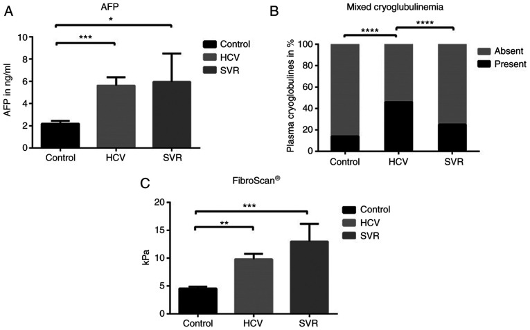
The plasma levels of AFP between groups are illustrated in Fig. 1A. The plasma levels of AFP in the HCV and SVR groups were significantly increased when compared to the control group (5.79±0.7 vs. 2.4±0.3, P=0.0009 and 5.9±2.5 vs. 2.4±0.27, P=0.042, respectively). No difference in AFP was observed between the HCV and SVR group [5.79±0.73 vs. 5.9±2.5, P>0.05 (NS, not significant)]. The presence of mixed cryoglobulinemia in plasma among the groups is shown in Fig. 1B. We observed a statistically increased incidence of mixed cryoglobulinemia in the HCV group when compared to both the control group (46 vs. 14%, P<0.0001) and SVR group (46 vs. 25%, P<0.0001).
Figure 1.
AFP, mixed cryoglobulinemia and Fibroscan® dynamics between groups. (A) Serum AFP, (B) incidence of mixed cryoglobulinemia and (C) liver fibrosis using FibroScan® were assessed in healthy volunteers (Control), Hepatitis C virus-infected patients with detectable viral load (HCV) and hepatitis C virus-infected patients 12 weeks after sustained viral response (SVR). Bars are mean ± SD. *P<0.05, **P<0.01, ***P<0.001 and ****P<0.0001. AFP, α-fetoprotein.
The Fibroscan® results are revealed in Fig. 1C. We identified significantly increased fibrosis in both the HCV and SVR groups when compared to the control group: 9.8±0.9 vs. 4.5±0.37, P=0.001 and 13±3.2 vs. 4.5±0.37, P=0.0006, respectively. No significant difference was observed between the HCV and SVR groups (9.8±1 vs. 13±3.2, NS).
AFP depends mainly on serum AST, GGT, and AP
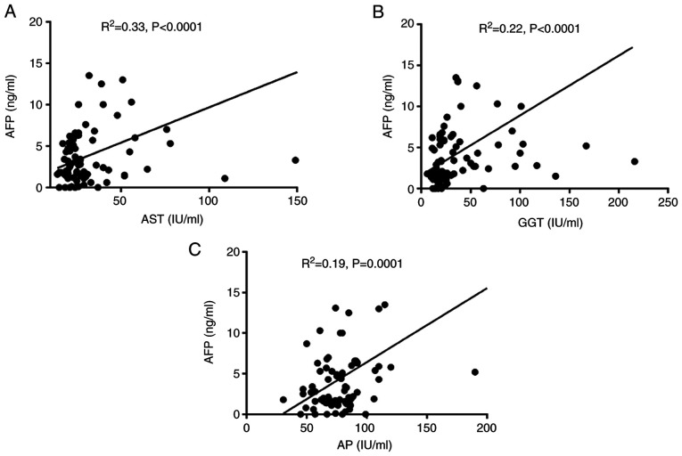
By means of a linear correlation model between AFP and all other biochemical and sonographic variables, we observed statistically significant minor positive correlations between AFP and AST (R2=0.33, P<0.0001), GGT (R2=0.22, P<0.0001), and AP (R2=0.19, P=0.0001) in the HCV-infected patients (HCV and SVR group respectively) (Fig. 2A-C). All other variables had either negligible (R2<0.15) or no correlative effect (P>0.05, NS).
Figure 2.
Correlations between serum AFP and (A) aspartate aminotransferase (AST), (B) γ-glutamyl transferase (GGT) and (C) alkaline phosphatase (AP) in the enrolled HCV-infected patients (HCV and SVR group respectively). R2 represents the squared correlation coefficient. AFP, α-fetoprotein; SVR, sustained viral response; HCV, hepatitis C virus.
Discussion
α-fetoprotein (AFP) is a well-known diagnostic tool for hepatitis C virus (HCV) patients when associated with hepatocellular carcinoma (HCC). AFP is, in fact, a protein present in high concentrations in a wide range of diseases, having a liver origin in the fetus (13,29). The cut-off level of AFP in predicting HCC is, however, controversial. Values between 17.4 ng/ml for early-stage HCC, to 200 ng/ml for recurrent HCC after liver transplantation are advocated (30,31). Considering that AFP stratifies the risk of developing HCC in patients with HCV, we observed that AFP values were statistically higher in patients with HCV unrelated to viral load or the presence of sustained viral response (SVR) after therapy in patients without HCC. Although we detected plasma AFP values under the cut-off values advocated in the literature as highly sensitive for HCC, our study reveals a controversial mean of this protein in HCV patients in the absence of HCC. Moreover, AFP could be an important outcome marker in hepatitis C, even if the patients are not yet diagnosed with HCC, regardless of the clinical response to therapy. The persistence of plasmatic AFP in patients with HCV and SVR suggests that the likelihood of developing HCC in the future despite long-term response to antiviral therapy could be a significant clinical issue. Conversely, a decreased clearance of AFP after SVR could be advocated. According to Chen et al, a longer follow-up period after SVR might influence the plasmatic levels of AFP (32).
Additionally, we observed a statistically significant direct correlation between AFP and aspartate transaminase (AST). Thus, AFP could quantify, indirectly, the magnitude of HCV-induced liver cytolysis. Our data are in accordance with Chen et al, whose study identified the predictive value of AFP to therapy response and the role of AST elevation as an independent risk factor for plasma AFP presence (32). Considering that cholestasis syndrome [high alkaline phosphatase (AP), γ-glutamyl transferase (GGT)] could be an independent factor for HCC in patients with viral hepatitis, especially in those with cirrhosis, the cumulative biochemical evidence of low to moderate plasmatic AFP and elevated GGT and AP could represent a future biochemical algorithm for identifying patients with HCV at risk of developing HCC (33). There are, however, inconsistent data, regarding patients with HCV in the absence of cirrhosis and their risk to develop HCC, when low to moderate levels of AFP are associated with high plasmatic GGT and FA levels. We identified a strong correlation between AFP levels, GGT, and FA as predictive variables in HCV patients without cirrhosis or HCC. The clinical significance should be, however, further explored.
Regarding mixed cryoglobulinemia, we observed a statistically significant increase in the HCV group compared to the control group. Mixed cryoglobulinemia in HCV infection is an important qualitative serologic tool of viral replication and HCV-related renal injury (2,22). Thus, we observed the presence of mixed cryoglobulinemia in patients with active disease conditions, unrelated to viral load. Moreover, patients with SVR showed a decreased incidence of plasmatic mixed cryoglobulinemia, confirming the role of this diagnostic tool in the qualitative assessment of disease activity. According to Landau et al, MC in the context of SVR is more an exception (34).
Furthermore, patients with HCV-related glomerulonephritis were not included in the present study. According to Santoriello et al, the presence of membranoproliferative glomerulonephritis is typically secondary to HCV but not to HBV and the presence of mixed cryoglobulinemia correlates with this renal condition (35).
Regarding Fibroscan® examination, our study results are in accordance with the literature. We detected an increased fibrosis score in HCV patients when compared to healthy volunteers. The fibrosis degree was, however, the same between the HCV and the SVR group. This paradox could be explained due the longer time until fibrosis resolution could be present in HCV patients after SVR (36). There are controversial data in the literature regarding the reversibility of liver fibrosis secondary to HCV by means of Fibroscan®. Some data advocate a relatively slow resolution of fibrosis in time after SVR. According to studies, the resolution could occur 1 year after SVR and is less predictive of successful therapy compared to liver biopsy (36,37). Furthermore, it is likely that earlier stages of liver fibrosis are more easily reversible than the later stages (38).
In conclusion, our study opens a new research pathway in quantifying HCV-related liver lesions by means of modern serologic biomarkers and sonographic assessment including AFP, MC, and Fibroscan® in the absence of a diagnosed neoplastic state.
Acknowledgements
The authors would like to thank Ileana Constantinescu for her help with blood test analysis.
Funding Statement
Funding: No funding was received.
Availability of data and materials
Due to confidentiality reasons data generated or analyzed during this study are not included in this published article.
Authors' contributions
TI conributed to all of the following aspects including design of the study, collection and analysis of the data, writing and reviewing of the manuscript. SIs, SIo and MDT wrote and reviewed the manuscript, analyzed the data, and contributed to the study design. EM, DGB, and AT performed the literature search and selected the studies to be included. LI coordinated and designed the study. All authors read and approved the manuscript and agree to be accountable for all aspects of the research in ensuring that the accuracy or integrity of any part of the work are appropriately investigated and resolved.
Ethics approval and consent to participate
This study was approved by the local Ethics Committee of Fundeni Clinical Institute (Romania), approval no. 48358/01.10.2019. Written informed consent was obtained from all patients prior to publication.
Patient consent for publication
Not applicable.
Competing interests
The authors declare that they have no competing interests.
References
- 1.Petruzziello A, Marigliano S, Loquercio G, Cozzolino A, Cacciaputi C. Global epidemiology of hepatitis C virus infection: An up-date of the distribution and circulation of hepatitis C virus genotypes. World J Gastroenterol. 2016;22:7824–7840. doi: 10.3748/wjg.v22.i34.7824. [DOI] [PMC free article] [PubMed] [Google Scholar]
- 2.Schamberg NJ, Lake-Bakaar GV. Hepatitis C virus-related mixed cryoglobulinemia: Pathogenesis, Clinica manifestations, and new therapies. Gastroenterol Hepatol (NY) 2007;3:695–703. [PMC free article] [PubMed] [Google Scholar]
- 3.Timofte D, Dragos D, Balcangiu-Stroescu AE, Tanasescu MD, Gabriela Balan D, Avino A, Tulin A, Stiru O, Ionescu D. Infection with hepatitis C virus in hemodialysis patients: An overview of the diagnosis and prevention rules within a hemodialysis center (Review) Exp Ther Med. 2020;20:109–116. doi: 10.3892/etm.2020.8606. [DOI] [PMC free article] [PubMed] [Google Scholar]
- 4.Balcangiu-Stroescu AE, Tanasescu MD, Diaconescu AC, Raducu L, Constantin AM, Balan DG, Tarmure V, Ionescu D. Cardiovascular comorbidities, inflammation and serum albumin levels in a group of hemodialysis patients. Rev Chim Buchar. 2018;69:926–929. [Google Scholar]
- 5.Dustin LB. Innate and adaptive immune responses in chronic HCV infection. Curr Drug Targets. 2017;18:826–843. doi: 10.2174/1389450116666150825110532. [DOI] [PMC free article] [PubMed] [Google Scholar]
- 6.Suceveanu AI, Stoian AP, Mazilu L, Voinea F, Hainarosie R, Diaconu CC, Pituru S, Nitipir C, Badiu DC, Ceausu I, Suceveanu AP. Interferon-free therapy is not a trigger for hepatocellular carcinoma in patients with chronic infection with hepatitis C virus. Farmacia. 2018;66:904–908. [Google Scholar]
- 7.Marinescu I, Schenker RA, Stovicek PO, Marinescu D, Ciobanu CF, Papacocea SI, Manea MC, Papacocea RI, Manea M, Chirita R, Ciobanu AM. Biochemical factors involved in the unfavorable evolution of prostate cancer. Rev Chim. 2019;70:3343–3347. [Google Scholar]
- 8.Papacocea MT, Badarau IA, Radoi M, Papacocea IR. The predictive role of biochemical plasma factors in patients with severe traumatic brain injuries. Rev Chim Buchar. 2019;70:1754–1757. [Google Scholar]
- 9.Sheridan DA, Aithal G, Alazawi W, Allison M, Anstee Q, Cobbold J, Khan S, Fowell A, McPherson S, Newsome PN, et al. Care standards for non-alcoholic fatty liver disease in the United Kingdom 2016: A cross-sectional survey. Frontline Gastroenterol. 2017;8:252–259. doi: 10.1136/flgastro-2017-100806. [DOI] [PMC free article] [PubMed] [Google Scholar]
- 10.Sagnelli E, Santantonio T, Coppola N, Fasano M, Pisaturo M, Sagnelli C. Acute hepatitis C: Clinical and laboratory diagnosis, course of the disease, treatment. Infection. 2014;42:601–610. doi: 10.1007/s15010-014-0608-2. [DOI] [PubMed] [Google Scholar]
- 11.Fierbinteanu-Braticevici C, Papacocea R, Tribus L, Baicus C. Role of 13C methacetin breath test for non invasive staging of liver fibrosis in patients with chronic hepatitis C. Indian J Med Res. 2014;140:123–129. [PMC free article] [PubMed] [Google Scholar]
- 12.Christiansen M, Høgdall CK, Andersen JR, Nørgaard-Pedersen B. Alpha-fetoprotein in plasma and serum of healthy adults: Preanalytical, analytical and biological sources of variation and construction of age-dependent reference intervals. Scand J Clin Lab Invest. 2001;61:205–215. doi: 10.1080/003655101300133649. [DOI] [PubMed] [Google Scholar]
- 13.Spătaru RI, Enculescu A, Popoiu MC. Gruber-Frantz tumor: A very rare pathological condition in children. Rom J Morphol Embryol. 2014;55:1497–1501. [PubMed] [Google Scholar]
- 14.Chen CH, Lin ST, Kuo CL, Nien CK. Clinical significance of elevated alpha-fetoprotein (AFP) in chronic hepatitis C without hepatocellular carcinoma. Hepatogastroenterology. 2008;55:1423–1427. [PubMed] [Google Scholar]
- 15.Timofte D, Mandita A, Balcangiu-Stroescu AE, Balan D, Raducu L, Tanasescu MD, Diaconescu A, Dragos D, Cosconel CI, Stoicescu SM, Ionescu D. Hyperuricemia and cardiovascular diseases-clinical and paraclinical correlations. Rev Chim. 2019;70:1045–1046. [Google Scholar]
- 16.Timofte D, Dragoș D, Balcangiu-Stroescu AE, Tănăsescu MD, Gabriela Bălan D, Răducu L, Tulin A, Stiru O, Ionescu D. Abdominal aortic calcification in predialysis patients: Contribution of traditional and uremia-related risk factors. Exp Ther Med. 2020;20:97–102. doi: 10.3892/etm.2020.8607. [DOI] [PMC free article] [PubMed] [Google Scholar]
- 17.Timofte D, Ionescu D, Medrihan L, Mandita A, Rasina A, Damian L. Vascular calcification and bone disease in hemodialysis patient, assessment, association and risk factors. Nephrol Dial Transplant. 2007;22:325–326. [Google Scholar]
- 18.Gaman MA, Dobrica EC, Pascu EG, Cozma MA, Epingeac ME, Gaman AM, Pantea Stoian AM, Bratu OG, Diaconu CC. Cardio metabolic risk factors for atrial fibrillation in type 2 diabetes mellitus: Focus on hypertension, metabolic syndrome and obesity. J Mind Med Sci. 2019;6:157–161. [Google Scholar]
- 19.Emokpae MA, Adejumol BG, Abdu A, Sadiq NM. Serum alpha-fetoprotein level is higher in hepatitis C than hepatitis B infected chronic liver disease patients. Niger Med J. 2013;54:426–429. doi: 10.4103/0300-1652.126302. [DOI] [PMC free article] [PubMed] [Google Scholar]
- 20.Tai WC, Hu TH, Wang JH, Hung CH, Lu SN, Changchien CS, Lee CM. Clinical implications of alpha-fetoprotein in chronic hepatitis C. J Formos Med Assoc. 2009;108:210–218. doi: 10.1016/S0929-6646(09)60054-1. [DOI] [PubMed] [Google Scholar]
- 21.Di Bisceglie AM, Sterling RK, Chung RT, Everhart JE, Dienstag JL, Bonkovsky HL, Wright EC, Everson GT, Lindsay KL, Lok AS, et al. Serum alpha-fetoprotein levels in patients with advanced hepatitis C: Results from the HALT-C trial. J Hepatol. 2005;43:434–441. doi: 10.1016/j.jhep.2005.03.019. [DOI] [PubMed] [Google Scholar]
- 22.Abdoul H, Mallet V, Pol S, Fontanet A. Serum alpha-fetoprotein predicts treatment outcome in chronic hepatitis C patients regardless of HCV genotype. PLoS One. 2008;3(e2391) doi: 10.1371/journal.pone.0002391. [DOI] [PMC free article] [PubMed] [Google Scholar]
- 23.Ozkok A, Yildiz A. Hepatitis C virus associated glomerulopathies. World J Gastroenterol. 2014;20:7544–7554. doi: 10.3748/wjg.v20.i24.7544. [DOI] [PMC free article] [PubMed] [Google Scholar]
- 24.Morra E. doi: 10.1182/asheducation-2005.1.368. Cryoglobulinemia. Hematology Am Soc Hematol Educ Program: 368-372, 2005. [DOI] [PubMed] [Google Scholar]
- 25.Gamil M, Alboraie M, El-Sayed M, Elsharkawy A, Asemn N, Elbaz T, Mokey M, Abbas B, Mehrez M, Esmat G. Novel scores combining AFP with non-invasive markers for prediction of liver fibrosis in chronic hepatitis C patients. J Med Virol. 2018;90:1080–1086. doi: 10.1002/jmv.25026. [DOI] [PubMed] [Google Scholar]
- 26.Fierbinteanu-Braticevici C, Baicus C, Tribus L, Papacocea R. Predictive factors for nonalcoholic steatohepatitis (NASH) in patients with nonalcoholic fatty liver disease (NAFLD) J Gastrointestin Liv Dis. 2011;20:153–159. [PubMed] [Google Scholar]
- 27.Martinot-Peignoux M, Stern C, Maylin S, Ripault MP, Boyer N, Leclere L, Castelnau C, Giuily N, El Ray A, Cardoso AC, et al. Twelve weeks posttreatment follow-up is as relevant as 24 weeks to determine the sustained virologic response in patients with hepatitis C virus receiving pegylated interferon and ribavirin. Hepatology. 2010;51:1122–1126. doi: 10.1002/hep.23444. [DOI] [PubMed] [Google Scholar]
- 28.Lucero C, Brown RS Jr. Noninvasive measures of liver fibrosis and severity of liver disease. Gastroenterol Hepatol (NY) 2016;12:33–40. [PMC free article] [PubMed] [Google Scholar]
- 29.He Y, Lu H, Zhang L. Serum AFP levels in patients suffering from 47 different types of cancers and noncancer diseases. Prog Mol Biol Transl Sci. 2019;162:199–212. doi: 10.1016/bs.pmbts.2019.01.001. [DOI] [PubMed] [Google Scholar]
- 30.Yoo T, Lee KW, Yi NJ, Choi YR, Kim H, Suh SW, Jeong JH, Lee JM, Suh KS. Peri-transplant change in AFP level: A useful predictor of hepatocellular carcinoma recurrence following liver transplantation. J Korean Med Sci. 2016;31:1049–1054. doi: 10.3346/jkms.2016.31.7.1049. [DOI] [PMC free article] [PubMed] [Google Scholar]
- 31.Ahn DG, Kim HJ, Kang H, Lee HW, Bae SH, Lee JH, Paik YH, Lee JS. Feasibility of α-fetoprotein as a diagnostic tool for hepatocellular carcinoma in Korea. Korean J Intern Med. 2016;31:46–53. doi: 10.3904/kjim.2016.31.1.46. [DOI] [PMC free article] [PubMed] [Google Scholar]
- 32.Chen TM, Huang PT, Tsai MH, Lin LF, Liu CC, Ho KS, Siauw CP, Chao PL, Tung JN. Predictors of alpha-fetoprotein elevation in patients with chronic hepatitis C, but not hepatocellular carcinoma, and its normalization after pegylated interferon alfa 2a-ribavirin combination therapy. J Gastroenterol Hepatol. 2007;22:669–675. doi: 10.1111/j.1440-1746.2007.04898.x. [DOI] [PubMed] [Google Scholar]
- 33.Yang JG, He XF, Huang B, Zhang HA, He YK. Rule of changes in serum GGT levels and GGT/ALT and AST/ALT ratios in primary hepatic carcinoma patients with different AFP levels. Cancer Biomark. 2018;21:743–746. doi: 10.3233/CBM-170088. [DOI] [PubMed] [Google Scholar]
- 34.Landau DA, Saadoun D, Halfon P, Martinot-Peignoux M, Marcellin P, Fois E, Cacoub P. Relapse of hepatitis C virus-associated mixed cryoglobulinemia vasculitis in patients with sustained viral response. Arthritis Rheum. 2008;58:604–611. doi: 10.1002/art.23305. [DOI] [PubMed] [Google Scholar]
- 35.Santoriello D, Pullela NK, Uday KA, Dhupar S, Radhakrishnan J, D'Agati VD, Markowitz GS. Persistent hepatitis C Virus-associated cryoglobulinemic glomerulonephritis in patients successfully treated with direct-acting antiviral therapy. Kidney Int Rep. 2018;3:985–990. doi: 10.1016/j.ekir.2018.03.016. [DOI] [PMC free article] [PubMed] [Google Scholar]
- 36.Pan JJ, Bao F, Du E, Skillin C, Frenette CT, Waalen J, Alaparthi L, Goodman ZD, Pockros PJ. Morphometry confirms fibrosis regression from sustained virologic response to direct-acting antivirals for hepatitis C. Hepatol Commun. 2018;2:1320–1330. doi: 10.1002/hep4.1228. [DOI] [PMC free article] [PubMed] [Google Scholar]
- 37.Ilie M, Rusu M, Rosianu C, Neagu TP, Motofei IG, Bratu OG, Socea B, Stanescu AMA, Gherghiceanu F, Pantea Stoian A, et al. Ultrasound-guided biopsy in focal liver lesions. Arch Balk Med Union. 2018;53:364–368. [Google Scholar]
- 38.Wang JH, Changchien CS, Hung CH, Tung WC, Kee KM, Chen CH, Hu TH, Lee CM, Lu SN. Liver stiffness decrease after effective antiviral therapy in patients with chronic hepatitis C: Longitudinal study using FibroScan. J Gastroenterol Hepatol. 2010;25:964–969. doi: 10.1111/j.1440-1746.2009.06194.x. [DOI] [PubMed] [Google Scholar]
Associated Data
This section collects any data citations, data availability statements, or supplementary materials included in this article.
Data Availability Statement
Due to confidentiality reasons data generated or analyzed during this study are not included in this published article.


