Abstract
Background
Filamentous fungi are ubiquitous in plants, water, and soil. The predominant fungi that infect the human cornea include Fusarium and Aspergillus species. The onset of fungal endophthalmitis is indolent, and typically takes weeks to months to develop after corneal infection. We report a case of Fusarium infection complicating rheumatic keratitis that acutely progressed to endophthalmitis during intravenous tocilizumab therapy.
Case presentation
A 65-year-old female patient was referred to our department due to pain and decreased vision in her left eye. Slit-lamp examination showed a white focus on the upper peripheral cornea, hypopyon, anterior chamber fibrin formation, marked ciliary hyperemia, and whole corneal epithelial defects. As the corneal scraping smear was positive for filamentous fungi and Fusarium species were detected by aqueous humor polymerase chain reaction, anti-fungal therapy was started. Although the initial response to anti-fungal therapy was good, we observed corneal infiltration, worsening hypopyon, and vitreous opacity after tocilizumab infusion. Given that the infection continued to progress despite conservative therapy, we performed penetrating keratoplasty combined with vitrectomy. After removal of the white focus beneath the intraocular lens, a temporary corneal prosthesis was mounted and the dense vitreous opacity was removed. Finally, a frozen donor graft was sutured in place. The corneal infiltration, hypopyon, and vitreous opacity all disappeared after the operation.
Conclusion
The rapid progression of Fusarium keratitis to endophthalmitis in a patient who was receiving a regular infusion of tocilizumab demonstrates that ocular condition should be closely monitored during systemic tocilizumab administration due to increased risk of infection.
Keywords: Fusarium keratitis, Endophthalmitis, Tocilizumab, Electroretinogram, Case report
Background
Filamentous fungi are ubiquitous; they are present in plants, water, and soil. Fusarium species and Aspergillus species are the main fungi that infect the human cornea. The major route of infection is corneal trauma, and the most frequent cause of traumatic fungal endophthalmitis is filamentous fungal infection [1]. Contact lens wear, corneal transplantation, and steroid use have also been reported as causes of infection [2].
The clinical features of a typical filamentous fungal keratitis include a grayish-white ulcer accompanied by peripheral feathery corneal infiltrate, endothelial plaque formation, and hypopyon. Resistance to antifungal agents is frequently encountered and surgical treatment, including corneal transplantation, is sometimes required [3].
Severe fungal keratitis can lead to endophthalmitis [4, 5]. The onset of fungal endophthalmitis is typically indolent, and occurs weeks or months after corneal infection. We report a case of Fusarium infection complicating rheumatic keratitis that acutely progressed to endophthalmitis while the patient was receiving regular infusion of the anti-interleukin-6 (IL-6) receptor antibody tocilizumab to treat rheumatoid arthritis.
Case presentation
A 65-year-old female patient was referred to our department due to pain and decreased vision in her left eye. Her medical history included rheumatoid arthritis, for which she received monthly intravenous tocilizumab injections (8 mg/kg). There was no family history. She was followed by her previous doctor for secondary glaucoma and recurrent rheumatic keratitis. She had a prior history of bilateral cataract surgery.
The patient was aware of the pain in her left eye 10 days before admission. At this time, 0.1% betamethasone 4 times daily and 0.1% tacrolimus 2 times daily were continued for the treatment of rheumatic keratitis. Five days before the patient came to our department, slit-lamp examination at the previous clinic showed a white stromal infiltrate on the peripheral cornea at the 12 o’ clock position. Since exacerbation of rheumatic keratitis was suspected, oral prednisolone (10 mg) was started, dexamethasone was administered via subconjunctival injection, and 0.1% betamethasone administration was increased to 6 times daily.
Three days before admission to our department, when the patient first experienced decreased vision, the stromal infiltrate in her left eye worsened and a hypopyon emerged. At this point, microbial keratitis was suspected, and hourly topical 0.5% moxifloxacin, hourly 0.3% tobramycin, and systemic ceftriaxone (2 g every 24 h) were prescribed, whereas oral prednisolone was withdrawn and 0.1% betamethasone administration was reduced to 3 times daily.
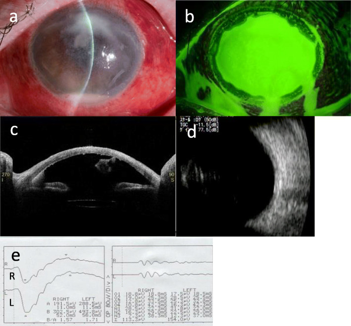
On initial examination in our department, her visual acuity in the left eye allowed her to count fingers at a distance of 0.3 ft. Slit-lamp examination showed a white focus on the upper peripheral cornea, hypopyon, anterior chamber fibrin formation, marked ciliary hyperemia, and whole epithelial defects in the left eye (Fig. 1a,b). Anterior segment optical coherence tomography (OCT; CASIA2, Tomey) also revealed hypopyon, anterior chamber fibrin formation, and keratic precipitates (Fig. 1c). As echography showed no evident vitreous opacity, we speculated that infection had not advanced to the posterior chamber at this point (Fig. 1d). The full-field electroretinogram (ERG) waveform had no amplitude reduction or latency delay, which suggested that retinal function was not impaired by the infection (Fig. 1e). She was emergently hospitalized for infectious keratitis, and anti-bacterial treatment was continued.
Fig. 1.
a Slit-lamp examination on the patient’s first visit. A white infiltrate on the upper cornea, hypopyon, anterior chamber fibrin formation, and ciliary hyperemia were observed. b A whole corneal epithelial defect was observed by fluorescein staining. c Anterior segment OCT with a CASIA2 showed a hypopyon, anterior chamber fibrin deposits, and keratic precipitates. d Echography showed no evident vitreous opacity. e The waveform of full-field ERG had no amplitude reduction or latency delay
The corneal scraping smear was positive for filamentous fungi and Fusarium species were detected by aqueous humor polymerase chain reaction (PCR). Antifungal susceptibility tests were performed and the MIC values were as follows: > 16 μg/ml for Micafungin, > 64 μg/ml for Flucytosine, > 64 μg/ml for Fluconazole, > 16 μg/ml for Miconazole, > 8 μg/ml for Itraconazole, 4 μg/ml for Voriconazole, and 2 μg/ml for amphotericin B. Thereafter, her treatment was shifted to anti-fungal therapy 2 days after hospitalization. The patient received topical 1% voriconazole hourly, 1% natamycin ointment 6 times daily, and intravenous liposomal amphotericin B injection (125 mg/day), whereas 0.1% betamethasone was replaced with 0.1% fluorometholone, and 0.1% tacrolimus was withdrawn. Adherence to topical medical therapy was checked by nursing staff by filling in the list of eyedrops.
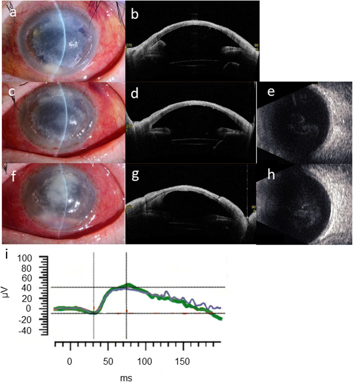
Two days after the onset of anti-fungal therapy, anterior inflammation improved, as characterized by a smaller corneal infiltrate and improved ciliary hyperemia; in addition, anterior segment OCT demonstrated a smaller hypopyon and anterior chamber fibrin deposits (Fig. 2a,b). However, after the patient’s monthly intravenous tocilizumab injection was administered 7 days after hospitalization (anterior chamber irrigation was performed on the same day), signs of recurrent inflammation were observed. Ciliary hyperemia markedly worsened, and vitreous opacity emerged on echography the day after tocilizumab injection (Fig. 2c–e).
Fig. 2.
a The corneal infiltrate shrank and ciliary hyperemia improved 2 days after the onset of anti-fungal therapy. b Anterior segment OCT demonstrated decreased hypopyon and anterior chamber fibrin deposition. c and d The day after intravenous tocilizumab injection, aggravation of ciliary hyperemia was noted, although the hypopyon decreased due to anterior chamber irrigation. e Vitreous opacity emerged on echography 2 days after tocilizumab infusion. f and g Slit-lamp examination and anterior segment OCT 10 days after tocilizumab infusion. The range of corneal infiltration increased and the hypopyon worsened. h Hyperechogenic foci on B-scan ultrasonography also worsened. i Reduction of both a-wave and b-wave amplitude was confirmed on full-field ERG 9 days after tocilizumab administration
As the initial response to anti-fungal therapy was good, and the vitreous opacity appeared to be a transient effect of tocilizumab application, therapy was continued without modification. However, 17 days after hospitalization, the degree of corneal infiltration, hypopyon, and vitreous opacity worsened, which suggested progression to endophthalmitis (Fig. 2f–h). In addition, both the a-wave and b-wave amplitude of the full-field ERG diminished, which suggested malfunction of the retina caused by advancement of the infection to the posterior segment (Fig. 2i). Given that the infection worsened in spite of conservative therapy, we determined that surgical treatment was required. After obtaining informed consent from the patient, penetrating keratoplasty (PKP) combined with vitrectomy was performed 19 days after hospitalization.
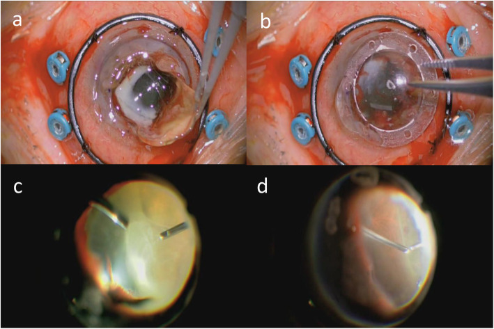
We used an intraocular infusion solution containing 10 mg/500 ml voriconazole. The host cornea was punched out with a 7-mm trephine. When the intraocular lens (IOL) was removed together with the lens capsule, a white focus ranging from the pupillary area to the back of the temporal iris became evident (Fig. 3a). After mounting the temporary corneal prosthesis (DORC, Fig. 3b), we performed a 25-gauge 4-port vitrectomy using the Constellation Vitrectomy System. The white focus in the pupillary area was removed during the anterior vitrectomy. Fundus examination revealed dense white vitreous opacity which was removed during the core vitrectomy and shaving (Fig. 3c). However, the focus in the vitreous had not adhered to the retina. A small white focus and dot hemorrhages were sparsely distributed on the peripheral retina (Fig. 3d). After removing the vitreous opacity completely, we removed the temporary corneal prosthesis and sutured in a frozen donor graft (7.75 mm in diameter). Pathological examination of the removed cornea by PAS and Grocott staining revealed filamentous fungi in the stroma.
Fig. 3.
a A white focus at the rear of the IOL became evident after the IOL was removed together with the lens capsule. b A temporary corneal prosthesis was mounted. c Fundus examination revealed dense white vitreous opacity. d Dot hemorrhages and white foci (not pictured) were sparsely distributed on the peripheral retina
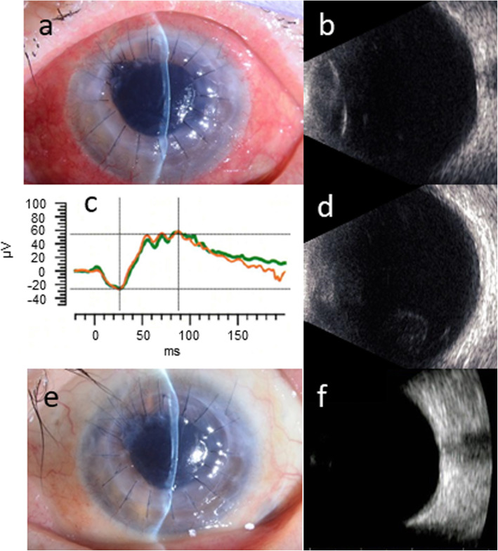
The hypopyon and corneal infiltration were completely absent 3 weeks after the operation (Fig. 4a). The vitreous opacity was nearly absent by echography, and the waveform of the full-field ERG normalized at the same time (Fig. 4b,c). Although mild vitreous opacity emerged 4 days after the monthly intravenous tocilizumab injection (3 weeks after the operation), it showed spontaneous remission (Fig. 4d). As signs of inflammation improved, the amphotericin B infusion was discontinued and topical voriconazole and natamycin were both reduced to 4 times daily 1 month after the operation. The patient was discharged 2 months after the operation.
Fig. 4.
a Hypopyon and corneal infiltration were both absent 3 weeks after the surgery. (b and c) Vitreous opacity was largely absent on B-scan ultrasonography, and the full-field ERG waveform acquired at the same time had no amplitude reduction or latency delay. d Although mild vitreous opacity was confirmed on ultrasonography 4 days after tocilizumab infusion, it spontaneously improved. e and f There were no signs of infection relapse in the slit-lamp examination, and no vitreous opacity on B-scan ultrasonography 4 months after the surgery
When she came to our department 4 months after the operation, the visual acuity of her left eye was 20/100. We observed neither relapse of infection nor vitreous opacity on echography (Fig. 4e,f). Optical PKP and IOL suture were performed 1 year after the initial therapeutic PKP combined with vitrectomy. The patient continues to visit our office (July 2020), and is doing well. The visual acuity of her left eye at her most recent visit was 30/100.
Discussion and conclusions
We report a case of Fusarium infection complicating rheumatic keratitis in a patient receiving regular infusions of tocilizumab for rheumatoid arthritis; the infection took an atypical course and acutely progressed to endophthalmitis. To our knowledge, this is the first report of a case of Fusarium keratitis which acutely progressed to endophthalmitis during tocilizumab infusion.
The patient’s initial response to anti-fungal therapy was good; however, vitreous opacity rapidly emerged after the first tocilizumab infusion. Mild vitreous opacity also emerged after the second tocilizumab administration. Although we cannot draw any definitive conclusions, these observations suggest that tocilizumab may have been the aggravating factor for the fungal infection.
Although progression of fungal keratitis is generally indolent, the inflammation in the present case progressed acutely. There are several case reports of Fusarium keratitis progressing to endophthalmitis, but few studies dealing with large populations. One study included 159 Fusarium keratitis patients [4], of whom 10 patients had infections that progressed to endophthalmitis. In those cases, hypopyon and endothelial plaques emerged 10 to 40 days after corneal infiltration. Compared with the previously published cases, the present case took an extremely acute course, as hypopyon emerged only two days after corneal infiltration appeared. Several factors—including epithelial defect due to rheumatoid arthritis-associated corneal ulceration, steroid administration, and susceptibility to infection arising from tocilizumab infusion—may explain these observations.
One might speculate that steroid discontinuation could be the cause of ocular inflammation worsening. However, ocular inflammation mainly aggravated five days before the admission to our department, when oral prednisolone was started and the administration frequency of topical steroids was increased. Moreover, it did not acutely aggravate after three days before the visit to our department, when oral prednisolone was withdrawn and the administration frequency of topical steroids was decreased. Additionally, ocular inflammation improved two days after the onset of anti-fungal therapy, when the topical steroid was changed to a milder type. These observations suggest that steroid withdrawal is at least not the main factor for ocular inflammation aggravation.
The present case was characterized by acute progression to ophthalmitis during tocilizumab administration. Tocilizumab is an anti-IL-6 receptor antibody used for the treatment of autoimmune diseases, including rheumatoid arthritis. It mediates anti-inflammatory effects by inhibiting the binding of IL-6 to its receptor. As it also has an immunosuppressive effect, complications associated with its use include pneumonia (including Pneumocystis pneumonia) and tuberculosis.
Few studies have examined the relationship between fungal keratitis and IL-6. In a study which investigated the cytokine milieu in the tears of patients with fungal keratitis, higher levels of IL-6 were reported than in the tears of healthy individuals [6]. Application of inactivated Fusarium solani hyphae to cultured human corneal epithelial cells upregulated the secretion of IL-6 via activation of Toll-like receptors [7]. These studies indicate that IL-6 is upregulated in the anterior portion of the eye in response to fungal infection. However, IL-6 activity in this context is unknown. During infection with the bacterial pathogen Streptococcus aureus, application of IL-6 to the cornea of IL-6 knockout mice improved the symptoms of infection and reduced the number of bacteria to 1/3 the pre-treatment level [8]. Therefore, IL-6 may have anti-bacterial activity. IL-6 promotes elastase and free radical production by neutrophils, suggesting that it may inhibit bacterial infection via neutrophil activation. As neutrophils are the main cells that infiltrate the cornea in response to fungal infection, IL-6 inhibition by tocilizumab may aggravate inflammation through blockade of neutrophil activation.
To our knowledge, this is the first report of a case of fungal keratitis of which tocilizumab infusion was speculated to be an aggravating factor. There are some past studies describing tocilizumab as a risk factor for the onset and progression of systemic cryptococcosis and bacterial infection [9–11], so we speculate that progression of fungal keratitis is affected by tocilizumab infusion. However, the mechanisms of exacerbation and its incidence are not clear in the present case. Further study is needed to solve these problems.
In the present case, final visual acuity was relatively spared despite the acute course of the fungal infection. Past studies have indicated that Fusarium endophthalmitis developed from keratitis is associated with a poor prognosis, even after PKP. Therefore, early diagnosis and detection of endophthalmitis are needed, in combination with early surgical treatment to prevent the progression of endophthalmitis [4]. In the present case, culture of corneal scrapings and aqueous humor PCR were performed on the day of visit, which facilitated prompt diagnosis of Fusarium infection. PKP in combination with vitrectomy was performed at a relatively early stage, 10 days after vitreous opacity emerged on echography. We removed as much of the focus of infection as possible, using a temporary corneal prosthesis to improve visibility, before the infection progressed to the retina. In addition, intraocular drug migration was improved by vitreous removal. Our specific approach to treatment may be responsible for the recovery of the patient’s visual acuity.
One might say that a 10-day time-frame between Tocilizumab infusion and surgery is a considerable period of time for infection aggravation. We followed up this period by continuing conservative therapy, as the treatment seemed to be effective and fungal infection generally takes an indolent course. However, endophthalmitis worsened considerably at this relatively short period of time. The patient’s prognosis might have been better if the surgery had been performed as soon as possible, and this would be our future challenge.
Our experience suggests that tocilizumab infusion may make patients more vulnerable to ocular infection. Therefore, patients should be closely monitored for signs of ocular inflammation during tocilizumab administration, especially as rapid detection and thorough treatment may contribute to the preservation or recovery of visual acuity.
Acknowledgements
Not applicable.
Abbreviations
- ERG
electroretinogram
- IL-6
interleukin 6
- IOL
intraocular lens
- OCT
optical coherence tomography
- PCR
polymerase chain reaction
- PKP
penetrating keratoplasty
Authors’ contributions
YM acquired the clinical information and drafted the manuscript. TS performed the surgery, acquired the clinical information, and reviewed the manuscript. KM performed the surgery. KN critically revised and corrected the manuscript. All authors read and approved the final manuscript.
Funding
There are no sources of funding associated with this work.
Availability of data and materials
All data generated and analyzed during this study are included in this article.
Declarations
Ethics approval and consent to participate
This study followed the tenets of the Declaration of Helsinki. Written informed consent was obtained from the participant.
Consent for publication
Written informed consent was obtained from the patient for publication of this case report and any accompanying images. A copy of the written consent is available for review by the editor of this journal.
Competing interests
KM is a member of the editorial board of this journal.
Footnotes
Publisher’s Note
Springer Nature remains neutral with regard to jurisdictional claims in published maps and institutional affiliations.
References
- 1.Silva RA, Sridhar J, Miller D, Wykoff CC, Flynn HW., Jr Exogenous fungal endophthalmitis: an analysis of isolates and susceptibilities to antifungal agents over a 20-year period (1990-2010) Am J Ophthalmol. 2015;159(2):257–264. doi: 10.1016/j.ajo.2014.10.027. [DOI] [PubMed] [Google Scholar]
- 2.Alfonso EC, Cantu-Dibildox J, Munir WM, Miller D, O’Brien TP, Karp CL, et al. Insurgence of Fusarium keratitis associated with contact lens wear. Arch Ophthalmol. 2006;124(7):941–947. doi: 10.1001/archopht.124.7.ecs60039. [DOI] [PubMed] [Google Scholar]
- 3.Klont RR, Eggink CA, Rijs AJMM. Wesseling P, Verweij. Successful treatment of Fusarium keratitis with cornea transplantation and topical and systemic voriconazole. Clin Infect Dis. 2005;40:110–112. doi: 10.1086/430062. [DOI] [PubMed] [Google Scholar]
- 4.Dursun D, Fernandez V, Miller D, Alfonso EC. Advanced fusarium keratitis progressing to endophthalmitis. Cornea. 2003;22(4):300–303. doi: 10.1097/00003226-200305000-00004. [DOI] [PubMed] [Google Scholar]
- 5.Rosenberg KD, Flynn HW, Jr, Alfonso EC, Miller D. Fusarium endophthalmitis following keratitis associated with contact lenses. Ophthalmic Surg Lasers Imaging. 2006;37(4):310–313. doi: 10.3928/15428877-20060701-08. [DOI] [PubMed] [Google Scholar]
- 6.Vasanthi M, Prajna NV, Lalitha P, Mahadevan K, Muthukkaruppan V. A pilot study on the infiltrating cells and cytokine levels in the tear of fungal keratitis. Indian J Ophthalmol. 2007;55(1):27–31. doi: 10.4103/0301-4738.29491. [DOI] [PubMed] [Google Scholar]
- 7.Jin X, Qin Q, Tu L, Zhou X, Lin Y, Qu J. Toll-like receptors (TLRs) expression and function in response to inactivate hyphae of Fusarium solani in immortalized human corneal epithelial cells. Mol Vis. 2007;13:1953–1961. [PMC free article] [PubMed] [Google Scholar]
- 8.Hume EBH, Cole N, Garthwaite LL, Khan S, Willcox MDP. A protective role for IL-6 in staphylococcal microbial keratitis. Invest Ophthalmol Vis Sci. 2006;47(11):4926–4930. doi: 10.1167/iovs.06-0340. [DOI] [PubMed] [Google Scholar]
- 9.Nishioka H, Takegawa H, Kamei H. Disseminated cryptococcosis in a patient taking tocilizumab for Castleman's disease. J Infect Chemother. 2018;24(2):138–141. doi: 10.1016/j.jiac.2017.09.009. [DOI] [PubMed] [Google Scholar]
- 10.Nishimoto N, Ito K, Takagi N. Safety and efficacy profiles of tocilizumab monotherapy in Japanese patients with rheumatoid arthritis: meta-analysis of six initial trials and five long-term extensions. Mod Rheumatol. 2010;20(3):222–232. doi: 10.3109/s10165-010-0279-5. [DOI] [PubMed] [Google Scholar]
- 11.Koike T, Harigai M, Inokuma S, Ishiguro N, Ryu J, Takeuchi T, Takei S, Tanaka Y, Sano Y, Yaguramaki H, Yamanaka H. Effectiveness and safety of tocilizumab: postmarketing surveillance of 7901 patients with rheumatoid arthritis in Japan. J Rheumatol. 2014;41(1):15–23. doi: 10.3899/jrheum.130466. [DOI] [PubMed] [Google Scholar]
Associated Data
This section collects any data citations, data availability statements, or supplementary materials included in this article.
Data Availability Statement
All data generated and analyzed during this study are included in this article.


