Abstract
We aimed to explore the application of three-dimensional (3D) printing technology with problem-based learning (PBL) teaching model in clinical nursing education of congenital heart surgery, and to further improve the teaching quality of clinical nursing in congenital heart surgery. In this study, a total of 132 trainees of clinical nursing in congenital heart surgery from a grade-A tertiary hospital in 2019 were selected and randomly divided into 3D printing group or traditional group. The 3D printing group was taught with 3D printed heart models combined with PBL teaching technique, while the traditional group used conventional teaching aids combined with PBL technique for teaching. After the teaching process, the 2 groups of nursing students were assessed and surveyed separately to evaluate the results. Compared to the traditional group, the theoretical scores, clinical nursing thinking ability, self-evaluation for comprehensive ability, and teaching satisfaction from the questionnaires filled by the 3D printing group were all higher than the traditional group. The difference was found to be statistically significant (P < .05). Our study has shown the 3D printing technology combined with the PBL teaching technique in the clinical nursing teaching of congenital heart surgery achieved good results.
Keywords: 3D printing technology, clinical nursing, congenital heart surgery, problem-based learning teaching
1. Introduction
There are dozens of congenital heart diseases, each with unique anatomy. These variations in anatomy lead to significant difference in pathophysiology, clinical presentations and management strategies. Therefore, it is very important that learners have certain knowledge and understanding of anatomy before learning the treatment and nursing of different kinds of congenital heart diseases. The authors all work at a congenital heart unit where all nursing students come to rotate to learn how to nurse children with congenital heart disease. We found clinical nursing trainees (referred to as “nursing students” hereafter) generally have lesser knowledge of the anatomy of the cardiovascular system and face certain difficulties in learning.[1,2] Traditional clinical teaching tools for congenital heart surgery are heart disease atlas, ultrasound pictures and normal human heart models among others. The lack of experience in practical cases leads to obstacles in anatomical structure reconstruction. Hence, it is difficult to combine their theoretical knowledge with the actual structure, which hinders the understanding, analysis, and problem-solving skills of nursing students.[3] It is seen that problem-based learning (PBL) teaching method, focuses on the students’ learning through real-world, complex problems. Thus, through solving practical problems together with other students in a group study setting, students can learn the knowledge behind the questions, so as to promote their independent problem-solving ability, autonomous learning ability, and lifelong learning skills.[4,5]
The three-dimensional (3D) printing technology is a technology based on digital image information that uses adhesive materials to construct objects layer-by-layer.[6–10] Recent literatures suggest that there have been an intensive application of 3D printing technology in clinical diagnosis, formulating surgical methods, and clinical teaching in the field of structural congenital heart disease.[1,7,11–20] However, research focusing on the use of 3D printing technology with PBL teaching model in teaching clinical nursing of congenital heart surgery is still relatively rare. Since January 2019, we combined 3D printing technology with PBL teaching mode in teaching clinical nursing in congenital heart surgery in selected typical cases of the atrial septal defect (ASD) and have achieved favorable results.
2. Materials and methods
2.1. Research subjects
In 2019, 132 nursing students of congenital heart disease surgery at a grade-A tertiary hospital were selected as research subjects. They came from 2 specialist colleges and 1 undergraduate college. Among them, 124 were female and 8 were male; 46 were undergraduate students and 86 were advanced college students. The nursing students were randomly divided into 2 groups: the 3D printing group and the traditional group, with each group containing 66 students (Table 1). The research subjects consented and agreed to participate in the research.
Table 1.
Demographics of all subjects.
| 3D printing group (n = 64) | Traditional group (n = 64) | P | |
| Age (yrs) | 20.3 | 20.1 | .13 |
| Education | 66 | 66 | .20 |
| Nursing school A (Diploma) | 18 | 18 | |
| Nursing school B (Bachelor) | 23 | 23 | |
| Nursing school C (Diploma) | 25 | 25 | |
| Male students in each group | 5 | 3 | .72 |
2.2. Methods
After approved by the institutional review board and written consent were obtained, this study used controlled experiments. The teaching process of combining 3D printing technology with the PBL model was used in the 3D printing group. Contrarily, traditional cardiology teaching aids combined with the PBL teaching model were used to teach the students in the traditional group.
2.3. Teaching implementation
From the branch of congenital heart diseases, ASD was selected as the learning content. The 2 groups of students were taught in the same teaching environment (such as the same teacher, the same assessment questions, equal learning hours, etc).
2.3.1. 3D printing group
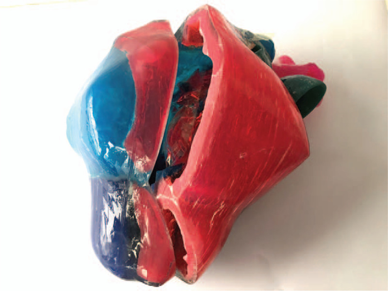
The teachers designed the related nursing problems based on a real case of ASD and provided a 3D printed heart model of that case to the group of nursing students 3 days before the class (Fig. 1). The teacher introduced the medical conditions to the students, assisted them with the medical assessment and raised questions using the 3D printed heart model combined with multimedia. The lesson was carried out in the form of small group of teamwork. Nursing students, through self-learning, data reviewing, and other ways of learning combined with 3D printed heart models, discussed, analyzed, and summarized the problems to answer related questions raised by teachers. The groups also produced lesson reports using PowerPoint slides; group representatives presented the results of the lessons with the use of 3D printed heart models and multimedia. The presentations included the definition of the ASD, its basic classification, medical imaging, pathophysiology, and other related knowledge. Furthermore, representatives also discussed their learnings on clinical manifestations, surgical methods, latest research trends, key points in perioperative nursing and psychological nursing etc. During the case discussion, teachers were responsible for guiding the students through highlighting the key points and challenges in the problems.
Figure 1.

A patient-specific ASD 3d printed heart model with resin.
2.3.2. Traditional group
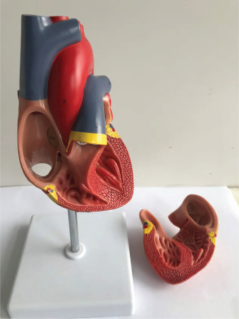
The teachers designed related nursing problems based on a real case of ASD and used conventional cardiology teaching model (Fig. 2) with multimedia to introduce students to the medical conditions and assist them in medical assessment. Subsequently, a series of questions were raised by the teachers in this group, based on real cases and employed the same PBL teaching method as the 3D printing group.
Figure 2.

A commercial normal heart model.
2.4. Teaching results evaluation
The assessment results were used as the objective indicators to evaluate the results, and the questionnaire surveys were used as the subjective indicator. Examinations of anatomy knowledge of congenital heart disease taken by nursing students upon admission were used as baseline data to understand their overall knowledge of the anatomy of cardiovascular system (Table S1, Supplemental Digital Content). After the class, the 2 groups were tested by theory assessments in-class (Table S2, Supplemental Digital Content). Two weeks after the class, the 2 groups of nursing students were further tested on their critical thinking ability (Table S3, Supplemental Digital Content). Additionally, they completed a Likert-type self-assessment questionnaire survey (Table S4, Supplemental Digital Content) about their comprehensive ability.
2.4.1. Theoretical assessment
The Relative knowledge of ASD assessment involves a quiz of 20 questions in both groups, 0.5 points for each question, and the total score is 10 points.
2.4.2. Questionnaire survey
2.4.2.1. Assessment on critical thinking ability
The Chinese version of California Critical Thinking Dispositions Inventory (CTDI-CV) was adopted as the assessment on critical thinking ability. This inventory consisted of 7 dimensions: truth-seeking, open-mindedness, analytical ability, systemic ability, the confidence in critical thinking, curiosity, and cognitive maturity. The whole inventory consisted of a total of 70 entries of which each dimension had 10 entries. Among the 70 entries, 30 were positive entries while the other 40 were negative. The approximate finishing time was 20 minutes. Its judgment and grading criteria were as follows: for positive entries, 6 points were awarded for “totally agree,” 5 points for “agree,” 4 points for “basically agree,” 3 points for “basically disagree,” 2 points for “disagree” and 1 for “totally disagree.” The grading scale for negative entries was opposite. The range of total score for CTDI-CV was 70 to 420 points. Of these, a score <280 points suggested a relatively weak critical thinking ability, a score of 280 to 349 points suggested a positive critical thinking ability, and a score ≥350, a very strong critical thinking ability. The range of score for each dimension was 10 to 60 points, in which a score of 30 to 39 points suggested a moderate level of the respective characteristic, a score of 40 to 49 points, a positive performance of that characteristic and ≥50 points suggested a very strong performance of that characteristic (Table S5, Supplemental Digital Content).
The study was conducted through focused distribution of anonymous questionnaire and an on-the-spot collection of the responses. A total of 132 questionnaires were distributed and 130 effective responses were collected. The rate of effective collection was 98.48%.
An anonymous questionnaire about self-assessed comprehensive ability was distributed amongst the 2 groups that collected data on the following 6 aspects: interest in learning about congenital heart disease, anatomical knowledge of the congenital heart disease, comprehension of the knowledge of congenital heart disease, comprehension of the knowledge of nursing congenital heart disease, confidence in nursing child patients with congenital heart disease and the level of satisfaction on the teaching methods. Students graded the aspects based on a 5-point Likert scale, where each survey item was scored from 1 to 5 points, respectively.
The study was conducted through a focused distribution of anonymous questionnaire and an on-the-spot collection of responses. A total of 132 questionnaires were distributed and 132 effective responses were collected. The rate of effective collection was 100%.
2.5. Statistical methodology
We conducted a pilot study involving 20 subjects with 10 in each group and Ability to understand the knowledge of congenital heart disease values in self-evaluation questionnaire was used to estimate sample size. The value was 3.8 and 4.4 for Traditional and 3D printing group, respectively. Taking α = 0.05 and 1 β = 0.9, a sample size of n = 60 per group was calculated using PASS 15.0 software (NCSS-PASS, Kaysville, UT). No loss was designed because no follow-up was involved in the study. Grouping was realized by randomly divided into 2 groups with Excel (Microsoft Corporation, Redmond, WA) random () function.
Stata 16.0 (Stata Corp, College Station, TX), a statistical software was used for data collation and analysis. Baseline categorical data were compared with Fisher exact or Chi-Squared tests as appropriate. The measurement of data was expressed by ( ± s), and the 2-tailed, paired t test was used for comparing the 2 groups. The self-evaluation of comprehensive ability and satisfaction level of nursing students on the teaching methods were tested by the Wilcoxon rank-sum test, where P < .05 indicated that the difference was statistically significant.
SAS 9.4 (SAS Institute, Cary, NC) and IBM SPSS AMOS 24.0 (Amos Development Corporation, Meadville, PA) were used to analyze the validity and reliability of the questionnaire survey. Cronbach α coefficient measured the reliability, and the reliability was evaluated by split-half reliability method. The split-half method (half-point) was used as an alternative to the test-retest reliability, by combining with the Cronbach α coefficient; the internal consistency of the questionnaire was tested by the 2. KMO (Kaiser–Meyer–Olkin) test and Bartlett test of sphericity were used to determine sample adequacy and appropriateness of data for factor analysis.
3. Result
3.1. Assessment of results
In terms of the admission examination of the anatomy knowledge of congenital heart disease, the 3D printing group scored 4.11 ± 1.40 and the traditional group scored 3.80 ± 1.24. The difference between the 2 groups was statistically insignificant (P > .05).
In the classroom assessment of theoretical knowledge, the 3D printing group scored 7.08 ± 1.66, and the traditional group scored 6.56 ± 1.17. The difference between the 2 groups was statistically significant (P < .05). The detailed results are shown in Table 2.
Table 2.
Comparison of baseline scores of congenital heart disease anatomy knowledge, scores of theoretical knowledge of atrial septal defect (ASD) after class between the 2 groups of nursing students (point, [ ± s]).
| 3D printing group | Traditional group | |||
| Observation indices | n = 66 | n = 66 | t value | P value |
| Baseline scores of anatomical knowledge for congenital heart disease. | (4.11 ± 1.40) | (3.80 ± 1.24) | 1.32 | .09 |
| Scores of ASD class knowledge examination. | (7.08 ± 1.66) | (6.56 ± 1.17) | 2.08 | .02 |
3.2. Questionnaire results
3.2.1. The validity and reliability evaluation of the questionnaire
The Cronbach α for CTDI-CV was 0.90 and the Cronbach α value for each dimension ranged from 0.54 to 0.77. After calculation, the Cronbach α coefficient of the students’ satisfaction level questionnaire was 0.684, and the split-half reliability coefficient was 0.793. They indicated that the internal consistency of the questionnaire was satisfactory. The KMO test values for each dimension of CTDI-CV ranged from 0.74 to 0.87. and for Bartlett test of sphericity, P < .00 for all dimensions.
3.2.2. Nursing students’ self-evaluation of comprehensive ability and teaching satisfaction score
The nursing students in the 3D printing group scored better than the traditional group with respect to the learning interest in congenital heart disease knowledge, comprehension of congenital heart disease knowledge, comprehension of congenital heart disease nursing knowledge, confidence in nursing patients with congenital heart disease, and the satisfaction level on teaching methods. The difference was statistically significant (P < .05). The detailed results are shown in Table 3.
Table 3.
Comparison of the self-evaluation of comprehensive ability and the teaching satisfaction score of the 2 groups (point, [ ± s]).
| 3D printing group | Traditional group | |||
| Observation indices | n = 66 | n = 66 | z value | P value |
| Interest in learning about congenital heart disease. | (4.44 ± 0.66) | (4.18 ± 0.70) | 2.18 | .03 |
| Anatomy knowledge in the cardiovascular system. | (4.41 ± 0.61) | (4.11 ± 0.50) | 3.32 | <.01 |
| Ability to understand the knowledge of congenital heart disease. | (4.44 ± 0.64) | (4.06 ± 0.52) | 3.79 | <.01 |
| Ability to understand the nursing knowledge of congenital heart disease. | (4.48 ± 0.56) | (4.24 ± 0.56) | 2.49 | .01 |
| Confidence in nursing child patients with congenital heart disease. | (4.45 ± 0.61) | (4.18 ± 0.58) | 2.71 | <.01 |
| Satisfaction with teaching methods. | (4.50 ± 0.59) | (4.23 ± 0.67) | 3.36 | .02 |
3.2.3. The comparison of the assessment of the results of critical thinking ability of the 2 groups of nursing students
The nursing students in the 3D printing group scored better than the traditional group with respect to critical thinking ability in all 7 dimensions, including truth-seeking, open-mindedness, analytical ability, systemic ability, the confidence in critical thinking, curiosity, and cognitive maturity and overall points. The difference was statistically significant (P < .05). The detailed results are shown in Table 4.
Table 4.
The comparison of the results of critical thinking ability assessment of the 2 groups of nursing students [point, ([ ± s]).
| 3D Printing group | Traditional group | |||
| Observation indices | n = 64 | n = 66 | T value | P value |
| Total Score | (299.98 ± 1.20) | (282.61 ± 0.67) | 12.586 | <.01 |
| Truth-seeking | (42.00 ± 0.58) | (40.67 ± 0.29) | 2.046 | .02 |
| Open-mindedness | (43.73 ± 0.52) | (40.83 ± 0.26) | 4.913 | <.01 |
| Analytical ability | (42.59 ± 0.42) | (39.95 ± 0.23) | 5.449 | <.01 |
| Systemic ability | (44.56 ± 0.27) | (40.30 ± 0.23) | 11.926 | <.01 |
| Confidence in thinking | (44.38 ± 0.24) | (41.16 ± 0.28) | 8.878 | <.01 |
| Curiosity | (42.21 ± 0.36) | (40.73 ± 0.42) | 2.690 | <.01 |
| cognitive Maturity | (40.52 ± 0.45) | (38.97 ± 0.35) | 2.716 | <.01 |
4. Discussion
4.1. 3D printing combined with PBL teaching method is more suitable for the current clinical teaching of congenital heart surgery
The anatomical structure of the cardiovascular system is complex and constructing its spatial structure requires solid anatomical knowledge, spatial understanding, and thinking ability.[21] The medical schools in China are facing a shortage of cadaver resources,[2,22] which is one of the probable reasons why the nursing students generally have a low level of anatomy knowledge and a major obstacle in obtaining the knowledge of congenital heart disease and subsequently, nursing. In addition, the traditional teaching tools of congenital heart surgery have a rough texture, poor simulation, and lack variations. These tools cannot meet the currently increasing teaching demands of clinical nursing.[5,6] It is of great significance in better visualization of the anatomical structure and better presentation of the pathophysiology, classification, treatment methods, and nursing key points of congenital heart diseases to nursing students. For a hollow irregular organ like the heart, 3D printing technology can allow the nursing students to observe its anatomical structure from multiple angles, so as to enhance their understanding of complex heart deformities, which make up for the limits of traditional teaching tools and insufficient physical teaching resources.[23,24]
On the other hand, the PBL teaching method is student-focused. It emphasizes the mode of teaching by implementing the learning content into the problem scenarios, solving practical problems through cooperating with group members and turning the students from the passive learners into the active ones. This teaching model can stimulate the ability of nursing students to discover, think, and solve problems in typical clinical cases, and enhance their learning interest.[25]
This study shows that the theoretical assessment results and results of critical thinking ability of the 3D printing group after class were higher than the traditional group. We use the PBL model to teach knowledge in a planned and systematic manner, and through analysis, evaluation, reasoning and other skills to promote the reflection and critical thinking ability of nursing students, so that they can respond to the complex clinical nurse-patient environment in future work with best possible decision. Moreover, the use of 3D printed heart enables nursing students to have a deep understanding of the anatomical characteristics of the heart with congenital heart diseases, and stimulates the curiosity of nursing students, thereby enhancing self-confidence and making it easier to obtain a sense of accomplishment.
The results suggest that the tools for the teaching of clinical nursing for congenital heart surgery need to be visual, with 3D space, and closer to the practical scenarios of the disease. Therefore, 3D printing technology combined with the PBL teaching method is more suitable for teaching clinical nursing in congenital heart surgery.
4.2. 3D printing combined with PBL teaching method can help improve the teaching quality for clinical nursing trainees
The thickness of the thinnest layer of the 3D heart model can be less than 1 millimeter with an accuracy error of less than 1%. It has the advantages of precision and individualization, and it avoids the misunderstanding and learning deviation during the students’ learning process.[3] For clinical nursing teaching case provided by this study, using 3D-printed heart model clarified the following issues for clinical nursing trainees: the location and size of the ASD, the relationship between the ASD, atrioventricular valves and large vessels, the spatial structural relationship of the cardiovascular system, hemodynamic abnormalities caused by pathological factors, the impact of the disease on the size and structure of the left and right atria and ventricles, as well as other issues.
Combining 3D printing and PBL teaching method to teach clinical nursing in congenital heart disease surgery, and formulating questions related to the disease before the classes have various advantages for the nursing students. They can search relevant literature and materials to make their learning more practical. Initiative learning helps raise the interest of nursing students, cultivate their ability to learn independently, ability to discover problems and solve problems, and develop their creative thinking ability.[12] This study shows that the scores of nursing students in the 3D-printing group in both the after class theoretical assessments and critical thinking ability assessment were higher than those in the traditional group. Furthermore, for 3 self-evaluation indicators including knowledge of cardiovascular system anatomy, comprehension of congenital heart disease, and comprehension of congenital heart disease nursing knowledge, the scores of the 3D-printing group were also higher than the traditional group. The results suggest that the learning about surgical nursing in congenital heart disease and the understanding of its anatomical knowledge are complementary to each other. Thus, 3D printing technology combined with the PBL teaching method helps nursing students understand the anatomical knowledge of congenital heart disease.
4.3. 3D printing combined with PBL teaching mode helps to improve the satisfaction level of nursing students with the teaching procedure
The 3D printed heart model is detachable, cuttable, and measurable. It can help nursing students to visually form a relatively complete understanding of the theoretical system of the anatomy of congenital heart disease. It avoids the drawbacks of traditional teaching such as the incomprehensible confusion of nursing students on the pathological anatomy of different types of congenital heart disease, hemodynamic changes, and nursing key points. Furthermore, it strengthens the understanding of the anatomical details of the disease, which improves their learning ability and confidence. At the same time, the use of 3D printing technology can strengthen the communication and interaction between teachers and nursing students. Through this technology, teachers can dynamically understand the learning level of nursing students, to integrate the teaching resources accordingly. When the learning interest of the nursing students are fully utilized, it can help to cultivate the independent-thinking and problem-solving ability of nursing students, leading the nursing students to develop scientific mindsets. The results of this study show that the 3D printing group scored higher than the traditional group in teaching satisfaction. The difference was statistically significant (P < .05). Thus, it can be said that the application of 3D printing technology in teaching clinical nursing in congenital heart surgery can improve teaching satisfaction and allow nursing students to better understand the lesson content.
5. Conclusion
The use of 3D printing combined with PBL teaching method can improve the quality and satisfaction of teaching clinical nursing in congenital heart disease. It is, therefore, worth promoting its application. However, 3D printing also has its own shortcomings. The most significant one is that 3D printing is currently over-priced, and the use of it will greatly increase the cost of teaching. It is believed that with further development of 3D printing technology, the cost of printing will decrease, and it will be widely used in clinical teaching.
Author contributions
Conceptualization: Hui Tan, Xicheng Deng, Shayuan Ouyang.
Formal analysis: Hui Tan, Xicheng Deng.
Investigation: Hui Tan, Erjia Huang, Shayuan Ouyang.
Methodology: Hui Tan, Xicheng Deng, Shayuan Ouyang.
Project administration: Xicheng Deng, Shayuan Ouyang.
Resources: Shayuan Ouyang.
Writing – original draft: Hui Tan, Shayuan Ouyang.
Writing – review & editing: Hui Tan, Erjia Huang, Xicheng Deng, Shayuan Ouyang.
Supplementary Material
Supplementary Material
Supplementary Material
Supplementary Material
Supplementary Material
Footnotes
Abbreviations: 3D = three-dimensional, ASD = atrial septal defect, PBL = problem-based learning.
How to cite this article: Tan H, Huang E, Deng X, Ouyang S. Application of 3D printing technology combined with PBL teaching model in teaching clinical nursing in congenital heart surgery: a case-control study. Medicine. 2021;100:20(e25918).
This study was approved by the Institutional Review Board and written consent was obtained.
The authors have no funding and conflicts of interests to disclose.
The datasets generated during and/or analyzed during the current study are available from the corresponding author on reasonable request.
Supplemental digital content is available for this article.
Subjects were from 3 different nursing schools pursing either diploma or bachelor.
References
- [1].Kim MS, Hansgen AR, Carroll JD. Use of rapid prototyping in the care of patients with structural heart disease. Trends Cardiovasc Med 2008;18:210–6. [DOI] [PubMed] [Google Scholar]
- [2].Olivieri L, Krieger A, Chen MY, et al. 3D heart model guides complex stent angioplasty of pulmonary venous baffle obstruction in a Mustard repair of D-TGA. Int J Cardiol 2014;172:e297–8. [DOI] [PubMed] [Google Scholar]
- [3].Biglino G, Capelli C, Koniordou D, et al. Use of 3D models of congenital heart disease as an education tool for cardiac nurses. Congenit Heart Dis 2017;12:113–8. [DOI] [PubMed] [Google Scholar]
- [4].Preeti B, Ashish A, Shriram G. Problem Based Learning (PBL) - an effective approach to improve learning outcomes in medical teaching. J Clin Diagn Res 2013;7:2896–7. [DOI] [PMC free article] [PubMed] [Google Scholar]
- [5].Albarrak AI, Mohammed R, Abalhassan MF, et al. Academic satisfaction among traditional and problem based learning medical students A comparative study. Saudi Med J 2013;34:1179–88. [PubMed] [Google Scholar]
- [6].Bullock P, Dunaway D, McGurk L, et al. Integration of image guidance and rapid prototyping technology in craniofacial surgery. Int J Oral Maxillofac Surg 2013;42:970–3. [DOI] [PubMed] [Google Scholar]
- [7].Rengier F, Mehndiratta A, von Tengg-Kobligk H, et al. 3D printing based on imaging data: review of medical applications. Int J Comput Assist Radiol Surg 2010;5:335–41. [DOI] [PubMed] [Google Scholar]
- [8].Michalski MH, Ross JS. The shape of things to come: 3D printing in medicine. JAMA 2014;312:2213–4. [DOI] [PubMed] [Google Scholar]
- [9].Tuomi J, Paloheimo KS, Vehvilainen J, et al. A novel classification and online platform for planning and documentation of medical applications of additive manufacturing. Surg Innov 2014;21:553–9. [DOI] [PubMed] [Google Scholar]
- [10].Malik HH, Darwood AR, Shaunak S, et al. Three-dimensional printing in surgery: a review of current surgical applications. J Surg Res 2015;199:512–22. [DOI] [PubMed] [Google Scholar]
- [11].Shiraishi I, Yamagishi M, Hamaoka K, et al. Simulative operation on congenital heart disease using rubber-like urethane stereolithographic biomodels based on 3D datasets of multislice computed tomography. Eur J Cardiothorac Surg 2010;37:302–6. [DOI] [PubMed] [Google Scholar]
- [12].Costello JP, Olivieri LJ, Krieger A, et al. Utilizing Three-Dimensional Printing Technology to Assess the Feasibility of High-Fidelity Synthetic Ventricular Septal Defect Models for Simulation in Medical Education. World J Pediatr Congenit Heart Surg 2014;5:421–6. [DOI] [PubMed] [Google Scholar]
- [13].Kim MS, Hansgen AR, Wink O, et al. Rapid prototyping: a new tool in understanding and treating structural heart disease. Circulation 2008;117:2388–94. [DOI] [PubMed] [Google Scholar]
- [14].Su W, Xiao Y, He S, et al. Three-dimensional printing models in congenital heart disease education for medical students: a controlled comparative study. BMC Med Educ 2018;18:178. [DOI] [PMC free article] [PubMed] [Google Scholar]
- [15].Valverde I, Gomez G, Gonzalez A, et al. Three-dimensional patient-specific cardiac model for surgical planning in Nikaidoh procedure. Cardiol Young 2015;25:698–704. [DOI] [PubMed] [Google Scholar]
- [16].Bramlet M, Olivieri L, Farooqi K, et al. Impact of Three-Dimensional Printing on the Study and Treatment of Congenital Heart Disease. Circ Res 2017;120:904–7. [DOI] [PMC free article] [PubMed] [Google Scholar]
- [17].Kurenov SN, Ionita C, Sammons D, et al. Three-dimensional printing to facilitate anatomic study, device development, simulation, and planning in thoracic surgery. J Thorac Cardiovasc Surg 2015;149:973–9. e971. [DOI] [PubMed] [Google Scholar]
- [18].Chae MP, Rozen WM, McMenamin PG, et al. Emerging Applications of Bedside 3D Printing in Plastic Surgery. Front Surg 2015;2:25. [DOI] [PMC free article] [PubMed] [Google Scholar]
- [19].Lim KH, Loo ZY, Goldie SJ, et al. Use of 3D printed models in medical education: A randomized control trial comparing 3D prints versus cadaveric materials for learning external cardiac anatomy. Anat Sci Educ 2016;9:213–21. [DOI] [PubMed] [Google Scholar]
- [20].Paige JT, Garbee DD, Kozmenko V, et al. Getting a head start: high-fidelity, simulation-based operating room team training of interprofessional students. J Am Coll Surg 2014;218:140–9. [DOI] [PubMed] [Google Scholar]
- [21].Drake RL, McBride JM, Lachman N, et al. Medical education in the anatomical sciences: the winds of change continue to blow. Anat Sci Educ 2009;2:253–9. [DOI] [PubMed] [Google Scholar]
- [22].Winkelmann A. Anatomical dissection as a teaching method in medical school: a review of the evidence. Med Educ 2007;41:15–22. [DOI] [PubMed] [Google Scholar]
- [23].Meier LM, Meineri M, Qua Hiansen J, et al. Structural and congenital heart disease interventions: the role of three-dimensional printing. Neth Heart J 2017;25:65–75. [DOI] [PMC free article] [PubMed] [Google Scholar]
- [24].Cantinotti M, Valverde I, Kutty S. Three-dimensional printed models in congenital heart disease. Int J Cardiovasc Imaging 2017;33:137–44. [DOI] [PubMed] [Google Scholar]
- [25].White SC, Sedler J, Jones TW, et al. Utility of three-dimensional models in resident education on simple and complex intracardiac congenital heart defects. Congenit Heart Dis 2018;13:1045–9. [DOI] [PubMed] [Google Scholar]
Associated Data
This section collects any data citations, data availability statements, or supplementary materials included in this article.


