Abstract
Objectives:
To reassess the long-term camouflage effects of resin infiltration (Icon, DMG, Hamburg, Germany) of white spot lesions (WSL) and sound adjacent enamel (SAE) achieved in a previous trial. The null hypothesis was tested that there were no significantly different CIE-L*a*b*-ΔE-values between WSL and SAE areas of assessment after at least 24 months (T24) compared to those at baseline (T0).
Materials and Methods:
Of twenty subjects who received previous resin infiltration treatment of nteeth = 111 nonrestored, noncavitated postorthodontic WSL after multibracket treatment during a randomized controlled trial and were contacted 20 months after baseline, eight subjects (trial teeth nteeth = 40; m/f ratio 1/7; age range (mean; SD) 12–17 [15.25; 2.12] years); response rate: 40%) were available for follow-up after at least 24 months (T24). CIE-L*a*b* differences between summarized color and lightness values (ΔEWSL/SAE) of WSL and SAE were assessed using a spectrophotometer and compared to baseline data assessed prior to infiltration (T0), and those after 6 (T6), and 12 (T12) months using paired t tests at a significance level of α = 5%.
Results:
T24 assessments were performed after a mean 33.86 (SD: 8.64; Min: 24; Max: 45) months following T0. Mean (SD) ΔEWSL/SAE units of available teeth were 8.76 (5.33) at baseline; 5.5 (2.75) at T6; 5.2 (2.41) at T12; and 5.57 (2.6) at T24. Comparisons of T6, T12, and T24 with T0 yielded highly significant differences, whereas T6–T24 and T12–T24 differences were found to be not significant.
Conclusions:
Assimilation of infiltrated WSL to the color of adjacent enamel by resin infiltration is considered to be suitable for the long-term improvement in the esthetic appearance of postorthodontic WSL.
Keywords: White spot lesion, Resin infiltration, Durability of camouflage effect, CIE-L*a*b, in vivo
INTRODUCTION
The incidence of labial enamel decalcification or white spot lesions (WSL) during treatment with fixed orthodontic appliances has been reported to vary between 46% and 73%.1,2 Although cavitated lesions require invasive therapy, the choice of WSL treatment is based on the patient's individual esthetic demands. Remineralization by local fluoride application may arrest lesion progression3 and, in combination with tooth brushing abrasion, bring about some improvement in the appearance of WSL within the first few months following debonding,4–6 however, rarely to an extent that provides for an esthetically acceptable dentofacial appearance.7–9 In contrast, the technique of resin infiltration (Icon, DMG) of postorthodontic enamel decalcifications yields esthetically more satisfying results. A recent split-mouth randomized controlled trial (RCT) revealed that there was a significant and clinically relevant abatement of color- and lightness differences between infiltrated WSL and sound adjacent enamel, whereas there were no significant changes in untreated control-WSL in the same time.8 This camouflage effect was substantiated by the similar refractive index of infiltrated and sound adjacent enamel areas.10,11 However, there is a lack of information regarding the long-term color stability of resin infiltrates in vivo and the durability of esthetic concealment of infiltrated postorthodontic WSL achieved.
Therefore, it was the aim of this follow-up study to reassess those subjects who received infiltration of their postorthodontic WSL during the previous 6-month RCT8 in terms of the durability of achieved color and lightness assimilation between WSL and sound adjacent enamel areas (SAE). This is an update of an earlier 12-month follow-up of these subjects.12
The null hypothesis was tested that there were no significantly different CIE-Lab ΔE-values between WSL and SAE areas of assessments after least of 24 months (T24) compared to those at baseline (T0).
MATERIALS AND METHODS
This study was a second follow-up of the patients treated with WSL infiltration during a single-center, split-mouth controlled simple-randomized trial.8 To this end, all of the initial 20 subjects who received a resin infiltration (Icon, DMG) treatment of their postorthodontic WSL according to the producer's instruction sheet were repeatedly contacted by telephone and in writing after 20 months following infiltration. Full prior ethical approval from the University of Göttingen (Germany) Ethics Committee was obtained, and all patients and their guardians gave informed consent to take part in this study.
Intervention
Infiltration of decalcified areas was by enamel preconditioning using 15% HCL gel (Icon-Etch) and subsequent application of the drying solution (Icon-Dry). Frequencies of additional etching intervals were adjusted to individual lesion depths and surface hardness by visual control following each of the etch/dry intervals.8 As the original control quadrants were also infiltrated 6 months after the infiltration of the intervention quadrants, as part of an agreement made with the patients prior to starting the trial, control teeth were no longer available for assessment beyond this time point.
Subjects
The original RCT included subjects with multibracket-induced WSL and completed debracketing, with exclusion criteria of cavitated lesions, as well as filled, restored, and deciduous teeth.8 Trial participants were given the same type of toothbrushes (Oral-B Classic Care, Procter & Gamble, Cincinnati, Ohio) and dentifrices (elmex, GABA, Lörrach, Germany) for oral hygiene home care.
Of twenty original RCT subjects (nine males, 11 females, mean age: 15.5 years) who received resin infiltration treatment of nteeth = 111 nonrestored, noncavitated (postorthodontic) WSL after multibracket treatment at the Department of Orthodontics, University of Göttingen (Germany), eight subjects (trial teeth nteeth = 40; male / female ratio 1/7; age range (mean; SD) 12–17 (15.25; 2.12) years); response rate: 40%) were available for a 24-plus month follow-up (T24). Treatment details and subject characteristics are given in Table 1.
Table 1.
Details on Time Points and Intensity of Etching and Infiltration Treatment
| Subject # |
Subject's Primarily Assigned # [8] |
Sex |
Infiltrated Teeth (n) |
Total Etching Duration (min) |
Time Elapse Between Debonding And Infiltration (mo) |
Time Elapse Between Infiltration and Final Assessment (Rounded to Full Months) |
| 1 | 2 | f | 5 | 6 | 3 | 43 |
| 2 | 3 | f | 6 | 6 | 5 | 36 |
| 3 | 8 | f | 6 | 6 | 12 | 40 |
| 4 | 10 | m | 6 | 6 | 9 | 45 |
| 5 | 13 | f | 6 | 6 | 3 | 34 |
| 6 | 14 | f | 4 | 7 | 1 | 25 |
| 7 | 17 | f | 6 | 8 | 4 | 24 |
| 8 | 18 | f | 6 | 8 | 1 | 24 |
| Total: 45* | Mean (SD): 6.6 (0.92) | Mean (SD): 4.75 (3.9) | Mean (SD): 33.86 (8.64) |
Trial teeth available at compared time points: n = 40.
Method
The same technique as used in the previous RCT and the follow-up for tooth color and lightness assessments was repeated in the same way as reported previously: CIE (L*a*b*) enamel color and lightness data were collected by an intra-oral spectrophotometer (ShadePilot, DeguDent, Hanau-Wolfgang, Germany). The same operator (AE) as before carried out the measurements at the Department of Orthodontics, University of Göttingen, Germany. Two elements contributed to a high level of standardization of assessments during this in vivo trial. First, the system-immanent target caption was used to retrieve the exact locations of infiltrated WSLs and previously used SAE control areas. Thus, variations in the patient's head position during CIE-(L*a*b*) assessments were compensated for and did not have an erroneous impact on the measurements.13 Second, assessment distortion by variation in ambient light was avoided by performing measurements with the patient's lips closed.
Differences in WSL and SAE CIE (L*a*b*) enamel color and lightness data were assessed after at least 24 months (T24) and were compared to baseline assessments (T0) and 6- and 12-month results (T6, T12). T0, T6, and T12 analyses were repeated with the subjects available at this time point.
Statistical Analysis
Means and 95% confidence intervals of lightness and color parameters L*, a*, and b* of WSL and sound adjacent enamel were calculated, both separately and summarized by ΔE-values using the formula:
 |
Differences between ΔE-CIE-L*a*b* of WSL and SAE (ΔEWSL/SAE) were compared at specific time points (T0 (baseline), T6 (6 month), T12 (12 month), T24 (24+ month) using paired t tests with a significance level of α = 5%. Statistical analyses were carried out with Statistica 10 (StatSoft, Tulsa, Okla).
Error of the Method
Variance in assessments with the spectrophotometer and the results of both intra- and interoperator method error were determined on the basis of 10-times repeated, pretrial assessments8 by two assessors (AE, MK), and ranged from 0.16 units (L* value, upper incisor) to 0.82 units (a* value, upper incisor), thereby corroborating previous assessments of the accuracy of the same type of spectrophotometer.14
RESULTS
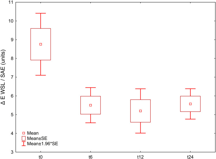
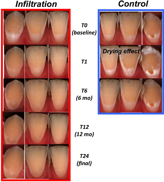
The mean time interval between baseline and T24 assessments was 33.86 (SD: 8.64; Min: 24; Max: 45) months (Table 1). There was a decrease in mean (SD) color and lightness differences for the available teeth by ΔEWSL/SAE units following infiltration. Segregated CIE-L*-, a*, and b* are given in Table 2. Summarized ΔEWSL/SAE differences were 8.76 (5.33) at baseline and decreased to 5.5 (2.75) at 6 months (T6) following infiltration, with nonsignificant changes after 12 months (T12; 5.2 (2.41), and after 24–45 months (T24; 5.57 (2.6) beyond that time point: Comparisons of T6, T12, T24, with T0 yielded highly significant differences, while T6–T24 and T12–T24 differences were found to be not significant (Table 3, Figure 1). An example of spectrophotometric recordings for one subject's lower incisors during the complete trial is given in Figure 2.
Table 2.
Descriptive statistics: CIE-L*-, a*, and b* values of infiltrated white spot lesion and sound adjacent enamel as well as WSL/SAE differences at the distinct time points. Data are dependent, ie, each value has been compared to its baseline valuea
| Time Points Compared: |
Area |
Parameter |
Mean ± SD |
Diff. |
P |
CI 95% |
||
| Time 1 |
Time 2 |
Time 1 |
Time 2 |
|||||
| T0 | T6 | WSL | L | 73.9 ± 4.84 | 70.96 ± 3.36 | 2.94 | <.001 | [1.57; 4.3] |
| T0 | T12 | WSL | L | 74.1 ± 3.83 | 71.01 ± 3.87 | 3.09 | .02 | [0.52; 5.66] |
| T0 | T24 | WSL | L | 73.94 ± 4.64 | 71.4 ± 3.44 | 2.5 | <.001 | [1.22; 3.79] |
| T0 | T6 | SAE | L | 73.62 ± 2.4 | 73.4 ± 1.84 | 0.2 | .5 | [−0.35; 0.75] |
| T0 | T12 | SAE | L | 73.57 ± 2.83 | 73.33 ± 1.97 | 0.24 | .63 | [−0.82; 1.29] |
| T0 | T24 | SAE | L | 73.5 ± 2.32 | 73.4 ± 1.8 | 0.1 | .64 | [−0.33; 0.52] |
| T0 | T6 | WSL-SAE | Diff L | 0.2 ± 4.57 | −2.5 ± 3.25 | 2.7 | <.001 | [1.47; 3.94] |
| T0 | T12 | WSL-SAE | Diff L | 0.53 ± 3.03 | −2.3 ± 3.17 | 2.85 | .006 | [0.93; 4.77] |
| T0 | T24 | WSL-SAE | Diff L | 0.44 ± 4.36 | −1.96 ± 3.32 | 2.4 | <.001 | [1.25; 3.56] |
| T0 | T6 | WSL | a | 6.1 ± 2.84 | 6.63 ± 2.49 | −0.53 | .2 | [−1.35; 0.28] |
| T0 | T12 | WSL | a | 6.14 ± 1.93 | 7.42 ± 1.67 | −1.28 | .009 | [−2.19; −0.37] |
| T0 | T24 | WSL | a | 6.09 ± 2.62 | 6.85 ± 2.44 | −0.76 | .04 | [−1.49; −0.03] |
| T0 | T6 | SAE | a | 5.13 ± 1.26 | 4.83 ± 1.39 | 0.3 | .049 | [0; 0.4] |
| T0 | T12 | SAE | a | 5.16 ± 1.28 | 4.6 ± 1.17 | 0.56 | .03 | [0.08; 1.04] |
| T0 | T24 | SAE | a | 5.00 ± 1.37 | 5.22 ± 1.2 | 0.22 | .09 | [−0.03; 0.47] |
| T0 | T6 | WSL-SAE | Diff a | 1.03 ± 2.93 | 1.88 ± 2.58 | −0.85 | .04 | [−1.65; −0.04] |
| T0 | T12 | WSL-SAE | Diff a | 0.97 ± 1.65 | 2.8 ± 1.741 | −1.84 | <.001 | [−2.45; −1.22] |
| T0 | T24 | WSL-SAE | Diff a | 0.87 ± 2.54 | 1.85 ± 2.76 | −0.98 | .007 | [−1.68; −0.28] |
| T0 | T6 | WSL | b | 15.69 ± 8.66 | 20.9 ± 3.92 | −5.21 | <.001 | [−7.69; −2.72] |
| T0 | T12 | WSL | b | 13.19 ± 4.22 | 22.27 ± 2.77 | −9.08 | <.001 | [−11.38; −6.79] |
| T0 | T24 | WSL | b | 15.79 ± 7.94 | 20.92 ± 3.84 | −5.13 | <.001 | [−7.21; −3.05] |
| T0 | T6 | SAE | b | 20.66 ± 3.25 | 22.15 ± 2.78 | −1.49 | <.001 | [−2.18; −0.8] |
| T0 | T12 | SAE | b | 20.64 ± 3.47 | 22.99 ± 2.38 | −2.35 | <.001 | [−3.68; −1.03] |
| T0 | T24 | SAE | b | 20.85 ± 3.18 | 22.18 ± 2.78 | −1.33 | <.001 | [−1.8; −0.86] |
| T0 | T6 | WSL-SAE | Diff b | −4.8 ± 7.94 | −1.16 ± 3.17 | −3.63 | .003 | [−5.91; −1.36] |
| T0 | T12 | WSL-SAE | Diff b | −7.45 ± 4.52 | −0.72 ± 2.8 | −6.73 | <.001 | [−8.48; −4.98] |
| T0 | T24 | WSL-SAE | Diff b | −5.06 ± 7.3 | −1.25 ± 3.48 | −3.8 | <.001 | [−5.76; −1.84] |
WSL indicates white spot lesion; SAE, sound adjacent enamel.
Table 3.
Intergroup (WSL, SAE) comparison: There was a decrease WSL/SAE differences by ΔEWSL/SAE following infiltration, and this decrease did not change significantly at the following assessment time points. P values indicate significance to previous assessments
| Time Points Compared |
Parameter |
Directly Compared Teeth, n |
Mean ± SD |
Diff. |
P |
CI 95% |
||
| Time 1 |
Time 2 |
Time 1 |
Time 2 |
|||||
| T0 | T6 | ΔEWSL/SAE | 33 | 9.12 ± 5.63 | 5.5 ± 2.76 | 3.61 | <.001 | [1.81; 5.41] |
| T0 | T12 | ΔEWSL/SAE | 16 | 8.58 ± 3.79 | 5.2 ± 2.41 | 3.38 | .01 | [0.81; 5.95] |
| T0 | T24 | ΔEWSL/SAE | 40 | 8.76 ± 5.34 | 5.57 ± 2.62 | 3.18 | <.001 | [1.64; 4.72] |
| T6 | T24 | ΔEWSL/SAE | 33 | 5.5 ± 2.76 | 5.57 ± 2.62 | −0.07 | .65 | [−0.32; 0.2] |
| T12 | T24 | ΔEWSL/SAE | 16 | 5.2 ± 2.41 | 5.57 ± 2.62 | −0.37 | .35 | [−0.67; 0.25] |
Figure 1.
t tests for summarized color- and lightness values (ΔE CIE-L*a*b* b) of the WSL vs SAE areas of the teeth available for this study yielded highly significant differences between baseline measurements and six, (T6) 12, (T12), and 24–45 months following infiltration (T24). Differences in ΔE-values between T6, T12, and T24 were found to be not significant.
Figure 2.
An example of spectrophotometric data for one subject's lower incisors during the complete trial, starting at baseline (T0), following infiltration (T1), to T24 final recordings after 24–45 months. In the case shown, recordings were performed after 24 months. This figure is an update of the spectrophotometer image series published earlier.12 Nontreated control teeth pictures are not available beyond 6 months after baseline, as these were then also infiltrated as part of an agreement with the patients.
DISCUSSION
The problem of postorthodontic WSL is most commonly tackled noninvasively by local fluoridation, leaving decalcified areas to be treated by tooth brushing abrasion.4–6,15 Although this may bring a slight optical abatement of WSL within the first 12 months following debracketing, it rarely provides for an esthetically acceptable improvement in dentofacial appearance.8,9,16 Resin infiltration of postorthodontic WSL has been shown to be more effective in achieving a clinically relevant camouflage effect with adjacent enamel, based on short- and mid-term studies, case reports and in vitro research.8,10–12,17,18 However, a common question raised by both patients and clinicians concerns the long-term durability of the results achieved. The current follow-up assessment of the lesions infiltrated in a previous RCT was conducted to add some long-term data to the topic of color stability of the resin infiltrant in vivo.
Null Hypothesis
There were highly significant color differences indicating an assimilation of WSL color to SAE appearance following infiltration which persisted for six months (T0–T6 assessments, Figure 1), and these camouflage effects persisted, without significant changes, after 24–45 months (T24). The null hypothesis of no significantly different CIE-L*a*b*-ΔE-values between WSL and SAE areas of assessments after at least 24 months (T24) compared to those at baseline (T0) was rejected (P < .001; Table 3).
Variation in Time Points for Final Follow-Up Assessments
This was the longest follow-up assessment of camouflage effects of infiltrated postorthodontic WSL carried out up until now. The subjects from a previous split-mouth RCT were contacted at 20 months following infiltration. Although it was the aim to reassess the infiltrated teeth at 24 months following the intervention, it turned out to be difficult to retrieve an adequate number of patients from the previous trial. Consequently, the time point for the eight subjects of this follow-up who agreed to participate varied and assessments were carried out after at least 24 months and up to 45 months (Table 1). Given a response rate of 40% and also the fact that ΔE CIE-L*a*b* values did not differ significantly (Table 3), with color and lightness alterations far below the clinical visibility threshold value of 3 ΔE CIE-L*a*b* units,19 it may be concluded, on the basis of the currently available data, that the esthetic camouflage results achieved by infiltration of decalcified enamel are stable for at least 24 months.
Numbers of Available Teeth
The participants in the original trial had received infiltration following debonding at an age range of 12 to 19 years (mean age 15.5 years). When they were contacted two years later, it became clear that a certain number of participants, especially older participants, had moved, resulting in some difficulties in recruiting participants. The eight subjects recruited for this follow-up after at least 24 months (T24) were aged 12–17 years (mean 15.25) by the time of infiltration, providing a total of 45 trial teeth, of which 40 were available for the comparison time points.
Adverse Effects
The patients did not report significant adverse events or side effects during the 24–45 month period following infiltration.
24-plus Month Changes in Segregated Color or Lightness Values
At T24, the WSL's lightness value L* was significantly reduced by a mean 2.5 units compared to baseline, whereas SAE areas showed almost exactly the baseline value at T24 (Table 2). CIE-a* values (red-green axis changes) of WSL and SAE did not change significantly between baseline and T24, although the WSL/SAE difference in CIE-a* showed a decrease of −0.98 units, indicating a minor change toward greenish appearance, which was, however, unlikely to be visible to the naked eye in a clinical setting.19 CIE-b* values (yellow-blue axis changes) of WSL (SAE) were decreased by −5.13 (−1.33), indicating a minor change from yellowish toward blueish appearance. WSL/SAE difference in CIE-b* were −3.8, indicating that this alteration may exceed the threshold value of clinical visibility.19
Total Effect on Summarized Color and Lightness Development after 24–45 Months
The camouflage effects achieved, as evident from the decrease in summarized ΔEWSL/SAE differences from a mean 8.76 units, at baseline, to a mean of 5.5 after 6 months were found to be stable without statistically significant or clinically relevant changes (T24; 5.57 units; Table 3, Figure 1).19 Therefore, WSL camouflage effects achieved by resin infiltration were considered to be stable in color and lightness, with no significant changes over at least 24 months.
Limitations
The limitation of this trial was the low participant response rate, as it was very hard to recruit eight of the original 20 subjects for this final follow-up, which produced a total of 40 teeth for assessment. As this took some time, the time points for the final assessments varied between 24 and 45 months. In addition, the eight patients were partly not congruent with the last nine patients in the 1-year follow-up, which suggests it is critical to compare the current data with the data collected following 12 months. The presented analyses were exploratory in nature and were based on the available subset of patients after 24 months. Therefore, no sample size calculation was applied. The numbers of teeth pairs that were directly compared at this time point are given in Table 3. Also, the impact of individual oral micro-environments such as potential smoking habits or consumption of beverages with staining potential was not screened by this follow-up study. However, this is the longest follow-up of infiltrated patients to date, with assessments up to 45 months after baseline. Since, for this sample, it will not be possible to reassess a sufficient number of the original trial patients, other research groups, in starting new trials on the subject, should adopt a separate control group instead of a split-mouth design, to judge, in the long term, the aging characteristics and color stability of infiltrated teeth in the esthetically relevant enamel area.
To achieve the best esthetic results for infiltration, it has previously been recommended to infiltrate early following debonding and to select this treatment option for more superficial lesions.8 With these limitations in mind, it was shown that the esthetic results, regardless of whether there was complete camouflage of the WSL, could be maintained for 24–45 months.
CONCLUSIONS
The assimilation of infiltrated WSL to the color of adjacent enamel by resin infiltration is considered suitable for long-term improvement in the esthetic appearance of postorthodontic WSL, as this camouflage effect did not change in a statistically significant or clinically relevant manner over a period of least of 24 months in vivo.
The longest observation time of infiltrated teeth achieved by this follow-up was 45 months.
The patients reported no important adverse events or side-effects during the 24–45-month period following infiltration.
ACKNOWLEDGMENTS
This study was supported financially by DMG, Hamburg, Germany. We also thank DeguDent, Hanau-Wolfgang, Germany for providing the spectrophotometer used in this trial. There were no restrictions with regard to the publication of the results.
REFERENCES
- 1.Richter AE, Arruda AO, Peters MC, Sohn W. Incidence of caries lesions among patients treated with comprehensive orthodontics. Am J Orthod Dentofacial Orthop. 2011;139:657–664. doi: 10.1016/j.ajodo.2009.06.037. [DOI] [PubMed] [Google Scholar]
- 2.Tufekci E, Dixon JS, Gunsolley JC, Lindauer SJ. Prevalence of white spot lesions during orthodontic treatment with fixed appliances. Angle Orthod. 2011;81:206–210. doi: 10.2319/051710-262.1. [DOI] [PMC free article] [PubMed] [Google Scholar]
- 3.Stahl J, Zandona AF. Rationale and protocol for the treatment of non-cavitated smooth surface carious lesions. Gen Dent. 2007;55:105–111. [PubMed] [Google Scholar]
- 4.Al-Khateeb S, Forsberg CM, de Josselin de Jong E, Angmar-Mansson B. A longitudinal laser fluorescence study of white spot lesions in orthodontic patients. Am J Orthod Dentofacial Orthop. 1998;113:595–602. doi: 10.1016/s0889-5406(98)70218-5. [DOI] [PubMed] [Google Scholar]
- 5.Fejerskov O, Nyvad B, Kidd EAM. Clinical and histological manifestations of dental caries. In: Dental Caries—The Disease and Its Clinical Management. Ames, Iowa: Blackwell Munksgaard; 2003;5:72–97. [Google Scholar]
- 6.Willmot DR. White lesions after orthodontic treatment: does low fluoride make a difference? J Orthod. 2004;31:235–242. doi: 10.1179/146531204225022443. [DOI] [PubMed] [Google Scholar]
- 7.Øgaard B, Rolla G, Arends J. Orthodontic appliances and enamel demineralization. Part 1. Lesion development. Am J Orthod Dentofacial Orthop. 1988;94:68–73. doi: 10.1016/0889-5406(88)90453-2. [DOI] [PubMed] [Google Scholar]
- 8.Knösel M, Eckstein A, Helms HJ. Durability of esthetic improvement following Icon resin infiltration of multibracket-induced white spot lesions compared with no therapy over 6 months: a single-center, split-mouth, randomized clinical trial. Am J Orthod Dentofacial Orthop. 2013;144:86–96. doi: 10.1016/j.ajodo.2013.02.029. [DOI] [PubMed] [Google Scholar]
- 9.Bock NC, Seibold L, Heumann C, Gnandt E, Röder M, Ruf S. Changes in white spot lesions following post-orthodontic weekly application of 1.25 per cent fluoride gel over 6 months-a randomized placebo-controlled clinical trial. Part I: photographic data evaluation. Eur J Orthod. 2017;39:134–143. doi: 10.1093/ejo/cjw060. [DOI] [PubMed] [Google Scholar]
- 10.Paris S, Meyer-Lueckel H. Masking of labial enamel white spot lesions by resin infiltration-a clinical report. Quintessence Int. 2009;40:713–718. [PubMed] [Google Scholar]
- 11.Rocha Gomes Torres C, Borges AB, Torres LM, Gomes IS, de Oliveira RS. Effect of caries infiltration technique and fluoride therapy on the colour masking of white spot lesions. J Dent. 2011;39:202–207. doi: 10.1016/j.jdent.2010.12.004. [DOI] [PubMed] [Google Scholar]
- 12.Eckstein A, Helms HJ, Knösel M. Camouflage effects following resin infiltration of postorthodontic white-spot lesions in vivo: one-year follow-up. Angle Orthod. 2015;85:374–380. doi: 10.2319/050914-334.1. [DOI] [PMC free article] [PubMed] [Google Scholar]
- 13.Knösel M, Attin R, Jung K, Brunner E, Kubein-Meesenburg D, Attin T. Digital image color analysis compared to direct dental CIE (L*,a*,b*) colorimeter assessment under different ambient conditions. Am J Dent. 2009;22:67–72. [PubMed] [Google Scholar]
- 14.Sluzker A, Knösel M, Athanasiou AE. Sensitivity of digital dental photo CIE L*a*b* analysis compared to spectrophotometer clinical assessments over 6 months. Am J Dent. 2011;24:300–304. [PubMed] [Google Scholar]
- 15.Wiegand A, Köwing L, Attin T. Impact of brushing force on abrasion of acid-softened and sound enamel. Arch Oral Biol. 2007;52:1043–1047. doi: 10.1016/j.archoralbio.2007.06.004. [DOI] [PubMed] [Google Scholar]
- 16.Øgaard B, Ten Bosch JJ. Regression of white spot enamel lesions. A new optical method for quantitative longitudinal evaluation in vivo. Am J Orthod Dentofacial Orthop. 1994;106:238–242. doi: 10.1016/S0889-5406(94)70042-7. [DOI] [PubMed] [Google Scholar]
- 17.Neuhaus KW, Graf M, Lussi A, Katsaros C. Late infiltration of post-orthodontic white spot lesions. J Orofac Orthop. 2010;71:442–447. doi: 10.1007/s00056-010-1038-0. [DOI] [PubMed] [Google Scholar]
- 18.Senestraro SV, Crowe JJ, Wang M, et al. Minimally invasive resin infiltration of arrested white-spot lesions: a randomized clinical trial. J Am Dent Assoc. 2013;144:997–1005. doi: 10.14219/jada.archive.2013.0225. [DOI] [PubMed] [Google Scholar]
- 19.Johnston WM, Kao EC. Assessments of appearance match by visual observation and clinical colorimetry. J Dent Res. 1989;68:819–822. doi: 10.1177/00220345890680051301. [DOI] [PubMed] [Google Scholar]


