Abstract
Objectives:
Major depressive disorder (MDD) is associated with dysregulations of leptin and tryptophan–kynurenine (Trp–Kyn) (TKP) pathways. Leptin, a pro-inflammatory cytokine, activates Trp conversion into Kyn. However, leptin association with down-stream Kyn metabolites in MDD is unknown.
Methods:
Fasting plasma samples from 29 acutely ill drug-naïve (n = 16) or currently non-medicated (⩾6 weeks; n = 13) MDD patients were analyzed for leptin, Trp, Kyn, its down-stream metabolites (anthranilic [AA], kynurenic [KYNA], xanthurenic [XA] acids and 3-hydroxykynurenine [3HK]), C-reactive protein (CRP), neopterin, body mass index (BMI), and insulin resistance (HOMA-IR). Depression severity was assessed by HAM-D-21.
Results:
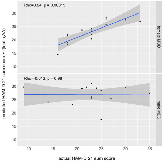
In female (n = 14) (but not in male) patients HAM-D-21 scores correlated with plasma levels of AA (but not other Kyn metabolites) (rho = −0.644, P = .009) and leptin (Spearman’s rho = −0.775, P = .001). Inclusion of AA into regression analysis improved leptin prediction of HAM-D from 48.5% to 65.9%. Actual HAM-D scores highly correlated with that calculated by formula: HAM-D = 34.8518−(0.5660 × leptin [ng/ml] + 0.4159 × AA [nmol/l]) (Rho = 0.84, P = .00015). In male (n = 15) (but not in female) patients leptin correlated with BMI, waist circumference/hip ratio, CRP, and HOMA-IR.
Conclusions:
Present findings of gender specific AA/Leptin correlations with HAM-D are important considering that AA and leptin are transported from plasma into brain, and that AA formation is catalyzed by kynureninase—the only TKP gene associated with depression according to genome-wide analysis. High correlation between predicted and actual HAM-D warrants further evaluation of plasma AA and leptin as an objective laboratory test for the assessment of depression severity in female MDD patients
Keywords: Major depressive disorder, anthranilic acid, leptin, gender, Hamilton depression rating scale, inflammation
Introduction
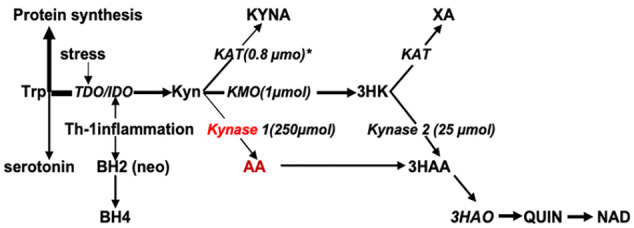
Dysregulations of leptin1 and serotonin-kynurenine (Kyn) pathways of tryptophan (Trp) metabolism2-4 are associated with major depressive disorder (MDD). Key enzymes of the Trp–Kyn pathway are activated by pro-inflammatory cytokines (Figure 1).5,6 Leptin is a pro-inflammatory cytokine,7 and was shown to up-regulate formation of Kyn from Trp.8 However, its association with down-stream Kyn metabolites in MDD has not been investigated.
Figure 1.

Tryptophan-Kynurenine pathway.
Abbreviations: 3HAA, 3-hydroxyanthranilic acid; 3HAO, 3-hydroxyanthranilate 3,4-dioxygenase; 3HK, 3-hydroxykynurenine; AA, anthranilic acid; BH2, 7,8-dihydroneopterin; BH4, tetrahydrobiopterin; KAT, kynurenine aminotransferase; KMO, kynurenine 3-monooxygenase; Kyn, kynurenine; KYNA, kynurenic acid; Kynase, kynureninase; NAD, nicotinamide adenine dinucleotide; neo, neopterin; QUIN, quinolinic acid; TDO/IDO, tryptophan- and indoleamine-2,3-dioxygenases; Trp, tryptophan; XA, xanthurenic acid.
*Kinetic Michaelis-Menten constants (µmol).
Leptin is a peptide hormone encoded by the obesity gene and is primarily produced by white adipose tissue. Leptin was initially discovered as factor which decreased appetite and increased energy utilization via interaction with hypothalamic and hippocampal leptin receptors.9 Further experimental studies revealed that selective deletion of leptin receptors10 or treatment with a pegylated leptin receptor antagonist elicited depression-like behavior that could be counteracted by leptin administration.11 Clinical studies found elevation of plasma leptin levels correlated with severity of depression in young adults with MDD.12
The serotonin-kynurenine hypothesis suggests that serotonin deficiency in MDD is caused by a shift of Trp metabolism from production of serotonin toward formation of Kyn and its metabolites.2-4 The formation of Kyn from Trp is catalyzed by either stress-activated hepatic tryptophan-2,3-dioxygenase 2 (TDO) or by extrahepatic indoleamine-2,3-dioxygenase 1 and 2 (IDO), which are induced by pro-inflammatory Th-1 type cytokines (Figure 1).5 Leptin belongs to the Th-1 cytokine family at structural and functional levels.7 and activates IDO, and, consequently, up-regulates Trp conversion into Kyn.8 This may imply that leptin activates down-stream metabolism of Kyn as well. However, up-regulation of IDO does not necessarily trigger activation of down-stream Kyn metabolism, which trifurcates into formation of 3-hydroxykynurenine (3HK), anthranilic (AA), and kynurenic (KYNA) acids (Figure 1). For example, up-regulation of IDO, induced by acute inflammation, elevated Kyn and KYNA, but not AA, plasma levels in COVID-19-infected patients.13 Furthermore, circulating AA levels were not elevated in subjects with obesity, a condition associated with chronic inflammation and increased leptin levels.14 On the other hand, serum levels of AA and KYNA (but not 3HK and Kyn) were elevated in leptin receptor-deficient Zucker fatty rats,15 and in conditions associated with decreased leptin levels such as weight loss induced by diet16 or bariatric surgery.17 Taken together, these findings suggest that leptin might interact with the Trp–Kyn pathway in MDD, and that studies of such an interaction should include evaluation of circulating levels of not only Trp and Kyn, but also of down-stream Kyn metabolites. The main goal of the present study was to assess the possible association of leptin and kynurenines plasma levels with the severity of depression in acutely depressed un-medicated MDD patients. Considering that plasma leptin levels is known to be much higher in females than males, we were interested to assess potential gender differences of leptin association with the studied variables.
Methods
Samples
Fasting plasma samples were obtained from 29 acutely ill patients with a DSM-IV diagnosis of MDD.18 MDD patients were drug-naïve (n = 16) or previously-treated but not receiving medication for ⩾6 weeks (n = 13). The investigation was carried out in accordance with the latest version of the Declaration of Helsinki. The study design was approved by the University of Magdeburg Review Board, and written informed consent was obtained after the nature of the procedures had been fully explained.
Laboratory and clinical measurements
Considering data on MDD (and leptin and kynurenines) associations with inflammation, obesity, and insulin resistance (IR),19 the following parameters were analyzed: (a) plasma levels of leptin, Trp, Kyn, and its down-stream metabolites (AA, KYNA, 3HK, and xanthurenic acid (XA), an immediate metabolite of 3HK); (b) neopterin, a biomarker of the activated human immune system, linked with Trp–Kyn pathway,20 and C-reactive protein (CRP), a non-specific marker of inflammation; (c) markers of obesity (body mass index [BMI], waist circumference/hip ratio [waist-hip-ratio]); a homoeostatic model assessment of insulin resistance (HOMA-IR); and age, cigarette consumption, and medication use histories. Leptin levels were determined by radioimmunoassay (DSL, Sinsheim, Germany). Kynurenines were evaluated by HPLC-mass spectrometry,15 CRP was analyzed by immunoturbidimetry (Cobas 8000 c701 modular analyzer, Roche Diagnostics; Basel, Switzerland), neopterin concentrations were determined by enzyme-linked immunosorbent assay (ELISA) (BRAHMS GmbH, Hennigsdorf, Germany). MDD was diagnosed according to DSM-IV, and severity of depression (HAM-D-21) and all other variables were assessed in the previous studies of the same patients.18,21
Data analysis
Data were analyzed using IBM SPSS Statistics version 25 software. Statistical significance was defined as P < .05, as trending toward statistical significance at P < .10. Most data were not normally distributed, as indicated by Shapiro–Wilk tests. Thus, non-parametric analyses were applied (Spearman’s rank correlation coefficient, Kruskal Wallis H-test and (post-hoc) Mann–Whitney U-test). Subsequent to correlation analyses, regression analysis was performed to identify variables (AA, leptin, age, BMI, smoking, drug-naïve vs unmedicated but previously ill) which allowed the best prediction of HAM-D scores in MDD patients. Bonferroni-correction was applied to control for multiple comparisons.
Results
Group and subgroup comparisons: gender differences
Plasma leptin levels were higher in females than in male patients (P = .010) (Table 1). On contrary, waist-hip-ratio ratio was significantly higher in male than female MDD (P = .001), while higher BMI and Trp levels in males than in females were trending toward statistical significance (P = .078 and P = .069, respectively). No other gender-related differences were observed in MDD patients.
Table 1.
Demographic data, leptin, and kynurenines measures for MDD patients.
| Female [n = 14] | Male [n = 15] | P (Bonferroni) | P (U-test) | |
|---|---|---|---|---|
| Age [years] | 47.50 [24.50;52.50] | 47 [31.00;54.00] | 1 | .678 |
| HAM-D | 23.00 [18.00;26.00] | 23.00 [19.00;25.00] | 1 | .843 |
| BMI [ kg/m2] | 22.23 [19.73;24.46] | 25.50 [23.57;26.58] | .078 | .020 |
| Waist-hip-ratio | 0.82 [0.76;0.86] | 0.92 [0.87;1.02] | .001 | <.001 |
| Leptin [ng/ml] | 6.370 [3.71;11.143] | 2.640 [0.010;5.000] | .010 | <.003 |
| HOMA-IR | 0.46 [0.36;0.71] | 0.45 [0.31;0.74] | 1 | .683 |
| CRP [μg/ml] | 0.60 [0.38;2.20] | 0.60 [0.50;2.60] | 1 | .809 |
| Neopterin [nmol/l] | 4.250 [3.900;4.550] | 4.200 [3.700;4.400] | 1 | .661 |
| Trp [μmol/l] | 54.40 [44.08;76.25] | 66.80 [59.70;78.90] | .069 | .217 |
| Kyn [μmol/l] | 1.895 [1.205;2.713] | 1.970 [1.630;2.360] | 1 | .793 |
| 3HK [nmol/l] | 14.00 [9.90;18.57] | 15.10 [9.52;19.00] | 1 | .949 |
| AA [nmol/l] | 18.35 [12.18.20.27] | 13.80 [11.00;16.80] | .687 | .172 |
| KYNA [nmol/l] | 19.35 [15.78;24.57] | 23.50 [18.60;29.90] | .575 | .144 |
| XA [nmol/l] | 9.365 [5.950;16.250] | 7.875 [5.867;12.825] | 1 | .671 |
Data are presented as median [quartile 1; quartile 3].
Significant differences are displayed in bold font.
Abbreviations: 3HK, 3-hydroxykynurenine; AA, anthranilic acid; BMI, body mass index; CRP, C-reactive protein; HAM-D, Hamilton depression rating scale; HOMA-IR, homoeostatic model assessment of insulin resistance ([Insulin (µU/ml) × Glucose (mmol/l)]/22.5); Kyn, kynurenine; KYNA, kynurenic acid; Trp, tryptophan; Waist-hip-ratio, waist circumference-hip ratio; XA, xanthurenic acid.
Correlations with Severity of depressionHAM-D scores of female MDD patients correlated inversely with plasma levels of leptin (P = .001), AA (P = .009) and CRP (P = .028) (Table 2). These correlations were absent in male MDD patients. HAM-D did not correlate with other studied parameters in both genders. Regression analysis showed that leptin alone explained 48.8% of HAM-D scores, while leptin in combination with AA predicted 65.9% of HAM-D scores in female MDD patients. HAM-D score in female MDD patients can be predicted using the following formula: HAM-D predicted = 34.8518–(0.5660 × leptin [ng/ml] + 0.4159 × AA [nmol/l]). Predicted HAM-D highly correlated with actual HAM-D (Rho = 0.84, P < .00015) (Figure 1). The other tested variables had no significant influence in this model. Regression analysis revealed no suitable model to predict HAM-D scores in male MDD patients (Figure 2).
Table 2.
Correlations between severity of depression (HAM-D) and studied variables.
| variables | HAM-D (males) Rho/P (n = 15) | HAM-D (females) Rho/P (n = 14) |
|---|---|---|
| BMI | 0.120/.669 | −0.192/.474 |
| Waist-hip-ratio | −0.026/.922 | −0.366/.179 |
| Leptin | −0.290/.23 | −0.755/.001 |
| HOMA-IR | −0.310/.260 | 0.07/.782 |
| CRP | −0.21/.45 | −0.563/.028 |
| Neopterin | −0.191/.494 | −0.169/.531 |
| Trp | −0.050/.863 | 0.029/.918 |
| Kyn | −0.22/.44 | −0.426/.112 |
| 3HK | −0.168 + .566 | −0.169/.531 |
| KYNA | 0.033 + .910 | −0.190/.496 |
| AA | 0.026/.928 | −0.644/.009 |
| XA | −0.065 + .847 | −0.066/.828 |
Spearman’s rank correlation coefficient test. Significant correlations (P < .05) are displayed in bold font.
Abbreviations: 3HK, 3-hydroxykynurenine; AA, anthranilic acid; BMI, body mass index; CRP, C-reactive protein; HAM-D, Hamilton depression rating scale.; HOMA-IR, homoeostatic model assessment of insulin resistance; Kyn, kynurenine; KYNA, kynurenic acid; Trp, tryptophan; WC, waist circumference; XA, xanthurenic acid.
Figure 2.

Correlation between actual and predicted HAM-D-21 sum scores in female and male MDD patients.
Leptin and AA plasma levels correlations with metabolic markers
Plasma leptin levels correlated with BMI, waist-hip-ratio, CRP, and HOMA-IR in male but not in female patients (Table 3). Plasma AA levels did not correlate with metabolic markers in both female and male patients.
Table 3.
Correlations of plasma anthranilic acid and leptin with metabolic markers.
| Anthranilic acid | Leptin | |||
|---|---|---|---|---|
| MDD (males) [n = 15] | MDD (females) [n = 14] | MDD (males) [n = 15] | MDD (females) [n = 14] | |
| HAM-D | 0.026/.928 | −0.644/.009 | −0.290/.293 | −0.755/.001 |
| BMI | −0.112/.702 | −0.131/.641 | 0.594/.019 | 0.462/.095 |
| Waist-hip ratio | 0.132/.638 | 0.249/.370 | 0.658/.005 | 0.249/.370 |
| HOMA-IR | −0.085/.770 | −0.004/.988 | 0.654/.008 | −0.085/.761 |
| CRP | 0.309/.280 | 0.226/.436 | 0.69/.0047 | 0.482/.068 |
| Neopterin | 0.024/.934 | 0.034/.904 | 0.111/.682 | −0.035/.896 |
| Leptin | 0.226/.436 | 0.203/.466 | Na | Na |
| AA | Na | Na | 0.226/.436 | 0.203/.466 |
Spearman’s rank correlation coefficient test. Significant correlations (P < .05) are displayed in bold font.
Abbreviations: 3HK, 3- hydroxykynurenine; AA, anthranilic acid; BMI, body mass index; CRP, C-reactive protein; HAM-D, Hamilton Depression Rating Scale; HOMA-IR, homoeostatic model assessment of insulin resistance; Kyn, kynurenine; KYNA, kynurenic acid; n.a., not applicable.; Trp, tryptophan; Waist-hip, waist circumference/hip; XA, xanthurenic acid.
There was no correlation between plasma leptin and AA levels in MDD as it was reported previously.21
Discussion
This study, although based on a relatively small sample size, is the first attempt to explore relationships between leptin and AA plasma levels with the severity of depression in MDD patients. Study participants were acutely depressed, drug-naïve or not medicated for at least 6 weeks before entering the study. There are 3 main findings of the study. First, there is an inverse correlation between AA (but not other metabolites of Trp–Kyn pathway) and the severity of depression in female (but not in male) MDD patients. Second, an improved prediction of HAM-D scores in female MDD patients was observed by combining leptin and AA in a regression model. Third, there was an inverse correlation between plasma leptin and CRP, BMI, waist-hip ration, and HOMA-IR in male (but not in female) patients.
AA had not attracted much of attention of the MDD researchers. The most recent meta-analysis of Trp–Kyn metabolism in MDD did not include AA at all.22 More recent studies did not find any differences between plasma AA levels in unmedicated,23 and medicated MDD patients in comparison with controls.24 There was no correlation between serum AA levels and HAM-D scores in depressed patients25 while rather weak (rho = −0.230) and marginally statistically significant (P = .050, Spearman’s rank test) correlation of HAM-D scores with plasma levels of AA (but not Kyn, 3HK, KYNA, and XA) were reported in medicated MDD patients before ECT treatment.26
AA formation from Kyn is catalyzed by Kynase. Under physiological conditions most of Kyn is metabolized into KYNA and 3-HK because Kynase has a 10-times lower affinity to Kyn than KAT and KMO (Figure 1). The present finding of an inverse correlation of AA (but not other Kyn metabolites) with HAM-D scores might be of importance considering that genome-wide analyses identified association of depressive symptoms with Kynase-1 (but not other Trp–Kyn pathway) genes.27 Notably, Sutphin et al28 discovered that RNAi knockdown of Kynase extended lifespan of Caenorhabditis elegans by >20%. This discovery implies negative correlation of Kynase activity, and, consequently, circulating AA levels, with aging, the condition highly associated with depression.
This is the first observation of gender-specific correlation of AA with HAM-D. The cause of such specificity warrants further investigation, especially in a context of 2:1 female: male ratio among MDD patients.
Leptin correlation with HAM-D had been observed in non-MDD patients.29 Inverse correlation between CSF leptin levels and Montgomery–Åsberg Depression Rating Scale scores were reported in MDD patients admitted after suicide attempts.30 However, the pro-inflammatory cytokines profile of suicide attempters is different from MDD without suicide attempts.31 Some32 but not all29 studies suggested that leptin correlation with HAM-D scores Influenced by BMI and other metabolic markers. The regression analysis of the present data found that leptin prediction HAM-D score did not depend on age, cigarette consumption, BMI, and waist-hip-ratio, as well as drug utilization history. In addition, BMI and waist-hip-ratio were significantly higher in male than female, while leptin plasma levels were, on contrary, higher in females than in male patients.
Gender was the main factor which affected the leptin correlation with HAM-D, as this was observed only in the case of female patients. In contrast, the correlations between leptin, metabolic (BMI, waist-hip-ratio, HOMA-IR) and inflammation (CRP) markers were observed only in male MDD patients. It is well known that levels of circulating leptin are significantly higher in women than in men, while administration of 17beta-estradiol increased leptin levels in healthy postmenopausal women,33 and female rats.34 Notably, co-localization of hypothalamic leptin and estrogen receptors may contribute to sexual dimorphism of plasma leptin correlations with metabolic and inflammatory biomarkers and the severity of depression.35
Therefore, our data point to a male-specific effect on leptin correlations with obesity, inflammation and IR biomarkers, and a female-specific association with the severity of depression. This suggests the different mechanisms for leptin involvement in mood and the metabolic/inflammation/IR regulated pathways.
One of the possible mechanistic explanations for the connection between AA and leptin might be that AA acts as an agonist to farnezoid X receptor (FXR).36 FXR is a nuclear hormone receptor, and regulates many genes involved in lipid and glucose metabolism.37 Activation of FXR up-regulates the gene that encodes leptin production.38 Therefore, elevation of AA might increase leptin production. This suggestion is in line with present finding of an inverse (but not positive) correlation of HAM-D with both leptin and AA, and with increased predictive strength of leptin correlation with HAM-D by inclusion of AA in the model. It is also in line with literature data on the anti-depressant effects of leptin.1
Peripherally originated AA might impact central mechanisms of mood regulation considering AA ability to penetrate blood-brain-barrier.5 Although AA is not known to directly affect any specific receptors, some data suggest that AA may modulate activity of NMDA receptors (NMDAR) that may mediate the antidepressant effect of some interventions (eg, ketamine infusion).39 NMDAR activity might be regulated by inhibitors of D-amino acids oxidase (DAAO), an enzyme catalyzing degradation of the NMDAR co-agonist, D-serine.40 Notably, administration of a single dose of one of DAAO inhibitor, benzoate, robustly elevates plasma AA levels in healthy volunteers.40 Considering that AA differs from benzoate by a single amino group, it might be interesting to explore AA effects on DAAO activity. On the other hand, AA could be converted into benzoate via deamination by gut microbiota.41 AA, therefore, may (directly or indirectly) inhibit DAAO, and, consequently, attenuate degradation of D-serine and contribute to NMDAR activation. Notably, both activation and inhibition of NMDAR might exert anti-depressant effects.39
Strong correlation of AA/leptin plasma levels with HAM-D scores suggests that AA/leptin might be a state or trait biomarker for MDD. Currently, subjective factors play a significant role in evaluation of the severity of depression (eg, mental status evaluation and self-reported scales), and, even in case of nominally “objective” scales, such as HAM-D and MADRS). Considering the importance of the assessment of depression severity for the selection of treatment modalities and for monitoring the efficacy of treatments, in particularly in clinical trials of new antidepressant medications, there is a need of objective tests based upon laboratory analysis. The present data on strong and highly significant correlation of AA/leptin plasma levels with HAM-D scores warrant further evaluation of plasma leptin and AA measurement as an objective laboratory test for the assessment of depression severity in female MDD patients.
Footnotes
Funding:The author(s) received no financial support for the research, authorship, and/or publication of this article.
Declaration of conflicting interests:The author(s) declared no potential conflicts of interest with respect to the research, authorship, and/or publication of this article.
Author Contributions: JS and GFO conceived the study. JS collected all samples from patients and clinical data. JR and DF performed the blood measures of kynurenines and neopterin. JS, HD and GFO obtained and analyzed the data and wrote the first version of the manuscript. HGB, DF, JR and PS reviewed the data analysis and contributed significantly to manuscript writing. PCG edited the final version of the manuscript and contributed to data interpretation. All authors approved this version for publication.
ORCID iD: Gregory F Oxenkrug  https://orcid.org/0000-0002-7193-9117
References
- 1. Lu XY, Kim CS, Frazer A, Zhang W. Leptin: a potential novel antidepressant. Proc Natl Acad Sci U S A. 2006;103:1593-1598. [DOI] [PMC free article] [PubMed] [Google Scholar]
- 2. Lapin IP, Oxenkrug GF. Intensification of the central serotoninergic processes as a possible determinant of the thymoleptic effect. Lancet. 1969;1:32-39. [DOI] [PubMed] [Google Scholar]
- 3. Lapin IP. Kynurenines as probable participants of depression. Pharmakopsychiatrie und Neuropsychopharmakologie. 1973;6:273-279. [DOI] [PubMed] [Google Scholar]
- 4. Oxenkrug G. Serotonin-kynurenine hypothesis of depression: historical overview and recent developments. Curr Drug Targets. 2013;14:514-521. [DOI] [PMC free article] [PubMed] [Google Scholar]
- 5. Schwarcz R, Bruno JP, Muchowski PJ, Wu HQ. Kynurenines in the mammalian brain: when physiology meets pathology. Nat Rev Neurosci. 2012;13:465-477. [DOI] [PMC free article] [PubMed] [Google Scholar]
- 6. Badawy AA-B, Guillemin G. The plasma [Kynurenine]/[Tryptophan] ratio and indoleamine 2,3-dioxygenase: time for appraisal. Int J Tryptophan Res. Published online August 21, 2019. doi: 10.1177/1178646919868978 [DOI] [PMC free article] [PubMed] [Google Scholar]
- 7. Lord GM. Leptin as a proinflammatory cytokine. Contrib Nephrol. 2006;151:151-164. [DOI] [PubMed] [Google Scholar]
- 8. Mangge H, Summers KL, Meinitzer A, et al. Obesity-related dysregulation of the tryptophan-kynurenine metabolism: role of age and parameters of the metabolic syndrome. Obesity (Silver Spring). 2014;22:195-201. [DOI] [PubMed] [Google Scholar]
- 9. Halaas JL, Gajiwala KS, Maffei M, et al. Weight-reducing effects of the plasma protein encoded by the obese gene. Science. 1995;269:543-546. [DOI] [PubMed] [Google Scholar]
- 10. Guo M, Huang TY, Garza JC, Chua SC, Lu XY. Selective deletion of leptin receptors in adult hippocampus induces depression-related behaviours. Int J Neuropsychopharmacol. 2013;16:857-867. [DOI] [PMC free article] [PubMed] [Google Scholar]
- 11. Macht VA, Vazquez M, Petyak CE, et al. Leptin resistance elicits depressive-like behaviors in rats. Brain Behav Immun. 2017;60:151-160. [DOI] [PubMed] [Google Scholar]
- 12. Syk M, Ellström S, Mwinyi J, et al. Plasma levels of leptin and adiponectin and depressive symptoms in young adults. Psychiatry Res. 2019;272:1-7. [DOI] [PubMed] [Google Scholar]
- 13. Thomas T, Stefanoni D, Reisz JA, et al. COVID-19 infection alters kynurenine and fatty acid metabolism, correlating with IL-6 levels and renal status. JCI Insight. 2020;5:pii:140327. [DOI] [PMC free article] [PubMed] [Google Scholar]
- 14. Theofylaktopoulou D, Midttun O, Ulvik A, et al. A community-based study on determinants of circulating markers of cellular immune activation and kynurenines: the Hordaland Health Study. Clin Exp Immunol. 2013;173:121-130. [DOI] [PMC free article] [PubMed] [Google Scholar]
- 15. Oxenkrug G, Cornicelli J, van der Hart M, Roeser J, Summergrad P. Kynurenic acid, an aryl hydrocarbon receptor ligand, is elevated in serum of Zucker fatty rats. Integr Mol Med. 2016;3:761-763. [PMC free article] [PubMed] [Google Scholar]
- 16. Strasser B, Berger K, Fuchs D. Effects of a caloric restriction weight loss diet on tryptophan metabolism and inflammatory biomarkers in overweight adults. Eur J Nutr. 2015;54:101-107. [DOI] [PubMed] [Google Scholar]
- 17. Christensen MHE, Fadnes DJ, Røst TH, et al. Inflammatory markers, the tryptophan-kynurenine pathway, and vitamin B status after bariatric surgery. PLoS One. 2018;13:e0192169. [DOI] [PMC free article] [PubMed] [Google Scholar]
- 18. Steiner J, Fernandes BS, Guest PS, et al. Glucose homeostasis in major depression and schizophrenia: a comparison among drug-naïve first-episode patients. Eur Arch Psychiatry Clin Neurosci. 2019;269:373-377. [DOI] [PubMed] [Google Scholar]
- 19. Marazziti D, Rutigliano G, Baroni S, Landi P, Dell’Osso L. Metabolic syndrome and major depression. CNS Spectr. 2014;19:293-304. [DOI] [PubMed] [Google Scholar]
- 20. Geisler S, Mayersbach P, Becker K, Schennach H, Fuchs D, Gostner JM. Serum tryptophan, kynurenine, phenylalanine, tyrosine and neopterin concentrations in 100 healthy blood donors. Pteridines. 2015;26:31-36. [Google Scholar]
- 21. Steiner J, Bernstein HG, Guest P, Summergrad P, Oxenkrug G. Plasma leptin correlates with anthranilic acid in schizophrenia but not in major depressive disorder. Eur Neuropsychopharmacol. 2020;41:167-168. [DOI] [PubMed] [Google Scholar]
- 22. Ogyua K, Kuboa K, Nodaa Y, et al. Kynurenine pathway in depression: a systematic review and meta-analysis. Neurosci Biobehav Rev. 2018;90:16-25. [DOI] [PubMed] [Google Scholar]
- 23. Colle R, Masson P, Verstuyft C, et al. Peripheral tryptophan, serotonin, kynurenine, and their metabolites in major depression: a case-control study. Psychiatry Clin Neurosci. 2020;74:112-117. [DOI] [PubMed] [Google Scholar]
- 24. Pompili M, Lionetto L, Curto M, et al. Tryptophan and kynurenine metabolites: are they related to depression? Neuropsychobiology. 2019;77:23-28. [DOI] [PubMed] [Google Scholar]
- 25. Sakurai M, Yamamoto Y, Kanayama N, et al. Serum metabolic profiles of the tryptophan-kynurenine pathway in the high risk subjects of major depressive disorder. Sci Rep. 2020;10:1961. [DOI] [PMC free article] [PubMed] [Google Scholar]
- 26. Guloksuz S, Arts B, Walter S, et al. The impact of electroconvulsive therapy on the tryptophan-kynurenine metabolic pathway. Brain Behav Immun. 2015;48:48-52. [DOI] [PubMed] [Google Scholar]
- 27. Arnau-Soler A, Macdonald-Dunlop E, Adams MJ, et al.; Generation Scotland, Major Depressive Disorder Working Group of the Psychiatric Genomics Consortium. 2019 Genome-wide by environment interaction studies of depressive symptoms and psychosocial stress in UK Biobank and Generation Scotland. Transl Psychiatry. 2019;9:14. [DOI] [PMC free article] [PubMed] [Google Scholar]
- 28. Sutphin GL, Backer G, Sheehan S, et al.; the Cohorts for Heart and Aging Research in Genomic Epidemiology (CHARGE) Consortium Gene Expression Working Group. Caenorhabditis elegans orthologs of human genes differentially expressed with age are enriched for determinants of longevity. Aging Cell. 2017;16:672-682. [DOI] [PMC free article] [PubMed] [Google Scholar]
- 29. Lawson EA, Miller KK, Blum JI, et al. Leptin levels are associated with decreased depressive symptoms in women across the weight spectrum, independent of body fat. Clin Endocrinol (Oxf). 2012;76:520-525. [DOI] [PMC free article] [PubMed] [Google Scholar]
- 30. Westling S, Ahrén B, Träskman-Bendz L, Westrin AJ. Low CSF leptin in female suicide attempters with major depression. Affect Disord. 2004;81:41-48. [DOI] [PubMed] [Google Scholar]
- 31. Janelidze S, Mattei D, Westrin Å, Träskman-Bendz L, Brundin L. Cytokine levels in the blood may distinguish suicide attempters from depressed patients. Brain Behav Immun. 2011;25:335-339. [DOI] [PubMed] [Google Scholar]
- 32. Morris AA, Ahmed Y, Stoyanova N, et al. The association between depression and leptin is mediated by adiposity. Psychosom Med. 2012;74:483-488. [DOI] [PMC free article] [PubMed] [Google Scholar]
- 33. Castelo-Branco C, Palacios S, Vázquez F, et al. Effects on serum lipid and leptin levels of three different doses of norethisterone continuously combined with a fixed dose of 17beta-estradiol for nasal administration in postmenopausal women: a controlled, double-blind study. Fertil Steril. 2007;88:383-389. [DOI] [PubMed] [Google Scholar]
- 34. Fungfuang W, Terada M, Komatsu N, Moon C, Saito TR. Effects of estrogen on food intake, serum leptin levels and leptin mRNA expression in adipose tissue of female rats. Lab Anim Res. 2013;29:168-173. [DOI] [PMC free article] [PubMed] [Google Scholar]
- 35. Diano S, Kalra SP, Sakamoto H, Horvath TL. Leptin receptors in estrogen receptor-containing neurons of the female rat hypothalamus. Brain Res. 1998;812:256-259. [DOI] [PubMed] [Google Scholar]
- 36. Merk D, Gabler M, Gomez RC, et al. Anthranilic acid derivatives as novel ligands for farnesoid X receptor (FXR). Bioorg Med Chem. 2014;22:2447-2460. [DOI] [PubMed] [Google Scholar]
- 37. Forman BM, Goode E, Chen J, et al. Identification of a nuclear receptor that is activated by farnesol metabolites. Cell. 1995;81:687-693. [DOI] [PubMed] [Google Scholar]
- 38. Xin XM, Zhong MX, Yang GL, Peng Y, Zhang YL, Zhu W. GW4064, a farnesoid X receptor agonist, upregulates adipokine expression in preadipocytes and HepG2 cells. World J Gastroenterol. 2014;20:15727-15735. [DOI] [PMC free article] [PubMed] [Google Scholar]
- 39. Chan SY, Matthews E, Burnet PWJ. On or off?: modulating the N-methyl-D-aspartate receptor in major depression. Front Mol Neurosci. 2017;9:169. [DOI] [PMC free article] [PubMed] [Google Scholar]
- 40. Lennerz B, Vafai SB, Delaney NF, et al. Effects of sodium benzoate, a widely used food preservative, on glucose homeostasis and metabolic profiles in humans. Mol Genet Metab. 2015;114:73-79. [DOI] [PMC free article] [PubMed] [Google Scholar]
- 41. Balderas-Hernández VE, Treviño-Quintanilla LG, Hernández-Chávez G, Martinez A, Bolívar F, Gosset G. Catechol biosynthesis from glucose in Escherichia coli anthranilate-overproducer strains by heterologous expression of anthranilate 1,2-dioxygenase from Pseudomonas aeruginosa PAO1. Microb Cell Fact. 2014;13:136. [DOI] [PMC free article] [PubMed] [Google Scholar]


