Summary
The intestinal marker CDX2 has recently been found to stain a small percentage of primary prostate adenocarcinomas, but little is known of its expression in metastatic prostate cancers. We present a case of metastatic prostate adenocarcinoma that stained for CDX2 and highlight the confusion this may create when evaluating a carcinoma of unknown primary.
Key words: Prostate adenocarcinoma, CDX2, Immunohistochemistry
Letter to Editor
Dear Editor-in-Chief,
Immunohistochemical expression of CDX2, a marker of intestinal epithelial cell differentiation which is expressed in most primary and metastatic colorectal adenocarcinomas, has recently been detected in a small percentage of prostate acinar adenocarcinomas 1. A higher rate of positivity has been found in prostatic adenocarcinomas with intestinal differentiation 2. However, metastatic deposits of prostate cancer have all tested negative for this marker except in one study where a lymph node metastasis and a brain metastasis both showed weak (1+) positivity for CDX2 2. Another study reported CDX2-positivity in a rectal metastasis, but it is unclear whether this was a true metastasis or direct extension from the prostate into the rectum 3.
We report a case of metastatic prostatic adenocarcinoma in a lymph node demonstrating prominent CDX2 immunoreactivity, thereby providing evidence of the potential for misdiagnosis during the evaluation of a metastasis of unknown primary origin.
A 68-year-old man presented with progressive back pain. He underwent a computed tomography scan which revealed bulky retroperitoneal lymphadenopathy extending to the left pelvic and inguinal regions, as well as innumerable sclerotic foci in various bones. Laboratory tests were significant for serum prostate-specific antigen (PSA) of 351 ng/ml. A needle core biopsy of a left inguinal node was performed.
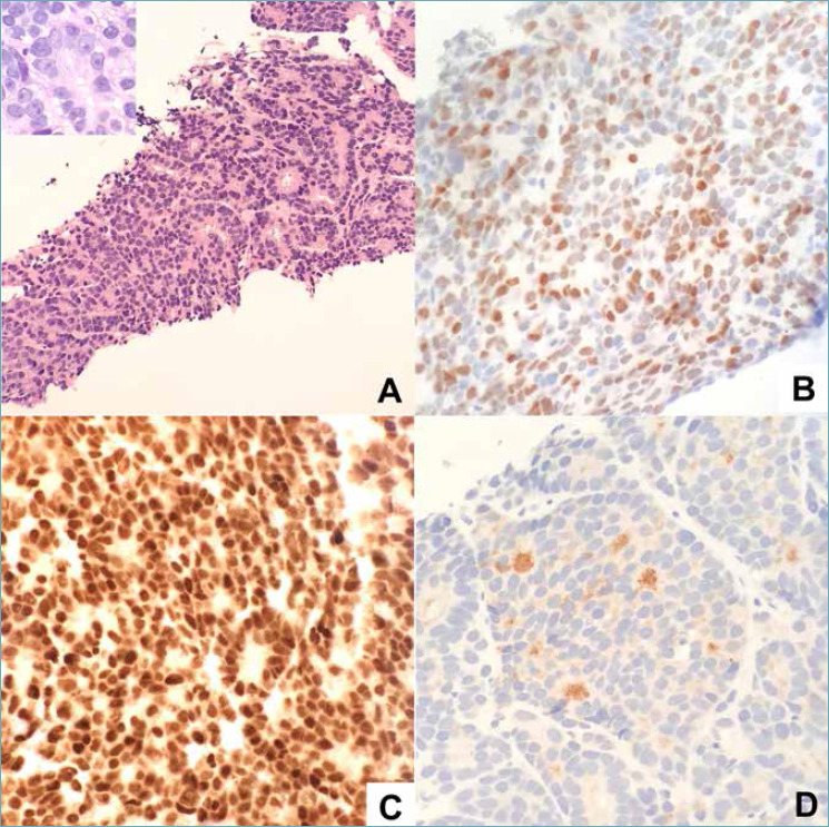
Histological examination of the biopsy revealed a diffuse proliferation of neoplastic epithelial cells arranged in small acinar formations (Fig. 1A). The cells had medium-sized nuclei which often had prominent nucleoli. Immunohistochemistry (IHC) revealed tumour cells that were positive for PSA, NKX3.1, and CDX2, and were negative for TTF-1. The nuclear staining for CDX2 was of moderate intensity and involved 50% of the tumor cells (Fig. 1B), while the nuclear staining for NKX3.1 was diffuse and strong (Fig. 1C). PSA was mainly positive in the acinar lumens with only focal cytoplasmic staining (Fig. 1D). Based on the histologic findings and IHC results, as well as clinical information, the tumor was diagnosed as metastatic prostatic adenocarcinoma despite the aberrant CDX2 expression. Several days later, a needle core biopsy of the prostate was performed and revealed a CDX2-positive Gleason score 4 + 3 = 7 acinar type adenocarcinoma.
Prostatic adenocarcinoma and colorectal carcinoma are two of the most common tumors in males. Given the difference in treatment and prognosis between these two tumors, it is important to obtain an accurate diagnosis in cases of metastasis of unknown origin or even in cases of locally advanced prostate or colorectal carcinoma with secondary involvement of the rectum or prostate, respectively. A list of morphologic features and IHC markers is used in such cases to resolve their primary origin.
The usual acinar adenocarcinoma of prostate is composed of microacini lined by cuboidal to low columnar cells with round nuclei often displaying a large nucleolus. On the other hand, many colorectal carcinomas reveal large irregular glands lined by pleomorphic columnar cells surrounding a lumen filled with dirty necrosis. Since cases of morphologic overlap between these two tumors do occur, one must often resort to ancillary tools such as IHC 4. PSA has been demonstrated to be a prostate tissue specific marker, but its reactivity may be lost in a significant number of high grade and metastatic prostatic adenocarcinomas 4. In this regard, NKX3.1 has proven to be of great value since it will stain the nuclei of PSA-negative prostatic adenocarcinoma cells in metastatic sites.
CDX2 expression has been documented within epithelial cells of the alimentary tract from the duodenum to the rectum. It is expressed not only in tumors of the colorectum and small intestine, but also in most gastric carcinomas as well as neoplasms with mucinous differentiation of various organs 5. In cases of prostatic adenocarcinomas, early studies reported no CDX2 staining. More recent studies have documented positive staining, with a 5% rate in primary acinar tumors and a 30% rate in primary tumors with mucinous features 1,2. In contrast, cases of CDX2-positive metastases of prostatic adenocarcinoma are vanishingly rare, and when positive, the staining has been graded as focal and of weak intensity. Our case is unique in that it exemplifies a case of metastatic prostatic adenocarcinoma wherein the tumour cells show moderately intense staining in a significant number of cells, thus potentially creating the risk of misclassifying the metastasis as intestinal in origin.
In conclusion, our case brings awareness to the possibility of CDX2-positivity in metastatic prostatic adenocarcinoma. Knowledge of such an uncommon occurrence will aid in preventing a false-positive diagnosis of metastatic gastrointestinal carcinoma during the histologic evaluation of a metastasis of unknown primary origin.
Figures and tables
Fig. 1.ABCD.

Metastatic CDX2-positive prostate adenocarcinoma, hematoxylin and eosin (H&E) and immunostains. (A) H&E (low power) with nuclear features in inset (high power); (B) CDX2 (high power); (C) NKX3.1 (high power); (D) PSA (high power).
Footnotes
CONFLICT OF INTEREST STATEMENT
None declared.
References
- 1.Herawi M, De Marzo A, Kristiansen G, et al. Expression of CDX2 in benign tissue and adenocarcinoma of the prostate. Hum Pathol 2007;38;72-8. https://doi.org/10.1016/j.humpath.2006.06.015 10.1016/j.humpath.2006.06.015 [DOI] [PubMed] [Google Scholar]
- 2.Leite KRM, Mitteldorf C, Srougi M, et al. Cdx2, cytokeratin 20, thyroid transcription factor and prostate-specific antigen expression in unusual subtypes of prostate cancer. Ann Diagn Pathol 2008;12;260-6. https://doi.org/10.1016/j.anndiagpath.2007.11.001. 10.1016/j.anndiagpath.2007.11.001 [DOI] [PubMed] [Google Scholar]
- 3.Nwankwo N, Mirrakhimov AE, Zdunek T, et al. Prostate adenocarcinoma with a rectal metastasis. BMJ Case Rep 2013;2013. https://doi.org/10.1136/bcr-2013-009503. 10.1136/bcr-2013-009503 [DOI] [PMC free article] [PubMed] [Google Scholar]
- 4.Owens CL, Epstein JI, Netto GJ. Distinguishing prostatic from colorectal adenocarcinoma on biopsy samples: the role of morphology and immunohistochemistry. Arch Pathol Lab Med 2007;131;599-603. https://doi.org/10.1043/1543-2165(2007)131[599:DPFCAO]2.0.CO;2 10.1043/1543-2165(2007)131[599:DPFCAO]2.0.CO;2 [DOI] [PubMed] [Google Scholar]
- 5.Saad RS, Ghorab Z, Khalifa MA, Xu M. CDX2 as a marker for intestinal differentiation: its utility and limitations. World J Gastrointest Surg 2011;27;159-66. https://doi.org/10.4240/wjgs.v3.i11.159. 10.4240/wjgs.v3.i11.159 [DOI] [PMC free article] [PubMed] [Google Scholar]


