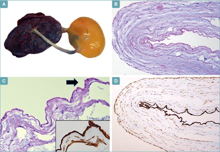
Fig. 1.

A) Macroscopic appearance the placenta and umbilical cord. A large cystic mass of 10 cm in greatest diameter can be appreciated in the umbilical cord close to the placental insertion. B) On histological examination, the cystic mass consisted of a single layer of eosinophilic cells. Myxoid degeneration of the cystic wall can also be appreciated. C) Higher magnification of the cyst reveals a single layer of amnionic epithelial cells with flattened or cuboidal morphology (arrow). The epithelial origin of the cyst is confirmed by positive immunostain for pan-cytokeratin antibody (clone AE1/AE3) (inset; D).
