
Figure 6. Ferroptosis induction correlates with p53 activation and better clinical responses to RT in cancer patients.
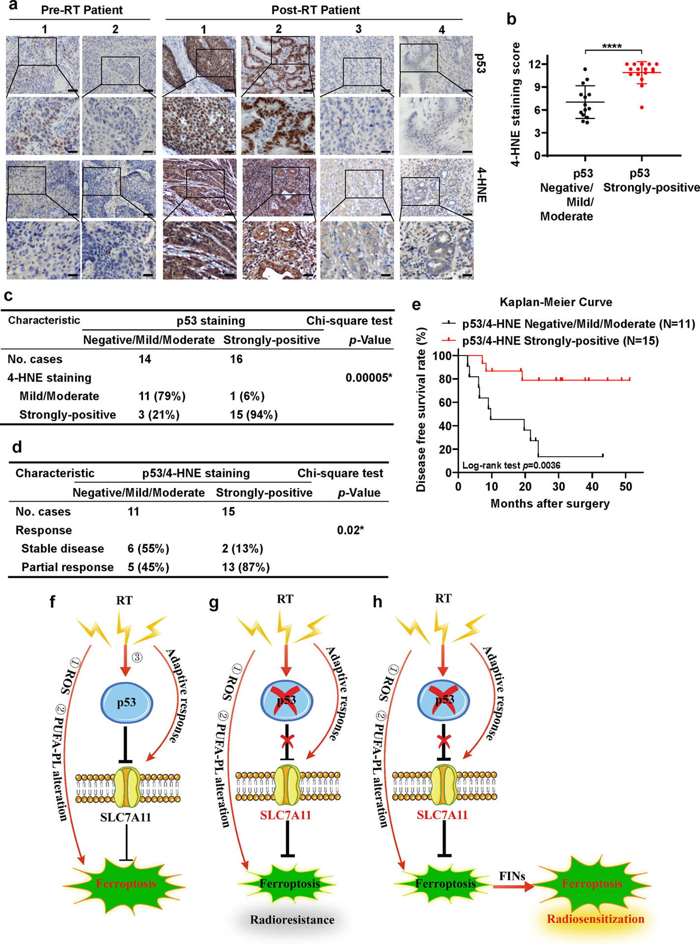
a Representative p53 and 4-HNE IHC staining images in esophageal cancer patient samples before and after radiotherapy. Scale bars represent 50 μm/20 μm.
b IHC scores for 4-HNE staining in p53 negative/mild/moderate and p53 strongly-positive esophageal cancer patient samples after radiotherapy. Error bars are mean ± SD from six randomly selected magnification fields. P values calculated by 2-tailed unpaired Student’s t-test.
c Correlation between 4-HNE and p53 staining in patients with esophageal cancer after radiotherapy. P values calculated by Chi-squared test.
d Correlation between radiotherapy response and p53/4-HNE combination staining in patients with esophageal cancer after radiotherapy. P values calculated by Chi-squared test.
e Kaplan–Meier survival curves for esophageal cancer patients stratified by p53 and 4-HNE staining after radiotherapy. P values calculated by log-rank test.
f-h The working model depicting the roles and mechanisms of ferroptosis in p53-mediated radiosensitization. See Discussion for detailed description.