Figure 1.

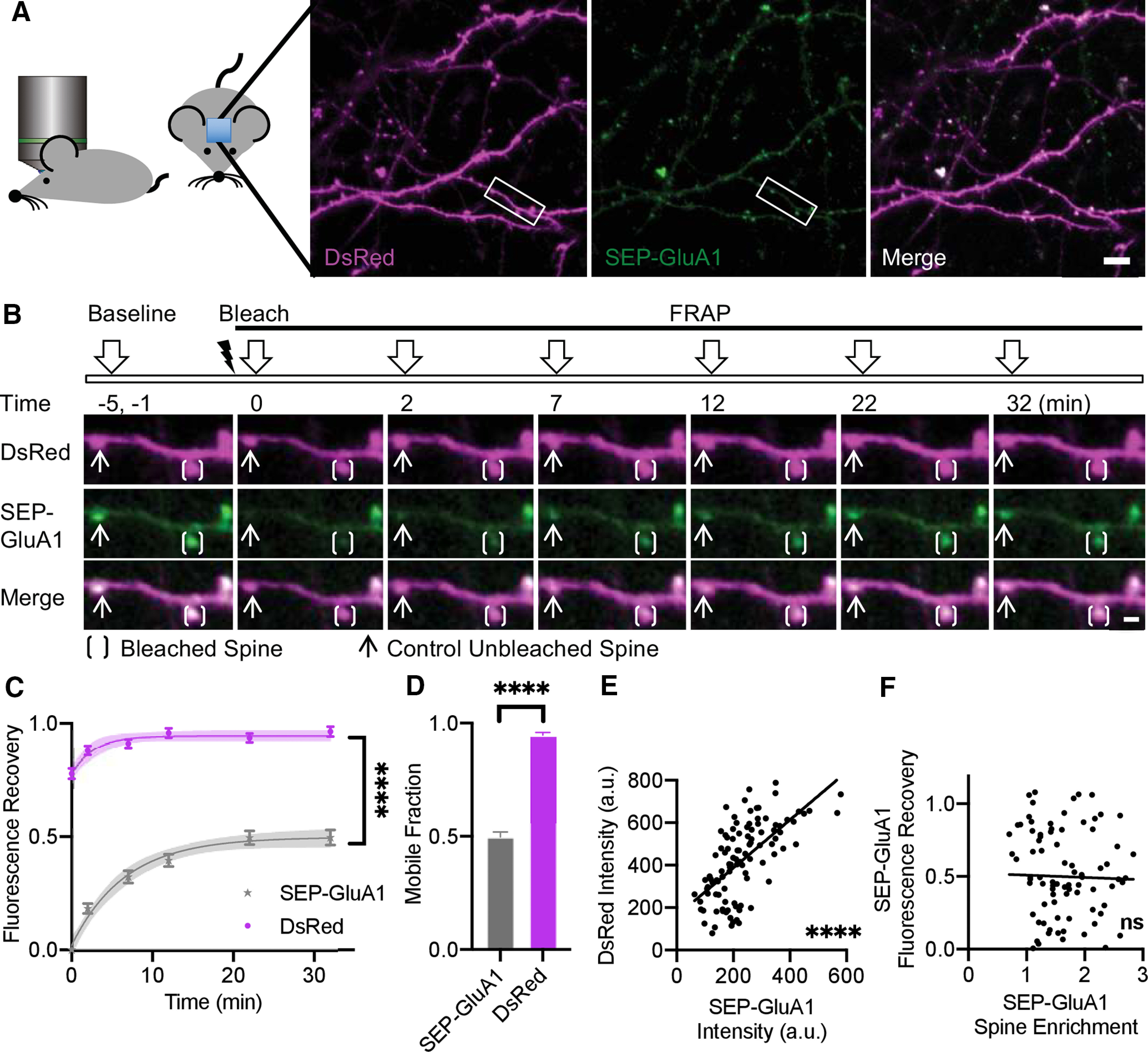
In vivo FRAP shows mobile and immobile fractions of GluA1 in cortical neurons. A, Schematic of experimental approach using two-photon imaging of cranial windows in live mice. Representative images of maximum intensity projection (MIP) of 3D z-stack of L5 visual cortex neurons expressing DsRed cell fill (magenta), SEP-GluA1 (green), and myc-GluA2. Scale bar: 10 μm. Area of interest indicated corresponding to panel B. B, Representative MIP image of dendrite with a bleached (bracket) and an unbleached (arrow) spine at indicated time points. Scale bar: 2 μm. C, Fluorescence recovery of SEP-GluA1 versus DsRed cell fill in spines (multifactorial ANOVA; Extended Data Figures 1-3, 1-4). Time points were fitted with an exponential curve indicated by solid line with 95% CI in shaded area. D, Mobile fraction defined by maximum of fluorescence recovery calculated as Ymax of fitted exponential curve displayed in panel C (t test). E, Correlation between initial raw intensity of DsRed cell fill and SEP-GluA1 per spine, Pearson’s correlation r2 = 0.41. F, Correlation between recovered fraction measured at 32 min postbleach and spine enrichment, Pearson’s correlation r2 = 0.0006. C–F, n = 104 spines, 4 mice, error bars indicate SEM; ****p < 0.0001, ns = not significant. See also Extended Data Figures 1-1, 1-2.
