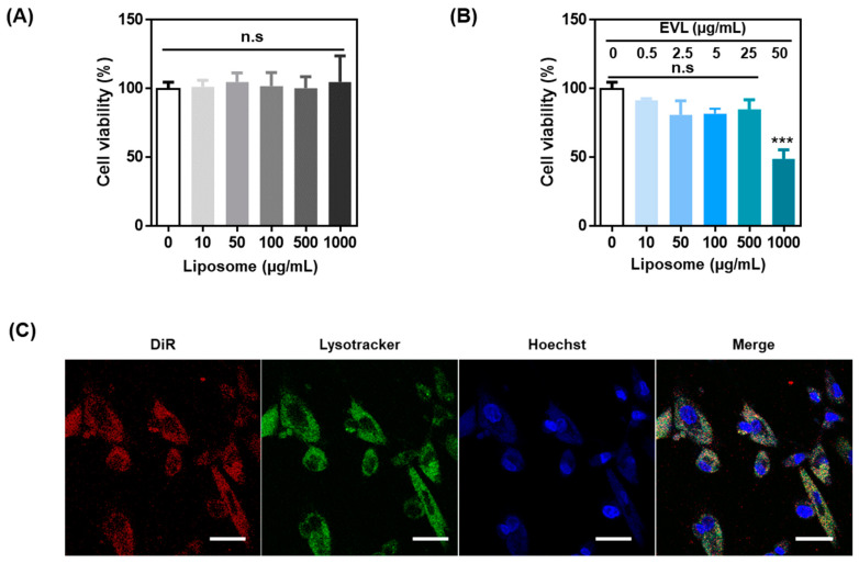
Figure 3.
In vitro study using liposome and EVL-loaded liposome. Cell viability of (A) liposome and (B) EVL-loaded liposome. (C) Confocal images for interaction between DiR-labeled liposome and HCASMCs. The lysosome and nucleus of HCASMCs were stained by lysotracker (green) and Hoechst (blue), respectively (Scale bars equal to 40 μm). Values are presented as mean ± SD (n = 3) and statistical significance was obtained with one-way analysis of ANOVA with Tukey’s multiple comparison post-test (* p < 0.05; ** p < 0.01; *** p < 0.001). HCASMCs: Human Coronary Artery Smooth Muscle Cells.

