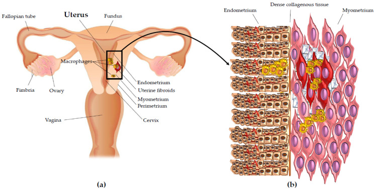
Figure 4.
Macrophages in uterine fibroids. (a) Illustration of uterus showing the macrophage density in uterine fibroids pathology. (b) Enlargement of the detail showing the macrophage density in uterine fibroids pathology. Macrophages (yellow in the figure) predominantly localize inside uterine fibroids (red in the figure) and in the myometrium tissue (pink in the figure) next to them. Autologous distant myometrium shows low levels of macrophage infiltration. The macrophage density is higher also in the endometrium (brown in the figure) next to uterine fibroids than in the autologous endometrium far from uterine fibroid nodules. The extracellular matrix (ECM) around and within the uterine fibroids is also represented (blue net in the figure). The blood vessels within endometrium are also represented (red lines in the figure).

