Abstract
BACKGROUND AND PURPOSE: T1-weighted spin-echo imaging has been widely used to study anatomic detail and abnormalities of the brain; however, the image contrast of this technique is often poor, especially at low field strengths. We tested a new pulse sequence, T1-weighted fluid-attenuated inversion recovery (FLAIR), which provides good contrast between lesions, surrounding edematous tissue, and normal parenchyma at low field strengths and at acquisition times comparable to those of T1-weighted spin-echo imaging.
METHODS: Thirteen patients with brain lesions underwent T1-weighted spin-echo and T1-weighted FLAIR imaging during the same imaging session. T1-weighted spin-echo and T1-weighted FLAIR images were compared on the basis of four quantitative (lesion-white matter [WM] contrast-to-noise ratio [CNR], lesion-CSF CNR, gray matter-WM CNR, and WM-CSF CNR) and three qualitative criteria (conspicuousness of lesions, image artifacts, and overall image contrast).
RESULTS: CNRs obtained with T1-weighted FLAIR were comparable but statistically superior to those obtained with T1-weighted spin-echo imaging. In general, T1-weighted FLAIR and T1-weighted spin-echo imaging produced comparable image artifacts. Conspicuousness of lesions and the overall image contrast were judged to be superior on T1-weighted FLAIR images.
CONCLUSION: T1-weighted FLAIR imaging may be a valuable alternative to conventional T1-weighted imaging, because the former technique offers superior image contrast at low field strengths and comparable acquisition times.
T1-weighted spin-echo images are widely used to study the anatomic detail and pathologic abnormalities of the brain. Image contrast obtained with MR imaging is superior to that obtained with CT; however, the former technique does not provide sufficient image contrast to differentiate white matter (WM) from gray matter (GM). This problem most commonly arises at low field strengths. Inversion recovery (IR) is a pulse sequence that provides superior T1-weighted image contrast to that of spin-echo imaging (1). IR, however, is not the first choice for T1-weighted imaging, because it requires a longer acquisition time than does spin-echo imaging.
Recently, the fast spin-echo (FSE) sequence has shown great promise for reducing acquisition times with T2-weighted imaging. FSE, however, has not been used clinically to acquire T1-weighted contrast-enhanced images, because the placement of early echoes in the center of k space to achieve a short TEeff can lead to image artifacts such as image blurring (2–4).
The fast inversion recovery (FIR) pulse sequence, presented herein, couples an IR preparation pulse to an FSE readout with interleaved rotary data acquisition and a TI suitable for suppressing signal intensity from CSF. This sequence enables a shorter acquisition time from that of a previously reported FIR sequence (5). Herein, we refer to this pulse sequence as T1-weighted fluid-attenuated inversion recovery (FLAIR).
The purpose of this study was to compare T1-weighted FLAIR image contrast with that of T1-weighted spin-echo images obtained at the same low field strength, at comparable acquisition times, and during the same imaging session.
Methods
Pulse Sequences
All examinations were performed on a 0.2-T MR system (Signa Profile [software version 7.7]; GE-Yokogawa Medical Systems, Tokyo, Japan) equipped with gradients that had a maximum slew rate of 17 T/m/s, a gradient strength of 10 mT/m, and a standard quadrature head coil.
The MR imaging protocol consisted of a T1-weighted FLAIR sequence (TR/TE/TI/NEX, 970/29/450/2; field of view, 240 mm × 240 mm; matrix, 256 × 192; receive bandwidth, 6.7 kHz; section thickness, 6 mm; intersection gap, 2 mm; acquisition time, 6 minutes 26 seconds) and T1-weighted spin-echo sequence (TR/TE/NEX, 500/14/3; field of view, 240 mm × 240 mm; matrix, 256 × 192; receive bandwidth, 7.8 kHz; section thickness, 6 mm; intersection gap, 2 mm; acquisition time, 5 minutes 58 seconds). These parameters are used at our institution as the standard intracranial imaging protocol. As detailed above, field of view, spatial resolution, section thickness, and intersection gap were the same for the T1-weighted FLAIR and T1-weighted spin-echo sequences. Eighteen axial images were obtained from both sequences.
Patient Characteristics
Thirteen patients (10 male, three female; mean age, 59 years [range, 27–78 years]) with brain disease underwent T1-weighted FLAIR and T1-weighted spin-echo imaging. Before MR examinations were performed, institutional review board approval was obtained and each patient provided written informed consent. The pathologic conditions of the patients included primary brain tumors (glioblastoma [n = 2], meningioma [n = 3], neuroma [n = 1]), metastatic brain tumors (lung cancer [n = 3], hepatoma [n = 2]), subacute infarction (n = 1), and meningitis (n = 1). Diagnoses were made on the basis of biopsy findings, clinical history, presentation, or follow-up imaging studies.
All patients underwent T1-weighted spin-echo and T1-weighted FLAIR imaging when IV administered gadolinium chelate (Prohance; Bracco-Eisai, Tokyo, Japan) reached a concentration of 0.2 mmol/kg. To avoid delays in contrast enhancement, sequences were randomly performed.
Image Analysis
Contrast on T1-weighted FLAIR images was compared with that of T1-weighted spin-echo images by using four quantitative and three qualitative criteria.
Quantitative Analysis.
Two of the quantitative criteria pertained to lesion characteristics: lesion-WM contrast-to-noise ratio (CNR) and lesion-CSF CNR. For each patient studied, lesions were measured on one to three sections from the same, randomly chosen level (ie, cerebellar level or basal ganglia level) for both pulse sequences. For these measurements, WM was defined as the normal brain parenchyma adjacent to the lesion that showed no edema or atrophy. Twenty-two lesions were measured on postcontrast images. The other two quantitative criteria, which were related to signals from normal tissue, were GM-WM CNR and WM-CSF CNR.
For all quantitative measurements, the mean signal intensity of the enhanced lesion, background, WM, GM, and CSF was measured from within regions of interest placed within the corresponding areas of the same section. The SD of noise was measured along the phase-encoding direction in regions outside the brain. Lesion-WM CNR was defined as the difference between the signals from the lesion and those from WM. Lesion-WM CNR was calculated by dividing the difference between the signals from the lesion and those from WM by the SD of image noise. Corresponding procedures were used to determine lesion-CSF CNR, GM-WM CNR, and WM-CSF CNR. Corresponding areas on T1-weighted FLAIR and T1-weighted spin-echo images were compared by use of relative CNR.
Contrast-to-background ratio was calculated by dividing the signals from a lesion by the SD of image noise, and values were classified into two groups according to whether T1-weighted FLAIR was performed before or after T1-weighted spin-echo imaging. An effect of delayed contrast medium administration was compared between the two groups from corresponding areas on T1-weighted FLAIR and T1-weighted spin-echo images by use of contrast-to-background ratio.
Statistical significance of quantitative data were determined by using a Wilcoxon signed rank test.
Qualitative Analysis.
Three qualitiative criteria were conspicuousness of lesions, presence of image artifacts, and overall image contrast. Three experienced neuroradiologists (M.H., T.O., K.U.) performed this qualitative analysis for all images obtained. All T1-weighted FLAIR images were compared with all T1-weighted spin-echo images. Conspicuousness of lesions, image artifacts, and overall image contrast were graded on a 5-point scale: one signified that T1-weighted FLAIR images were clearly inferior to T1-weighted spin-echo images; 2, T1-weighted FLAIR images were slightly inferior to T1-weighted spin-echo images; 3, T1-weighted FLAIR images were comparable to T1-weighted spin-echo images; 4, T1-weighted FLAIR images were slightly superior to T1-weighted spin-echo images; and 5, T1-weighted FLAIR images were clearly superior to T1-weighted spin-echo images.
Results
In all patients, both T1-weighted spin-echo and T1-weighted FLAIR imaging were effective in showing lesions. As expected, T1-weighted FLAIR provided improved GM-WM CNRs and CSF-WM CNRs compared with those of T1-weighted spin-echo imaging. Another notable finding was the absence of contrast-enhancing blood vessels on T1-weighted FLAIR images, which is a typical T1-weighted FLAIR finding.
Quantitative Results
Quantitative lesion-related CNR, GM-WM CNR, and WM-CSF CNRs are summarized in Table 1. The T1-weighted FLAIR lesion-WM and lesion-CSF CNR values were comparable and statistically superior to those of the T1-weighted spin-echo images at a TI of 450 (P < .005). As expected, T1-weighted FLAIR images provided statistically superior image contrast between lesion and WM and lesion and CSF compared with corresponding T1-weighted spin-echo images because of the greater contrast provided by the IR technique. Moreover, T1-weighted FLAIR provided greater GM-WM and WM-CSF CNR than did T1-weighted spin-echo imaging. Overall, T1-weighted FLAIR images were judged superior on the basis of all criteria.
TABLE 1:
Quantitative results*
| Lesion-WM | Lesion-CSF | GM-WM | CSF-WM | |
|---|---|---|---|---|
| T1-weighted spin-echo CNR | 8.07 ± 4.84 | 30.6 ± 5.71 | 5.13 ± 2.14 | 22.4 ± 3.29 |
| T1-weighted FLAIR CNR | 14.1 ± 8.78 | 41.5 ± 9.10 | 9.10 ± 3.07 | 27.1 ± 4.26 |
Note.—WM signifies white matter; CSF, cerebrospinal fluid; GM, gray matter; CNR, contrast-to-noise ratio; and FLAIR, fluid-attenuated inversion recovery.
Values represent the mean ± SD.
Results of the contrast-to-background ratio between T1-weighted FLAIR and T1-weighted spin-echo imaging are summarized in Table 2. No significant difference in contrast-to-background ratio was seen between T1-weighted FLAIR performed before and T1-weighted FLAIR performed after T1-weighted spin-echo imaging (P < .05), which suggests that delays in contrast medium administration did not affect findings.
TABLE 2:
Contrast-to-background ratio*
| T1-weighted FLAIR Before T1-weighted/ Spin-Echo Imaging | T1-weighted FLAIR After T1-weighted/ Spin-Echo Imaging | |
|---|---|---|
| T1-weighted spin-echo imaging | 35.5 ± 7.18 | 37.5 ± 4.05 |
| T1-weighted FLAIR imaging | 41.6 ± 5.64 | 48.7 ± 11.4 |
Note.—FLAIR signifies fluid-attenuated inversion recovery.
Values represent the mean ± SD.
Qualitative Results
Results of the three qualitative comparisons between T1-weighted FLAIR and T1-weighted spin-echo imaging are summarized in Table 3. In general, T1-weighted FLAIR and T1-weighted spin-echo images produced comparable artifacts. John and colleagues (5) suggested that the FIR technique was inferior to T1-weighted spin-echo techniques because of flow-related artifacts (5). This type of artifact is typically seen on images of the posterior fossa and is caused by blood flow in the dural sinus, particularly the transverse sinus, when present. In this study, artifacts on T1-weighted FLAIR images did not interfere with image interpretation.
TABLE 3:
Qualitative results*
| Lesion Conspicuity | Image Artifact | Image Contrast | |
|---|---|---|---|
| Radiologist 1 | 3.90 ± 0.91 | 3.00 ± 0.46 | 4.25 ± 0.72 |
| Radiologist 2 | 4.20 ± 0.95 | 3.75 ± 0.64 | 3.95 ± 0.89 |
| Radiologist 3 | 3.55 ± 0.60 | 3.05 ± 0.39 | 3.95 ± 0.69 |
Values represent mean ± SD.
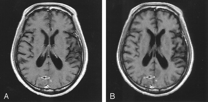
As presented in Table 3, all neuroradiologists judged the overall image contrast to be superior on T1-weighted FLAIR images compared with that on T1-weighted spin-echo images. Although the depiction of edema and areas of small ischemic changes is usually present on T2-weighted images, we considered the greater ability of T1-weighted FLAIR to depict surrounding edematous tissue in relation to enhancing lesions to be clinically valuable (Fig 1).
Fig 1.
MR images obtained in a 70-year-old man with metastatic brain tumor.
A, T1-weighted spin-echo image does not clearly show distinction between lesion (arrow) and surrounding edematous tissue.
B, T1-weighted FLAIR image clearly shows enhancing lesion (arrow).
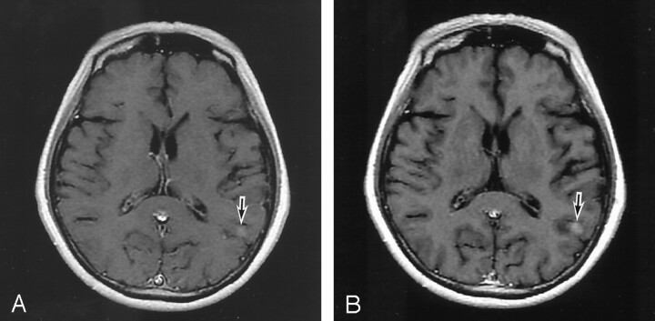
Qualitative analysis also suggested that T1-weighted FLAIR images showed lesions more clearly than did T1-weighted spin-echo imaging (Table 3). For example, in a patient with multiple metastatic tumors from a hepatoma, T1-weighted FLAIR imaging was more effective in depicting tumors than was T1-weighted spin-echo imaging (Fig 2).
Fig 2.
MR images obtained in a 60-year-old man with multiple metastatic brain tumors.
A, T1-weighted spin-echo image has poor lesion-WM CNR (arrow).
B, T1-weighted FLAIR image clearly shows enhancing lesion (arrow).
Discussion
Precontrast T1-weighted images acquired by conventional spin-echo techniques often have poor image contrast, especially at low field strengths. Therefore, T1-weighted spin-echo images have often been used in a comparable way to gadolinium-enhanced T1-weighted images to detect lesions in the brain rather than for the evaluation of anatomic structures. Our T1-weighted spin-echo images showed poor differentiation between GM and WM, and CSF signals were not sufficiently suppressed.
Previous investigators (5) have compared the T1-weighted FIR technique with T1-weighted spin-echo imaging; their results indicated that, of the two techniques, FIR provided superior contrast between CSF and WM, WM and GM, and enhancing lesions and WM at 1.5 T. These studies, however, reported on results obtained at high field strengths. The need for improved T1-weighted image contrast is greater at lower field strengths because of inferior image quality.
Before we conducted this study, we sought to find optimal parameters for the T1-weighted FLAIR sequence. Because the T1-weighted FLAIR sequence was required to image 18 sections with a 256 × 192 spatial resolution in a 5–7-minute acquisition time based on the T1-weighted spin-echo intracranial imaging protocol used at our institution, T1-weighted FLAIR images were acquired with two NEX compared with three NEX used for the T1-weighted spin-echo sequence and with an echo train length of two compared with that of one used for T1-weighted spin-echo images. It is impossible to change the echo train length to three or more, because image degradation increases with increased echo train length. We hope that future use of the T1-weighted FLAIR sequence includes more echoes per repetition. That would lead to shorter acquisition times. The T1-weighted FLAIR sequence described herein used a receive bandwidth of 6.7 kHz as compared with a receive bandwidth of 7.8 kHz used with the T1-weighted spin-echo sequence. The receive bandwidth used for the T1-weighted FLAIR sequence allowed an interecho space of 29.
When the TR was one one-fourth the TI value of CSF, the longitudinal magnetization recovered incompletely; we therefore shortened the TI value for CSF signal intensity suppression. We used a TI-TR combination of 450/970 for T1-weighted FLAIR, which allowed an 18-section acquisition with rotary data acquisition.
We also used an FIR technique with advanced data acquisition for the T1-weighted FLAIR sequence, which provided greater CNR than did conventional T1-weighted spin-echo imaging in all categories and at almost the same acquisition time with a 0.2-T MR system. These results suggest that FLAIR may be a useful T1-weighted technique.
The limitations of this investigation were the small number of patients and heterogeneous diseases studied. Our results suggest that T1-weighted FLAIR can provide greater CNR with clinically acceptable doses of contrast medium. For other concentrations of gadolinium-based contrast material, the increase in CNR between an enhancing lesion and WM on a T1-weighted FLAIR image remains unknown. Further investigation is needed before the T1-weighted FLAIR technique can become a criterion standard for intracranial T1-weighted imaging.
Conclusion
Overall image contrast provided by T1-weighted FLAIR at low field strength may be a valuable clinical tool in the armamentarium of intracranial T1-weighted imaging. Recent advances in MR imaging software and hardware allow greater flexibility in determining which T1-weighted sequence is most suitable for brain imaging and what pulse sequence should be used as a criterion standard.
Acknowledgments
We thank Noriko Hirasawa, Toru Hayasaka, and Kenji Suzuki of GE-Yokogawa Medical Systems for technical assistance and advice.
Footnotes
Presented at the 40th annual meeting of the American Society of Neuroradiology, Vancouver, B.C., May 11–14, 2002.
References
- 1.Bydder GM, Young IR. MR imaging: clinical use of the inversion recovery sequence. J Comput Assist Tomogr 1985;9:659–675 [PubMed] [Google Scholar]
- 2.Mulkern RV, Wong STS, Winalski C, Jolesz FA. Contrast manipulation and artifact assessment of 2D and 3D RARE sequences. Magn Reson Imaging 1990;8:557–566 [DOI] [PubMed] [Google Scholar]
- 3.Glover GH, Tkach JA, Shimakawa A. Reduction of non-equilibrium effects in RARE sequences (abstr). In: Book of Abstracts: Society of Magnetic Resonance in Medicine 1991. Berkeley, Calif: Society of Magnetic Resonance in Medicine,1991. , 1242
- 4.Hinks RS, Listerud J Approach to steady state in fast spin-echo imaging. In: Book of Abstracts: Society of Magnetic Resonance in Medicine 1991. Berkeley, Calif: Society of Magnetic Resonance in Medicine,1991. , 1235
- 5.John NR, Charlotte AH, John H, Clifford RJ, Rover CG, Stephen JR. T1-weighted MR imaging of the brain using a fast inversion recovery pulse sequence. J Magn Reson Imaging 1996;6:356–362 [DOI] [PubMed] [Google Scholar]


