Abstract
BACKGROUND AND PURPOSE: Encephaloduroarteriosynangiosis (EDAS) has become the main treatment for moyamoya disease, a chronically progressive cerebrovascular occlusive disease in children. We aimed to assess the utility of perfusion-weighted MR imaging for evaluating hemodynamic changes before and after EDAS.
METHODS: Thirteen patients with angiographically confirmed moyamoya disease who underwent EDAS were investigated, and results were compared with those of a control group (n = 5). Perfusion MR imaging was performed before and after EDAS by using a T2*-weighted contrast material-enhanced technique. Relative cerebral blood volume (rCBV) and time to peak enhancement (TTP) maps were calculated. Relative ratios of rCBV and TTP in the middle cerebral artery (MCA) and basal ganglia were measured and compared with those of the posterior cerebral artery (PCA). Changes in hemodynamic parameters between pre- and post-EDAS perfusion maps were investigated.
RESULTS: The mean rCBV ratio of MCA to PCA in the patient group was slightly higher than that in the control group, without statistical significance. All 13 patients showed a delayed TTP in the MCA before EDAS compared with the control group (P = .0006), and the TTP after EDAS was significantly reduced (P = .0002). In the basal ganglia, shortening of the TTP was demonstrated before EDAS, but no significant change was observed after EDAS.
CONCLUSION: Perfusion-weighted MR imaging can be applied for evaluating postoperative changes in cerebral blood flow in moyamoya disease. Shortening of the TTP in the MCA of the hemisphere operated on is a marker for the development of collateral circulation from the external carotid artery to the internal carotid artery.
Moyamoya disease is a chronically progressive cerebrovascular disease affecting the supraclinoid internal carotid arteries (ICAs) with prominent collateral arterial formation (1). Despite the fact that moyamoya disease is thought to affect primarily Japanese or Koreans, it also has been reported in other ethnic groups (2, 3). The etiology is still uncertain, and without proper treatment, children presenting with ischemic symptoms tend to show an aggravating natural course (4). Encephaloduroarteriosynangiosis (EDAS) is the treatment of choice because this procedure can reestablish cerebral perfusion by means of external carotid artery (ECA)-to-ICA bypass (5). Development of collateral vessels after surgery usually occurs in the middle cerebral artery (MCA) territory because of the distribution of the superficial temporal artery, which is the most commonly used supplier for EDAS. Although other advanced procedures for reconstruction of blood flow to the anterior cerebral artery (ACA) territory have been introduced recently (6–8), EDAS is still widely used in many institutes.
Conventional angiography is used to diagnose moyamoya disease (9), but single photon emission tomography (SPECT) is known to be the reference standard for evaluating the hemodynamic status of patients with moyamoya disease (10, 11). MR imaging also has been widely used in assessing moyamoya disease, because of its capacity to illustrate anatomic detail and the vascular architecture (12, 13). Recently, T2*-weighted perfusion MR imaging has been found to be effective in estimating cerebral hemodynamics in moyamoya disease (14–17). These diagnostic maneuvers can be used to assess local hemodynamic changes after ICA-to-ECA bypass surgery (18–20). However, there is a paucity of reports describing the efficacy of perfusion-weighted MR imaging in postoperative evaluation. The purpose of this study was to assess the utility of perfusion MR imaging in illustrating the hemodynamic changes of an ischemic brain in moyamoya disease before and after EDAS.
Methods
Thirteen patients with childhood moyamoya disease who underwent EDAS between July 1999 and March 2002 were evaluated by using perfusion-weighted MR imaging before and after surgery. The subjects consisted of eight female and five male patients, with ages ranging from 2 to 27 years (mean, 8.5 years). They received preoperative diagnostic evaluation with technetium-99m ethyl cysteinate dimer SPECT and conventional angiography. On cerebral angiograms, all 13 patients showed stenosis, either partial or complete obstruction, in the supraclinoid portion of the ICAs. All showed patent posterior cerebral arteries (PCAs) and various degrees of leptomeningeal collateral vessels (eg, less than Suzuki grade V [21]). EDAS was performed according to the patients’ clinical symptoms and angiographic findings. EDAS was performed in the left cerebral hemisphere in four patients, in the right cerebral hemisphere in three patients, and in both hemispheres in the remaining six patients.
All patients underwent preoperative perfusion-weighted MR imaging 3–7 days before EDAS and postoperative perfusion-weighted MR imaging 6–8 months after EDAS. Five children (three male, two female subjects; mean age, 9.3 years; age range, ) were included in the control group because healthy volunteers were not available for perfusion study. Age-matched control subjects could not be recruited because they would have needed sedation, and ethical considerations prohibited this. The control subjects were referred for brain imaging with the complaint of simple headache or psychiatric problem. They were found to have no central nervous system disease radiologically and at follow-up studies. Informed consent was received from all participants or the participants’ parents or legal guardian, and all procedures were performed under approval by the institutional board of clinical studies.
The MR examinations were conducted by using a 1.5-T system (Signa Horizon, GE Medical System, Milwaukee, WI, or Gyroscan Intera, Philips Medical Systems, Best, the Netherlands) with a quadrature head coil. Perfusion-weighted MR imaging was performed by using a single-shot gradient-echo echo-planar imaging sequence during an intravenous bolus injection of 0.2 mmol/kg gadopentetate dimeglumine (Magnevist; Schering AG, Berlin, Germany) with the following parameters: 1500/40/1 (TR/TE/excitations), 24-cm field of view, 5-mm section thickness with a 2-mm intersection gap, and 128 × 128 matrix. Six sections were chosen starting from the anteroposterior commisure line including the transthalamic level, and 40 dynamic perfusion images were obtained from each level. Contrast material was injected by using a power injector (Spectris; Medrad, Indianola, PA) after the fifth dynamic image, followed by flushing with normal saline, and image acquisition continued until all 40 phase images were obtained.
Data from the eight patients who were examined with the GE system were transferred to a personal computer, where the data were analyzed by using a program coded for Interactive Data Language (IDL 5.4 Win32; Research Systems Inc., Boulder, CO). The data of the other five patients examined with the Philips system were processed by Easyvision (Philips Medical Systems). For each pixel, the time-concentration or ΔR2* curve was obtained, which was calculated from the equation ΔR2*(t) = [ln(SI0/SI(t))], where SI0 is the average precontrast signal intensity and SI(t) is the signal intensity at time t. The relative cerebral blood volume (rCBV) was calculated pixel by pixel from the time-relaxation curve obtained by dynamic imaging. The time interval to peak enhancement (TTP) maps were also calculated. The regions of interest in the bilateral MCA territory, PCA territory, and basal ganglia were drawn (Fig 1) and contained more than 20 pixels. The rCBV ratios and TTP differences of the MCA and of the basal ganglia to the PCA territory were calculated on the pre- and postoperative perfusion MR images. Statistical analysis was done between patients and control group by using the Student’s t test for the independent samples. A paired t test was used to analyze the patient group for changes before and after EDAS. A P value of less than or equal to .05 was considered to indicate a statistically significant difference.
Fig 1.
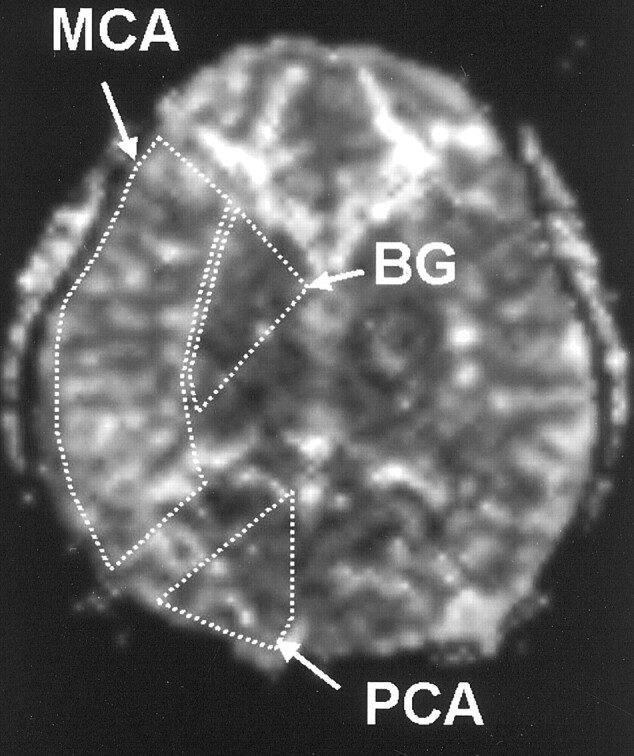
Regions of interest (MCA, PCA, and BG) are drawn on a TTP map. On the rCBV maps (not shown), the relative signal intensity ratios of the MCA and the BG to the PCA territory were obtained, whereas on the TTP maps the time differences in the MCA and BG were calculated and compared with that of the PCA territory.
Results
The measured rCBV and TTP values in the control and patient groups are listed in the Table. The mean rCBV ratio of the MCA to PCA territory in the control group was 1.35 ± 0.14 in the MCA territory and 0.79 ± 0.12 in the basal ganglia. The TTP in the MCA territory was identical to that of the PCA territory, whereas the TTP in the basal ganglia was shorter than that in the PCA territory.
rCBV Ratios and TTP Values in the Control Group and Patients
| Parameter | Control Group (n = 5) | Patient Group (n = 13) | P Value* |
|---|---|---|---|
| rCBV ratio | |||
| MCA to PCA | 1.35 ± 0.14 | 1.31 ± 0.66 | .950 |
| BG to PCA | 0.79 ± 0.12 | 0.78 ± 0.25 | .756 |
| TTP difference (sec) | |||
| MCA to PCA | 0 | 4.37 ± 2.25 | .0006 |
| BG to PCA | −0.76 ± 0.22 | −1.08 ± 1.99 | .765 |
Note.—Data are the mean ± SD.
Two-tailed t test.
The mean rCBV ratio of MCA to PCA territory was slightly higher than that in the control group, without statistical significance (P = .950, t test for independent samples, two-tailed). In the basal ganglia, no definite change in rCBV was observed in the patient group when compared with the control group (P = .756). The TTP delay was striking in the MCA territory of the affected hemisphere in the patient group, with an average 4.37 seconds delay compared with the control group (P = .0006). Three of 13 patients showed an asymmetric delay in the TTP in both MCA territories. This corresponded well with conventional angiography, which depicted a different degree of stenosis between both MCAs (Fig 2). In the basal ganglia, no significant TTP differences could be found between the two groups.
Fig 2.
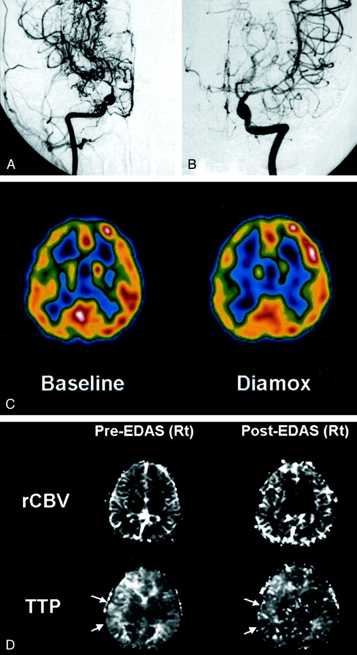
9-year-old boy with moyamoya disease.
A, Anteroposterior angiogram of the right ICA shows an occlusion of the supraclinoid portion, with development of multiple basal collateral vessels (moyamoya vessels).
B, Left ICA angiogram depicts mild changes of the luminal caliber in the left MCA, but with preserved patency. Left ACA shows severe narrowing.
C, Technetium-99m ethyl cysteinate dimer SPECT scan (left scan) reveals decreased perfusion in both hemispheres, more prominent on the right side. After the acetazolamide (Diamox) injection (right scan), there is no evidence of a significant perfusion reduction.
D, Perfusion MR images show delayed TTP in the right hemisphere before EDAS (arrows in left TTP map) and a signal intensity reduction after EDAS (arrows in right TTP map), which means a restoration of rapid flow at the surgical site. Note, rCBV maps show nothing significant.
After surgery, the rCBV ratio in the MCA territory decreased in the case of the initial rCBV increase, but the overall change in the rCBV value was not statistically significant (P = .281, paired t test, two-tailed) (Fig 3A). The rCBV in the basal ganglia also did not depict any changes after surgery (Fig 3B). The TTP in the MCA territory operated on was significantly shortened after surgery (P = .0002), whereas the TTP in the basal ganglia was not (Fig 4). TTP shortening was demonstrated as a marked signal intensity reduction in the calculated maps after EDAS (Fig 2D). One patient underwent additional postoperative perfusion MR imaging before a second surgery on the contralateral side. The second perfusion MR imaging examination was performed 1 month after EDAS on the left side, and the patient then underwent a right EDAS. On the second perfusion MR image, the TTP shortening of the operated hemisphere was not definite, which suggested that neovascularization was not established at that time. Six months later, the patient was again evaluated with perfusion MR imaging and conventional angiography. On the follow-up conventional angiogram, neovascularization and prominent vascular staining were demonstrated from the ECA to the frontoparietal cerebral cortex. The results agreed well with those of perfusion MR imaging, which depicted shortened TTP at the left MCA territory after surgery (Fig 5).
Fig 3.
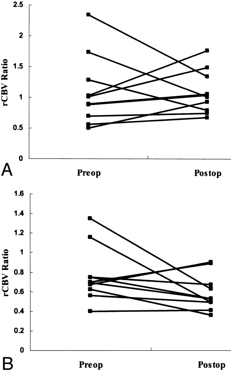
A and B, Graphs show changes in the rCBV ratios to the posterior circulation of the MCA territory (A) and the basal ganglia (B). The rCBV can be increased or decreased before (Preop) and after (Postop) surgery, which cannot characterize the rCBV patterns of moyamoya disease.
Fig 4.
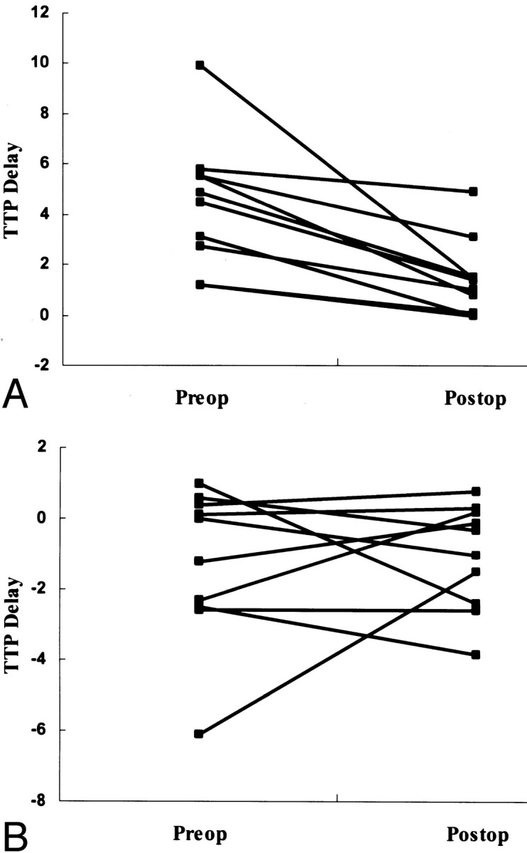
A and B, Graphs show changes in the TTP delay to the posterior circulation of the MCA territory (A) and basal ganglia (B). Note the markedly increased TTP in the MCA territory, with a significant reduction after surgery (Postop). There were no specific changes in the TTP values in the basal ganglia.
Fig 5.
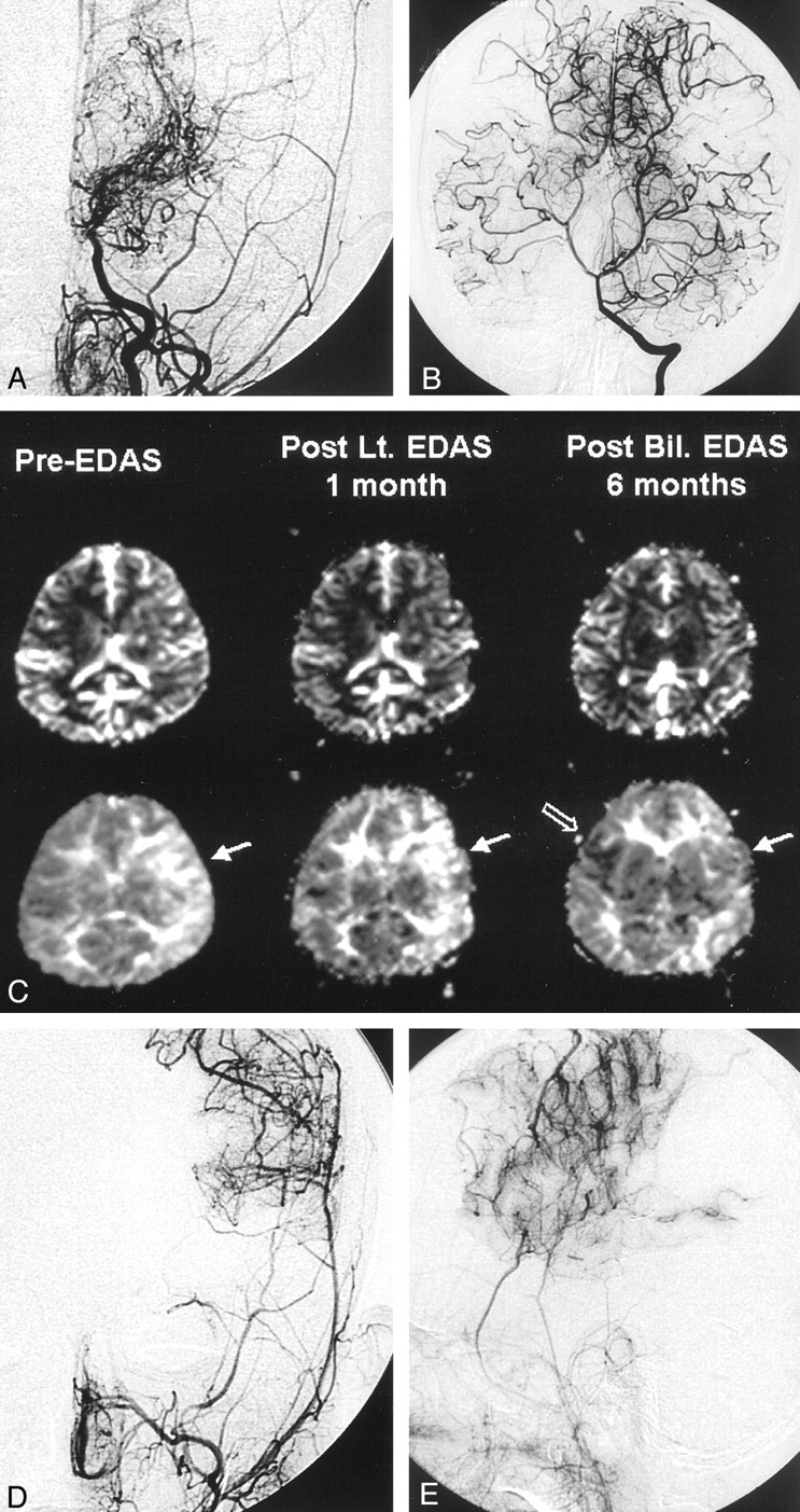
7-year-old boy with moyamoya disease.
A, Preoperative left carotid angiogram shows an occlusion of the supraclinoid ICA with basal collateral vessels. No transdural collateral vessels from the superficial temporal or middle meningeal arteries are seen.
B, Vertebral artery angiogram shows prominent leptomeningeal collateral vessels supplying the bilateral hemispheres.
C, Serial perfusion-weighted MR images (rCBV, top row; TTP, bottom row) were obtained because the patient underwent bilateral EDAS procedures with a 1-month interval. One-month follow-up perfusion MR image shows a mild reduction in the TTP delay (arrow in middle TTP map), which means neovascularization is beginning to develop. The last perfusion MR image (right TTP map) shows near-complete normalization of the TTP in the left hemisphere (solid arrow) and a partial TTP shortening on the right side (open arrow).
D and E, Postoperative 7-month follow-up left ECA angiograms show successful neovascularization from the middle meningeal and superficial temporal arteries.
Discussion
As moyamoya disease forms many collateral vessels owing to the occlusion of major proximal intracranial arteries, the key role of imaging diagnosis has been focused on detecting collateral circulation, although the disease may induce an infarct or hemorrhage. CT was found to be useful in detecting the important diagnostic signs such as arterial occlusions and the formation of collateral vessels (22). However, a definite diagnosis is made with conventional angiography (9), which can depict the characteristic basal collateral vessels resembling a “puff of smoke” (ie, moyamoya vessels). Conventional angiography provides a detailed status of collateral development and stenosis of major cerebral arteries. In children, the moyamoya vessels change through six stages: 1) carotid fork stenosis; 2) progressive carotid stenosis with initial moyamoya collateral vessels and dilatations of cerebral arteries; 3) dilatation of moyamoya collateral vessels and disappearance of ACAs and MCAs; 4) thinning of moyamoya vessels; 5) contraction of moyamoya vessels and disappearance of PCAs; and 6) intracerebral vessels perfused from the ECA and/or vertebrae. On the basis of this grading system, the moyamoya vessels of all patients in this study were below grade 5. MR angiography has also played a role in detecting the collateral arteries, although many institutes prefer conventional angiography as a primary diagnostic tool.
Alterations in the local cerebral hemodynamic status are more important in understanding and planning treatments for patients with moyamoya disease, which cannot be assessed with angiography alone. Scintigraphic methods, such as SPECT or positron emission tomography, are the reference standard for assessing cerebral hemodynamics because the tracer decay kinetics are well known, and the tracer distribution is proportional to the local cerebral blood flow. Previous studies have reported reduced regional cerebral blood flow in the frontal and temporal lobes and increased flow in the parietal and occipital lobes and the basal ganglia (10, 23). These flow changes reflect the course of the disease, which starts from the ICA, MCA, and extends to the PCA. The posterior circulation usually supplies the ischemic frontotemporal lobes via the leptomeningeal collateral vessels, whereas the deep nuclei are supplied from the multiple basal collateral vessels. Such findings can be detected with perfusion-weighted echo-planar MR imaging. Yamada et al (17) demonstrated a reduced rCBV and a TTP delay at the anterior circulation in the case of a stenotic PCA. In contrast, the current study showed a TTP delay in all patients, even those with a normal PCA, whereas the rCBV changes varied. Angiography showed normal calibers of the PCAs and prominent leptomeningeal collateral vessels in all subjects. An asymmetric TTP delay between both MCA territories was also observed in three of 10 patients, in whom a different degree of stenosis was clearly depicted at conventional angiography. The TTP delay of the anterior circulation was proportional to the severity of the ICA stenosis. Therefore, this is a universal finding of moyamoya disease on perfusion MR images and can reflect the severity of the disease.
The goal of surgical treatment in moyamoya disease is to reestablish cerebral blood flow to the ischemic regions. EDAS is widely used in childhood moyamoya disease because it is a simple procedure and has a low risk of ischemia during temporary blocking of the cerebral blood flow in a conventional superficial temporal artery-MCA anastomosis (4, 24). Most surgical procedures have focused on increasing the blood flow primarily in the MCA territory and do not directly benefit the ACA territory (8). We measured the perfusion changes of only the MCA territory because of the nature of the surgical procedure. If a modified procedure that can reestablish the ACA flow is chosen for investigation, perfusion measurement must be done in the ACA territory in the future.
After EDAS, the superficial temporal artery or the adjacent middle meningeal artery participates in forming collateral pathways and can be detected with conventional angiography (25) or MR angiography (20). The effectiveness of EDAS must be assessed not only by visualizing the neovascularization but also by monitoring improved perfusion status with other diagnostic techniques. In this study, an alteration of the temporal parameters of perfusion-weighted MR imaging (eg, delay of TTP in the affected territory and reduction of TTP delay after surgical treatment, which corresponded with the rapid staining of cerebral arteries from ECA anastomosis) was shown. All patients who showed a decreased TTP delay in this study showed a fairly good outcome after surgery. Therefore, a reduction of the TTP in the MCA territory can indirectly reflect the improved perfusion status, although an accurate interpretation of the TTP in cerebral hemodynamics is still unclear.
TTP maps or angiography cannot assess perfusion at the cellular level, because they depict only the capillary blood flow. A cellular extraction of radiotracers can reflect such microhemodynamics, and a complete evaluation of postoperative patients must include postoperative scintigraphic studies. Although our protocol did not include postoperative SPECT, clinical improvement and angiographic evidence are enough to support satisfactory cerebral perfusion. Moreover, as conventional MR imaging can be performed easily during the postoperative period, it can be used as a primary follow-up diagnostic tool for evaluating the surgical outcome.
All hemodynamic parameters of perfusion-weighted imaging were relative values; therefore, the differences between the MCA and PCA territories could not be compared. The arterial input function of the MCA territory was impossible to obtain because the MCA was not identified in moyamoya disease. Actually, the real arterial input function of the cerebrum could not be assessed owing to the nature of the disease.
Previous studies with perfusion MR imaging or SPECT used a cerebral-to-cerebellar ratio or region-to-mean hemispheric blood volume to calculate regional cerebral blood flow (17, 26, 27). In the current study, the authors compared MCA territorial blood flow to occipital lobe-PCA territory because all patients showed patent PCAs at angiography. We could not use the occipital lobe instead of cerebellum as a standard because posterior fossa images were poor with echo planar imaging sequences due to susceptibility artifacts by the skull base and mastoid air cells. If ECA-to-ICA anastomosis occurs, the cerebral hemodynamics must be changed, including the mean hemispheric blood flow and volume. Therefore, the method in this study of using PCA and its territorial blood volume as a standard may be more ideal than the mean hemispheric blood volume in the pre- and postoperative evaluation with normal PCA flow. However, it is clear whether one should use another standard hemodynamic parameter if the patient has a stenotic or occluded PCA.
Lack of normal control values from healthy volunteers is a limitation of this study. We used control data from patients with simple headache or psychiatric conditions because we could not perform dynamic bolus injection of MR contrast material in healthy children. However, the control group children were found to have no central nervous system disease radiologically or clinically on follow-up.
The relatively broad age range of 2–27 years is another limitation. It may raise the question as to whether age-related hemodynamic changes would affect outcome. To our knowledge, TTP or rCBV change according to aging in the childhood period has not been reported. Oyama et al (28) reported that cerebral blood flow gradually decreases with aging in moyamoya disease; however, their patient population ranged from 16 to 58 years. We also could not get proper data from a healthy population to get standard hemodynamic features of children. This remains an unsolved problem in this study, and future investigation must be performed.
In summary, TTP perfusion maps can depict the preoperative hemodyamic status of patients with moyamoya disease and postoperative changes as well. We postulate that perfusion MR imaging is a useful tool to evaluate cerebral blood flow in moyamoya disease.
Conclusion
Perfusion-weighted MR imaging can be applied for evaluating the postoperative changes in the cerebral blood flow in moyamoya disease. A shortening of the TTP in the MCA territory of the hemisphere operated on is a marker for the development of collateral circulation from the ECA to the ICA.
Acknowledgments
The authors wish to thank Sei Young Kim, MR technologist at the Severance Hospital, Yonsei University, for assistance with the MR examination and image preparation.
Footnotes
Supported by a grant from the Korean Radiological Foundation and HMP-99-N-01–0001 of the Ministry of Health and Welfare, Korea.
References
- 1.Suzuki J, Takaku A. Cerebrovascular “moyamoya” disease: a disease showing abnormal net-like vessels in base of brain. Arch Neurol 1969;20:288–299 [DOI] [PubMed] [Google Scholar]
- 2.Taveras JM. Multiple progressive intracranial arterial occlusions: a syndrome of children and young adults. AJR Am J Roentgenol 1969;106:235–268 [DOI] [PubMed] [Google Scholar]
- 3.Pecker J, Simon J, Guy G, Herry JF. Nishimoto’s disease: significance of its angiographic appearances. Neuroradiology 1973;5:223–230 [DOI] [PubMed] [Google Scholar]
- 4.Choi JU, Kim DS, Kim EY, Lee KC. Natural history of moyamoya disease: comparison of activity of daily living in surgery and non-surgery groups. Clinic Neurol Neurosurg 1997;99(suppl 2):S11–S18 [DOI] [PubMed] [Google Scholar]
- 5.Fujita K, Tamak N, Matsumoto S. Surgical treatment of moyamoya disease in children: which is the more effective procedure, EDAS or EMS? Childs Nerv Syst 1986;2:134–138 [DOI] [PubMed] [Google Scholar]
- 6.Iwama T, Hashimoto N, Miyake H, Yonekawa Y. Direct revascularization to the anterior cerebral artery territory in patients with moyamoya disease: report of five cases. Neurosurgery 1998;42:1157–1162 [DOI] [PubMed] [Google Scholar]
- 7.Matsushima T, Inoue TK, Suzuki SO, et al. Surgical techniques and the results of a fronto-temporo-parietal combined indirect bypass procedure for children with moyamoya disease: a comparison with the results of encephalo-duro-arterio-synangiosis alone. Clin Neurol Neurosurg 1997;99(suppl 2):S123–127 [DOI] [PubMed] [Google Scholar]
- 8.Kim SK, Wang KC, Kim IO, Lee DS, Cho BK. Combined encephalo-duro-arterio-synangiosis and bifrontal encephalo-galeo-(periosteal)-synangiosis in pediatric moyamoya disease. Neurosurgery 2002;50:88–96 [DOI] [PubMed] [Google Scholar]
- 9.Hasuo K, Tamura S, Kudo S, et al. Moyamoya disease: use of digital subtraction angiography in its diagnosis. Radiology 1985;157:107–111 [DOI] [PubMed] [Google Scholar]
- 10.Kuroda S, Houkin K, Kamiyama H, Abe H, Mitsumor K. Regional cerebral hemodynamics in childhood moyamoya disease. Childs Nerv Syst 1995;11:584–590 [DOI] [PubMed] [Google Scholar]
- 11.Miller JH, Khonsary A, Raffel C. The scintigraphic appearance of childhood moyamoya disease on cerebral perfusion imaging. Pediatr Radiol 1996;26:833–838 [DOI] [PubMed] [Google Scholar]
- 12.Yamada I, Matsushima Y, Suzuki S. Moyamoya disease: diagnosis with three-dimensional time-of-flight MR angiography. Radiology 1992;184:773–778 [DOI] [PubMed] [Google Scholar]
- 13.Yamada I, Suzuki S, Matsushima Y. Moyamoya disease: comparison of assessment with MR angiography and MR imaging versus conventional angiography. Radiology 1995;196:211–218 [DOI] [PubMed] [Google Scholar]
- 14.Tzika AA, Robertson RL, Barnes PD, et al. Childhood moyamoya disease: hemodynamic MRI. Pediatr Radiol 1997;27:727–735 [DOI] [PubMed] [Google Scholar]
- 15.Tsuchiya K, Inaoka S, Mizutani Y, Hachiya J. Echo-planar perfusion MR of moyamoya disease. AJNR Am J Neuroradiol 1998;19:211–216 [PMC free article] [PubMed] [Google Scholar]
- 16.Adams WM, Laitt RD, Li KL, Jackson A, Sherrington CR, Talbot P. Demonstration of cerebral perfusion abnormalities in moyamoya disease using susceptibility perfusion- and diffusion-weighted MRI. Neuroradiology 1999;41:86–92 [DOI] [PubMed] [Google Scholar]
- 17.Yamada I, Himeno Y, Nagaoka T, et al. Moyamoya disease: evaluation with diffusion-weighted and perfusion echo-planar MR imaging. Radiology 1999;212:340–347 [DOI] [PubMed] [Google Scholar]
- 18.Ohashi K, Fernandez-Ulloa M, Hall LC. SPECT, magnetic resonance and angiographic features in a moyamoya patient before and after external-to-internal carotid artery bypass. J Nucl Med 1992;33:1692–1695 [PubMed] [Google Scholar]
- 19.Touho H, Karasawa J, Ohnishi H. Preoperative and postoperative evaluation of cerebral perfusion and vasodilatory capacity with 99mTc-HMPAO SPECT and acetazolamide in childhood moyamoya disease. Stroke 1996;27:282–289 [DOI] [PubMed] [Google Scholar]
- 20.Yoon HK, Shin HJ, Lee M, Byun HS, Na DG, Han BK. MR angiography of moyamoya disease before and after encephaloduroarteriosynangiosis. AJR Am J Roentgenol 2000;174:195–200 [DOI] [PubMed] [Google Scholar]
- 21.Suzuki J, Takaku A, Kodama N, Sato S. An attempt to treat cerebrovascular moyamoya disease in children. Childs Brain 1975;1:193–206 [DOI] [PubMed] [Google Scholar]
- 22.Takahashi M, Miyauchi T, Kowada M. Computed tomography of moyamoya disease: demonstration of occluded arteries and collateral vessels as important diagnostic signs. Radiology 1980;134:671–676 [DOI] [PubMed] [Google Scholar]
- 23.Takeuchi S, Tanaka R, Ishii R, Tsuchida T, Kobayashi K, Arai H. Cerebral hemodynamics in patients with moyamoya disease: a study of regional cerebral blood flow by the 133Xe inhalation method. Surg Neurol 1985;23:468–474 [DOI] [PubMed] [Google Scholar]
- 24.Karasawa J, Touho H, Ohnishi H, Miyamoto S, Kikuchi H. Long-term follow-up study after extracranial-intracranial bypass surgery for anterior circulation ischemia in childhood moyamoya disease. J Neurosurg 1992;77:84–89 [DOI] [PubMed] [Google Scholar]
- 25.Yamada I, Matsushima Y, Suzuki S. Childhood moyamoya disease before and after encephalo-duro-arterio-synangiosis: an angiographic study. Neuroradiology 1992;34:318–322 [DOI] [PubMed] [Google Scholar]
- 26.Hoshi H, Ohnishi T, Jinnouchi S, et al. Cerebral blood flow study in patients with moyamoya disease evaluated by IMP SPECT. J Nucl Med 1994;35:41–43 [PubMed] [Google Scholar]
- 27.Yamada I, Murata Y, Umehara I, Suzuki S, Matsushima Y. SPECT and MRI evaluations of the posterior circulation in moyamoya disease. J Nucl Med 1996;37:1613–1617 [PubMed] [Google Scholar]
- 28.Oyama H, Niwa M, Kida Y. CBF change with aging in moyamoya disease [abstr]. J Neurosurg Sci 1998;42:33–36 [PubMed] [Google Scholar]


