

Abstract
Summary: The authors present 3 patients who underwent neuroendovascular procedures in which DynaCT produced by a flat-panel detector facilitated management of complications. As part of a combined CT/angiography suite, DynaCT offered the major advantage of immediate detection or exclusion of intracranial complication without patient transfer. The quality of cone-volume CT-generated images produced by DynaCT was sufficient to make a diagnosis.
For patients undergoing neuroendovascular procedures such as intracranial aneurysm coiling, earlier recognition and management of complications can improve clinical outcomes.1–3 When a complication occurs during a neuroendovascular procedure, the patient is usually transported emergently from the angiography suite to the nearest CT scanner to determine a diagnosis. This protocol can endanger the patient not only from the transport itself but also from the delay in critical decision making. To overcome this shortcoming, Siemens has developed a new combined angiography/CT suite that uses flat-panel detector (FD) technology for higher-resolution angiography that is also capable of producing improved cone-beam volume CT images. In this new suite, 3D rotational digital subtraction angiography (DSA) or cone-beam volume CT can be obtained interchangeably with the same FD C-arm motion without patient transfer.
Technique
A biplane flat-panel detector angiographic suite (AXIOM Artis FD Biplane Angiosuite with DynaCT; Siemens Medical Solutions, Erlangen, Germany) was used (Fig 1A). A DynaCT acquisition was obtained by using the following parameters: 20-second rotation; 0.4° increment; 1024 × 793 matrix in projections at zoom 0 after resampling; 217° total angle; ∼11°/s, ∼27 frames/s, system dose 1.2 μGy/frame, total of 538 projections. Images were automatically corrected for gain of the image intensifier during the acquisition. Image reconstruction was performed on a commercially available dedicated workstation (X-Leonardo with DynaCT; Siemens Medical Solutions). Advanced postprocessing for beam hardening, scatter, and truncation correction was performed. The volume dataset produced with typical voxel size 0.4 mm by X-Leonardo was interactively viewed/windowed/leveled and manipulated in orthogonal planes (Fig 1B). The time required from acquisition to 3D-image rendering was 3–5 minutes. Radiation dose to patients was similar to that of a conventional CT head dose, up to 60 mGy. Differentiation of soft tissue densities was approximately 10 HU.
Fig 1.
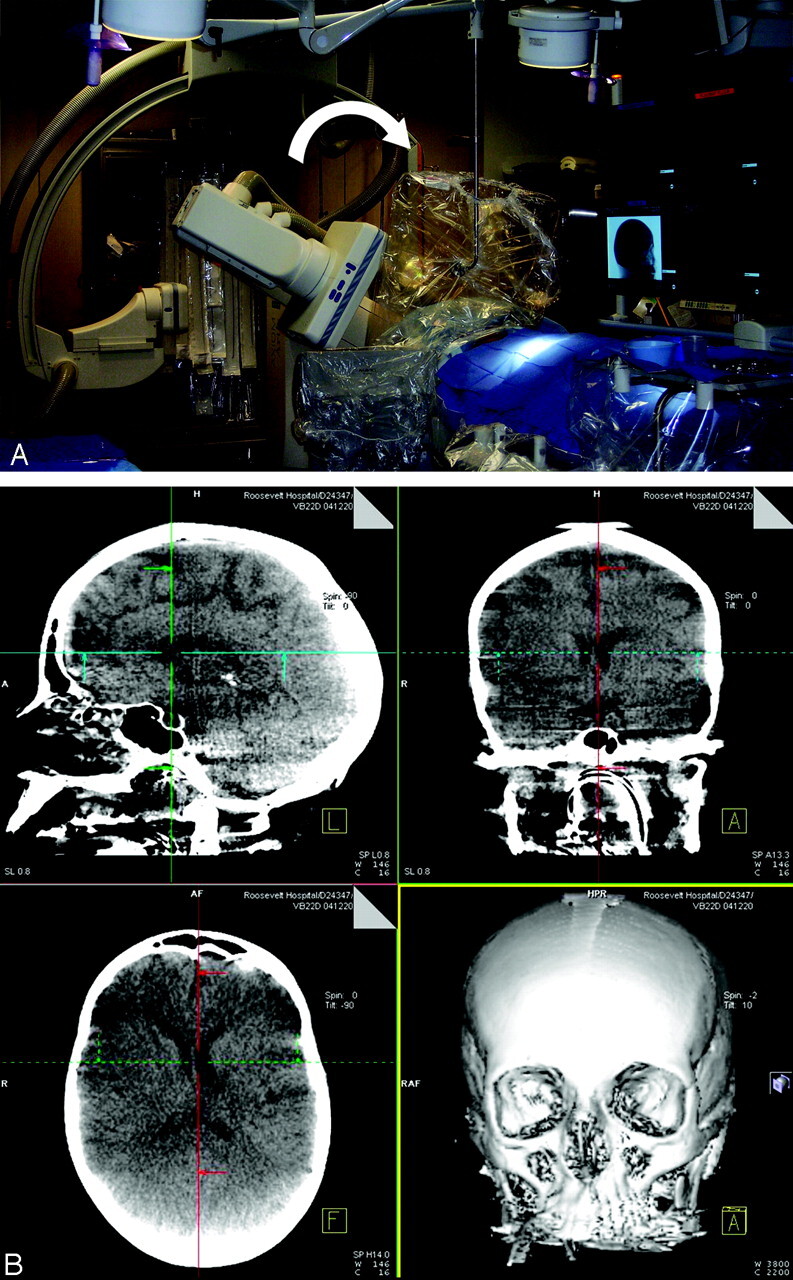
A, DynaCT acquisition demonstrating rotation of FD C-arm (curved arrow) during actual case. B, A screen shot of acquired orthogonal images immediately available to the user after acquisition and postprocessing.
Case Reports
Case 1
This 33-year old man, presenting with a grade III Hunt and Hess (H&H) subarachnoid hemorrhage (SAH), was treated for a ruptured left P2 posterior cerebral artery aneurysm. During the procedure, proximal to the aneurysm, vessel perforation occurred that was immediately treated with Guglielmi coil detachment. With the patient on the angiography table, DynaCT was then urgently performed, which, compared with preprocedure CT, demonstrated increased SAH and new intraventricular blood and acute hydrocephalus, despite the presence of a ventriculostomy catheter (Fig 2A, -B). In the angiography suite, another ventriculostomy catheter was emergently placed. The patient was transferred to the intensive care unit and on awakening from anesthesia, he was found to be neurologically unchanged.
Fig 2.

DynaCT was performed following vessel perforation (A ) and demonstrated increased ventriculomegaly compared with preoperative CT scan (B).
Case 2
This 51-year-old man, presenting with a grade I H&H SAH, underwent coiling of an anterior communicating artery aneurysm. After coiling, the contralateral right A2 became compromised secondary to thrombus. Treatment with 8 mg of intra-arterial abciximab (ReoPro) resulted in complete recanalization of the right A2 and distal anterior cerebral artery segments but not in aneurysm reopening after closure with coils. At the end of the procedure, the patient suddenly became hypotensive and was noted to be in ventricular fibrillation. Immediate resuscitation was begun. We suspected an aneurysm rehemorrhage and, as part of the resuscitation efforts, successfully placed an emergent ventriculostomy but without return of bloody CSF. Immediately after resuscitation efforts were stopped, postmortem DynaCT demonstrated no evidence of new hemorrhage or hydrocephalus when compared with the preoperative CT scan (Fig 3A, -B), which indicates a primary cardiac event.
Fig 3.

This patient experienced cardiac arrest and aneurysm rehemorrhage was suspected. After emergent ventriculostomy placement, however, DynaCT (A) demonstrated no new intracranial hemorrhage, which indicates a primary cardiac event. Note good catheter placement and interhemispheric air (arrow). Preoperative CT scan (B) is shown for comparison.
Case 3
This 33-year-old man presented with decreased left visual acuity, intermittent right hemiparesis, and hemiparesthesias from a spontaneous extracranial left internal carotid artery (ICA) dissection. His left visual acuity worsened because of decreased retinal perfusion. Cerebral angiogram confirmed left ICA dissection “string” sign of the distal cervical ICA extending into the proximal petrous segment. The left ICA dissection was stented, which improved ICA caliber and antegrade flow. Control cerebral angiography of the left ICA, however, demonstrated left-to-right shift of the anterior cerebral artery branches indicating mass effect consistent with probable reperfusion hemorrhage. After immediate reversal of anticoagulation, DynaCTshowed an unexpected left subdural hematoma and focal left occipitoparietal intraparenchymal hemorrhage (Fig 4A). Emergent medical and surgical treatment for increased intracranial pressure was initiated. A conventional CT scan was performed (Fig 4B) before the patient was transferred to the operating room.
Fig 4.

DynaCT (A) demonstrated unexpected subdural hematoma and occipitoparietal intraparenchymal hemorrhage. A conventional CT scan (B) was later obtained before patient transfer to the operating room, which revealed increased hemorrhage and mass effect.
Discussion
The images produced by DynaCT are not of the quality of conventional CT. Compared to non-FD cone-beam volume CT, DynaCT contrast images have improved spatial and resolution from lack of image distortion, higher detection dynamics, less sensitivity to overexposure, and higher efficiency.4 Despite initial concerns about inadequate image quality of the first-generation DynaCT images, our cases demonstrate that current DynaCT image quality is sufficient to make a diagnosis when a complication is suspected. In cases 1 and 3, in an operator- and time-convenient manner, while concurrently avoiding risky and time-consuming patient transfer, DynaCT enabled detection of intracranial hemorrhages and their extent within the angiography suite. Because DynaCT allowed earlier diagnosis and management of hemorrhagic complications, the extent of brain injury suffered by our patients may have been minimized.1–3 In case 2, a negative DynaCT reassured us that a suspected intracranial hemorrhage had not occurred. In case 1, after an emergent ventriculostomy was placed in the angiography suite, DynaCT immediately verified ventriculostomy placement. In the management of acute stroke, DynaCT can be performed to exclude intracranial hemorrhage before planned intra-arterial thrombolysis.5 Periprocedural DynaCT performed during brain or dural arteriovenous malformation embolization can detect obvious or occult hemorrhagic complications from vessel perforation or from venous penetration of embolic material or venous thrombosis.6,7 In head and neck embolization procedures using direct percutaneous access, in combination with biplane fluoroscopy, DynaCT can be used for more precise needle placement.
Conclusion
Within the angiography suite and without moving the patient, DynaCT enabled immediate detection or exclusion of intracranial hemorrhage and hydrocephalus, and its use facilitated the management of neuroendovascular complications.
Acknowledgments
The authors thank our outstanding radiology technologists who have worked tirelessly to improve DynaCT images: Mathew Joshua, Alex Eusebio, Danny Hom, Michael D. Lamon, and Virender Ahuja.
References
- 1.Horowitz MB, Crammond D, Balzer J, et al. Aneurysm rupture during endovascular coiling: effects on cerebral transit time and neurophysiologic monitoring and the benefits of early ventriculostomy: case report. Minim Invasive Neurosurg 2003;46:300–305 [DOI] [PubMed] [Google Scholar]
- 2.Cloft HJ, Kallmes DF. Cerebral aneurysm perforations complicating therapy with Guglielmi detachable coils: a meta-analysis. AJNR Am J Neuroradiol 2002;23:1706–1709 [PMC free article] [PubMed] [Google Scholar]
- 3.Levy E, Koebbe CJ, Horowitz MB, et al. Rupture of intracranial aneurysms during endovascular coiling: management and outcomes. Neurosurgery 2001;49:807–11; discussion 811–13 [DOI] [PubMed] [Google Scholar]
- 4.Akpek S, Brunner T, Benndorf G, et al. Three-dimensional imaging and cone beam volume CT in C-arm angiography with flat panel detector. Diagn Interv Radiol 2005;11:10–13 [PubMed] [Google Scholar]
- 5.Furlan A, Higashida R, Wechsler L, et al. Intra-arterial prourokinase for acute ischemic stroke: the PROACT II study: a randomized controlled trial: Prolyse in Acute Cerebral Thromboembolism. JAMA 1999;282:2003–11 [DOI] [PubMed] [Google Scholar]
- 6.Iwama, T, Yoshimura K, Keller E, et al. Emergency craniotomy for intraparenchymal massive hematoma after embolization of supratentorial arteriovenous malformations. Neurosurgery 2003;53:1251–58; discussion 1258–60 [DOI] [PubMed] [Google Scholar]
- 7.Picard L, Da Costa E, Anxionnat R, et al. Acute spontaneous hemorrhage after embolization of brain arteriovenous malformation with n-butyl cyanoacrylate. J Neuroradiol 2001;28:147–65 [PubMed] [Google Scholar]