We recently read the interesting article by Albayram et al (1), which concisely explained the anomalies of the vertebral arteries. We would like to present another rare case of bilateral vertebral arteries originating from the aortic arch. We incidentally diagnosed this condition on the basis of findings on a chest CT scan obtained in a patient with trauma. Our trauma chest CT protocol uses 8 × 2.5-mm detector configuration with a pitch of 1.35, allowing us to scan the chest in under 10 seconds. After the study, we reconstruct 1.25-mm-thick images at 1.25-mm intervals.
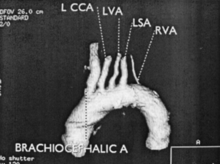
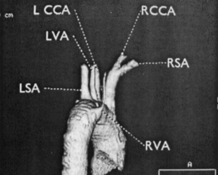
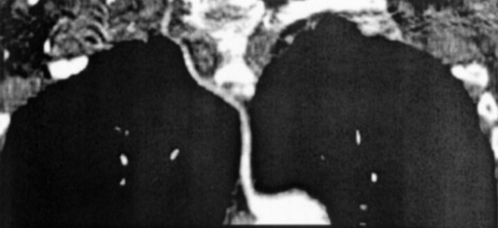
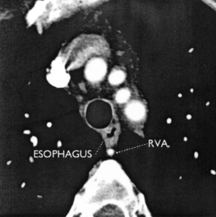
Both vertebral arteries originate from aortic arch (Figs 1 and 2). The left vertebral artery arises between the origins of the left common carotid and left subclavian arteries. An aberrant right vertebral artery arises from the descending aorta distal to the origin of the left subclavian artery, with a dilatation at the origin consistent with a Kommerel diverticulum (Figs 2 and 3), and it passes posterior to the esophagus (Fig 4). An aberrant right vertebral artery is a rare anomaly, and it can easily be distinguished from an aberrant right subclavian artery by the presence of the normal right subclavian artery (Fig 2) (2).
Fig 1.
CT angiogram, left anterior oblique projection. Three-dimensional volume-rendered image demonstrates the bilateral origin of the vertebral arteries.
Fig 2.
CT angiography, posterior projection. Three-dimensional volume-rendered image shows the posterior arch origin of an aberrant right vertebral artery. Note the Kommerel diverticulum at the origin.
Fig 3.
Curved planar reformatted image outlines the course of the aberrant right vertebral artery.
Fig 4.
Axial image at the level of left brachiocephalic vein shows the retroesophageal location of an aberrant right vertebral artery.
High-resolution multidetector-row CT angiography enables the display of images from different views. These can be useful as a roadmap in interventional and surgical therapeutic decision making, especially in patients with complex aortic arch anatomy.
References
- 1.Albayram S, Gailloud P, Wasserman BA. Bilateral arch origin of the vertebral arteries. AJNR Am J Neuroradiol 2002;23:455–458 [PMC free article] [PubMed] [Google Scholar]
- 2.Lemke AJ, Benndorf G, Liebig T, Felix R. Anomalous origin of the right vertebral artery: review of the literature and case report of the right vertebral artery origin distal to the left subclavian artery. AJNR Am J Neuroradiol 1999;20:1318–1321 [PMC free article] [PubMed] [Google Scholar]


