Abstract
Summary: We report a case of lumbar facet joint synovial cyst at L5-S1 with clinically significant radicular pain diagnosed by means of MR imaging. This cyst spontaneously resolved, as determined on follow-up MR images obtained 18 months later. The patient’s clinical symptoms substantially improved after conservative medical treatment.
Approximately 180 case reports of synovial cysts of the vertebral facet joints have been described in the literature. Of those, only two refer to spontaneously resolving intraspinal synovial cysts (1, 2).
Synovial cysts most often develop secondary to substantially degenerated facet joints and to both acute and chronic trauma. Synovial cysts of the lumbar vertebral facet joints may serve as sources of nerve root compression. The most common symptoms are painful radiculopathy (85%), neurogenic single-root or multiroot claudication (44%), sensory loss (43%), and motor weakness (27%) (3).
Case Report
A 58-year-old woman presented with a medical history notable for only minor daily joint stiffness of the hip and knee and pain that was relieved by nonsteroidal anti-inflammatory drugs (NSAIDs). Her present illness started insidiously 1 month before the first MR examination, with sciatic-like posterior pain and numbness and tingling in the right leg. These symptoms were not associated with any known injury. Physical examination showed unimpaired strength of the lower extremity, normal and symmetrical deep tendon reflexes, and normal muscle tone in both lower extremities. Her initial response to NSAIDs and rest was minimal.
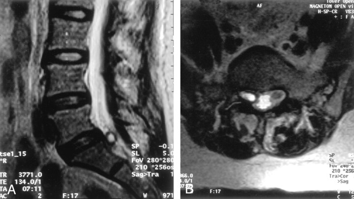
MR imaging of the lumbar spine performed 1 month after the onset of symptoms demonstrated facet osteoarthropathy with marked overgrowth of the facets but no remarkable canal stenosis at L5-S1. Grade I degenerative spondylolisthesis and mild bulging of the disk at L5-S1 were present, but these were present without notable canal stenosis. A synovial cyst was identified; this arose off the facet complex on the right at L5-S1 (Fig. 1) extending into the neural foramen and lateral recess of S1, causing compression at the right L5 and S1 nerve root. No disk disease was noted from T12 through L4-L5, and only mild facet osteoarthropathy was observed at L4-L5. with mild canal stenosis dorsolaterally.
Fig 1.
T2-weighted fast spin-echo MR images.
A, Sagittal image (TR/TE, 3771/134) demonstrates a synovial cyst extending into the spinal canal at the L5-S1 level.
B, Axial image (5866/134) through the L5-S1 level demonstrates the synovial cyst arising from the facet complex on the right, extending into the lateral recess of S1 on the right side, and causing compression of the S1 nerve root.
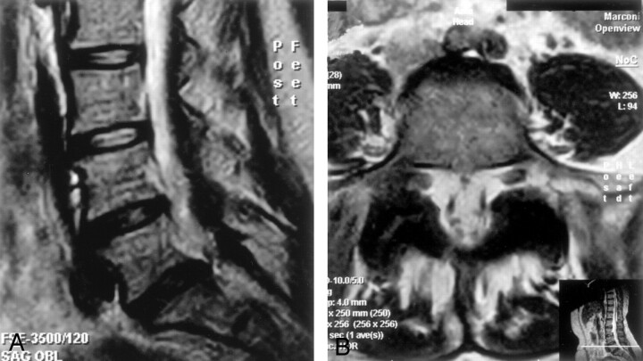
Conservative medical treatment was initiated and consisted of physical therapy (mainly stretching), NSAID therapy, and orthopedic bracing. The patient reported gradual but substantial improvement of both pain and sensory symptoms over the next 12 months. Follow-up MR imaging was performed 18 months later. The images demonstrated continued, mild bulging at L5-S1 and facet osteoarthropathy with marked overgrowth of facets without canal stenosis at L5-S1. The previously noted synovial cyst was no longer present (Fig 2).
Fig 2.
Follow-up T2-weighted fast spin-echo MR images demonstrate complete resolution of the previously noted synovial cyst. These follow-up images do not completely match the angulation or detail of the images in Figure 1. The follow-up examination was performed by using a low-field-strength (0.35-T) open magnet, and the images are less than optimal. The patient did not desire to enter a closed high-field-strength machine for additional studies.
A, Sagittal image (3500/120) through the lumbar spine.
B, Axial image through the L5-S1 lumbar spine (-10/15).
Discussion
Synovial cysts are most often described in extraspinal joints such as the knee, hip, and elbow. They are infrequently identified adjacent to the facet joints, and they account for an incidence of 0.002–0.8% depending on the institution and the diagnostic technique (2, 4). Among synovial cysts located in facet joints, 68.4% are found at L4-L5; 21.1%, at L5-S1; 5.2%, at L1-L2; and 5.2%, at L2-L3 (5). The incidence and location of synovial cysts at our institution is similar to that of the referenced studies. This was our first documented case of a spontaneously resolving synovial cyst of a facet joint.
Intraspinal synovial cysts generally result from facet joint arthrosis and degeneration that causes a capsular lesion and eventual herniation of the synovial membrane. Alternatively, synovial cysts can appear as complications of lumbar trauma, both a single major trauma and repeated microtraumatic events. Specifically, in this patient, the correlation between the synovial cyst of the facet joint and spondylolisthesis at the same lumbar level support the theory of functional instability (2) and repeated microtraumatic events leading to synovial cyst formation. The presence of spondylolisthesis in 33% of cases and facet joint hypermobilization in 60% of patients support the idea that hypermobility is an important etiologic factor in development of synovial cysts (6).
Explaining the spontaneous resolution of a lumbar synovial cyst is complicated. In this patient, who underwent conservative treatment with NSAIDs and mobilization, the synovial cyst resolved probably because of decreased inflammatory fluid production and a decrease in microtraumatic events. Another plausible explanation is that the physical therapy provided enough of a mechanical stimulus to cause the cyst to extrude its contents, followed by reabsorption of the cyst wall.
The management of symptomatic synovial cysts of the lumbar spine is controversial. No standards or guidelines have been established to lead the clinician toward one therapeutic technique or another. In this case, a conservative approach to treatment was implemented, starting with physical therapy (mainly stretching with and then without resistance to relieve perceived pain and tightness by patient), NSAID administration, and bracing. Clinical improvement noted by the patient and the physician encouraged the continued use of a conservative approach, which ultimately resulted in substantial improvement of the patient’s symptoms. This change was associated with complete resolution of the synovial cyst, as shown on the follow-up MR images. One explanation for incomplete symptomatic improvement even after spontaneous resolution of the intraspinal synovial cyst was the preexisting facet osteoarthropathy and the marked overgrowth of the facets at the same level as the previously noted synovial cyst. In addition, the grade I degenerative spondylolisthesis and the mild bulging of the L5-S1 disk probably contributed to the mild residual symptoms.
If conservative management had failed in this patient, other more invasive therapeutic interventions remained available; these included percutaneous cyst aspiration under CT control, methylprednisolone injections into the cyst under CT guidance, and cystectomy via a laminectomy or microneurosurgical flavectomy.
Conclusion
Although rare, synovial cyst formation and subsequent resolution in the clinical context of degenerated facet joints with or without acute or chronic trauma must be considered. Resolution of a lumbar synovial cyst at follow-up imaging can generally be attributed to decreased inflammatory fluid production and decreased microtraumatic events or mechanical irritation of the cyst wall that results in extrusion of cyst contents and resolution of the cyst capsule. Although no standard of care for treatment of a lumbar synovial cyst has been established, the general trend is for conservative management first followed by invasive treatment if conservative therapy fails.
References
- 1.Maezawa Y, Baba H, Uchida K, Furusawa N, Kubota C, Yoshizawa K. Spontaneous remission of a solitary intraspinal synovial cyst of the lumbar spine. Eur Spine J 2000;9:85–87 [DOI] [PMC free article] [PubMed] [Google Scholar]
- 2.Mercader J, Gomez J, Cardenal C. Intraspinal synovial cyst: diagnosis by CT-follow-up and spontaneous remission. Neuroradiology 1985;27:346–348 [DOI] [PubMed] [Google Scholar]
- 3.Lyons MK, Atkinson JL, Wharen RE, Deen HG, Zimmerman RS, Lemens SM. Surgical evaluation and management of lumbar synovial cysts: the Mayo Clinic experience. J Neurosurg 2000;93:53–57 [DOI] [PubMed] [Google Scholar]
- 4.Trummer M, Flaschka G, Tillich M, Homann CN, Unger F, Eustacchio S. Diagnosis and surgical management of intraspinal synovial cysts: report of 19 cases. J Neurol Neurosurg Psychiatry 2001;70:74–77 [DOI] [PMC free article] [PubMed] [Google Scholar]
- 5.Hsu KY, Zucherman JF, Shea WJ, Jeffrey RA. Lumbar intraspinal synovial and ganglion cysts (facet cysts): ten-year experience in evaluation and treatment. Spine 1995;20:80–89 [DOI] [PubMed] [Google Scholar]
- 6.Freidberg SR, Fellow T, Thomas CB, et al. Experience with symptomatic spinal epidural cysts. Neurosurgery 1994;34:989–993 [DOI] [PubMed] [Google Scholar]


