Abstract
Summary: We describe the 3D digital subtraction angiography (3D-DSA) image of extravasation from a rupturing aneurysm. This image clearly showed that the extravasation was coming from a pseudoaneurysm on an aneurysmal wall. To the best of our knowledge, this is the first demonstration of a 3D-DSA image of a rupturing aneurysm.
Although it is known that a fragile pseudoaneurysm forms at the rupture site on a ruptured irregularly shaped aneurysm (1), to the best of our knowledge clear angiographic demonstration of a rupture from the fragile pseudoaneurysm has not been reported. Recent development of 3D radiologic images makes it possible to demonstrate the fine structure of the cerebral arteries. We report here the first demonstration of a 3D rotational digital subtraction angiography (3D-DSA) image of extravasation from a pseudoaneurysm on an aneurysmal wall.
Case Report
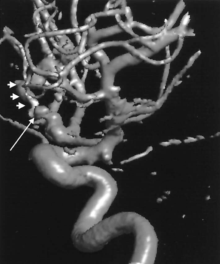
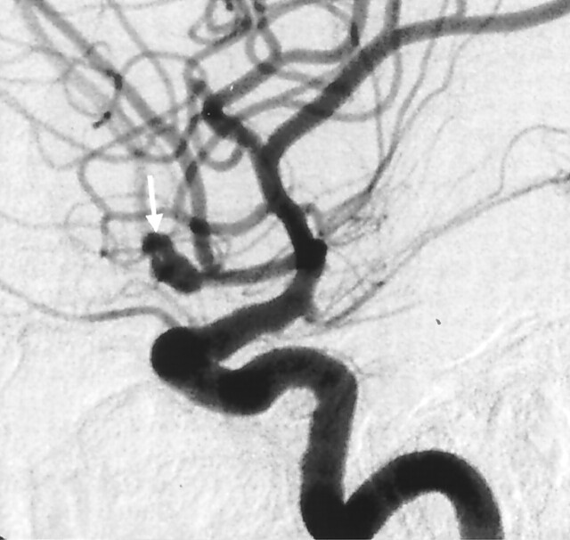
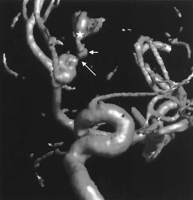
A 49-year-old woman was admitted to our department after complaining of severe headache, the cause of which was diagnosed as subarachnoid hemorrhage by CT scan. She was well oriented and showed no focal neurologic deficit. Angiography was performed following the CT scan. Angiography of the left internal carotid artery revealed an anterior communicating artery aneurysm, which had a small daughter aneurysm on its anterior wall (Fig 1). To clarify the exact anatomic relationship between the aneurysm neck and bilateral anterior cerebral arteries, which was thought to be useful preoperative or preembolization information, 3D-DSA (Integris Allura; Philips, Best, the Netherlands) was performed. During rotation of the C-arm, rebleeding of the aneurysm occurred. An extravasation of the contrast media from the daughter aneurysm was visible on the 3D-DSA image (Fig 2). The 3D-DSA image also revealed that the daughter aneurysm had a connection to the aneurysm dome with very narrow lumen (Fig 3). This finding suggested that the daughter aneurysm was not a true aneurysm, but a pseudoaneurysm located on the rupture site. After the rebleeding, the patient was in a comatose state. Therefore, she underwent emergency aneurysmal neck clipping and hematoma removal, and this was followed by ventricle-peritoneal shunt 5 weeks after the operation. She showed good postoperative recovery and returned to her home on foot with only slight mental impairment 8 weeks after the operation.
Fig 1.
Lateral view of the left internal carotid angiogram taken immediately before the rerupture shows a small daughter aneurysm on its anterior wall (arrow).
Fig 2.

3D-DSA image of the left internal carotid angiography during the rerupture of the aneurysm clearly demonstrates a jet of blood flow (small arrows) coming from the daughter aneurysm (large arrow).
Fig 3.
3D-DSA image of waters view reveals that the daughter aneurysm (small arrow) has connection to the aneurysm dome with very narrow space (large arrow). The asterisk indicates the extravasation.
Discussion
This is the first demonstration of an extravasation from rupturing aneurysm imaged by 3D-DSA. The jet of blood flow coming from the pseudoaneurysm was clearly shown. Nomura et al. suggested that a ruptured, irregularly shaped aneurysms might be accompanied by a fragile pseudo-aneurysm-like cavity located at the rupture point (1). This 3D-DSA image supported this hypothesis and clearly demonstrated that the fragile pseudoaneurysm was formed at a rupture site on the irregularly shaped aneurysm.
To ensure a clear image, greater volume of contrast media should be injected for a longer duration (3–4 mL/s for 5 seconds, for a total volume of 15–20 mL) for cerebral 3D-DSA than for usual cerebral angiography. It is well known that rebleeding in the acute stage is predominant within 6 hours from the initial subarachnoid hemorrhage (2, 3). The rebleeding rate during angiography within 6 hours after initial bleeding has been reported as 3.1–3.3% (4, 5), which is higher than the rebleeding rate during angiography beyond 6 hours. As the pressure wave of the injected contrast media may overcome the weak wall of the rupture site even during normal cerebral angiography (6), we should keep it mind that the rebleeding rate might be higher during cerebral 3D-DSA than during ordinary cerebral DSA for acutely ruptured aneurysms.
References
- 1.Nomura H, Kida S, Uhiyama N, et al. Ruptured irregularly shaped aneurysms: pseudoaneurysm formation in a thrombus located at the rupture site. J Neurosurg 2000;93:998–1002 [DOI] [PubMed] [Google Scholar]
- 2.Inagawa T, Kamiya K, Ogasawra H, et al. Rebleeding of ruptured intracranial aneurysms in the acute stage. Surg Neurol 1987;28:93–99 [DOI] [PubMed] [Google Scholar]
- 3.Yasui N, Suzuki A, Ohta H, et al. Rebleeding attack of the cerebral aneurysm: clinical significance of the early aneurysmal rebleeding. No Shinkei Geka 1985;13:61–68 [PubMed] [Google Scholar]
- 4.Komiyama M, Tamura K, Nagata Y, et al. Aneurysmal rupture during angiography. Neurosurgery 1993;33:798–803 [DOI] [PubMed] [Google Scholar]
- 5.Sampei T, Yasui N, Mizuno M, et al. Contrast medium extravasation during cerebral angiography for ruptured intracranial aneurysm: clinical analysis of 26 cases. Neurol Med Chir (Tokyo) 1990;30:1011–1015 [DOI] [PubMed] [Google Scholar]
- 6.Saitoh H, Hayakawa K, Nishimura K, et al. Intracranial blood pressure change during contrast medium injection. AJNR Am J Neuroradiol 1996;17:51–54 [PMC free article] [PubMed] [Google Scholar]


