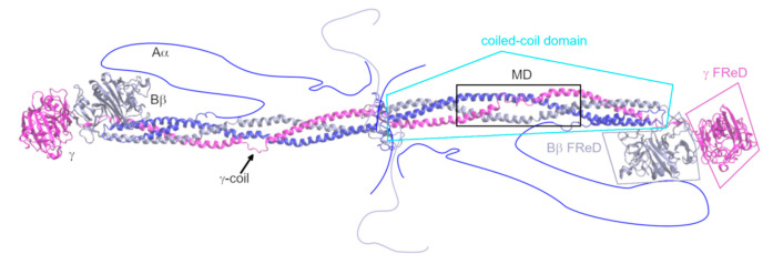
Figure 1.
Structure of fibrinogen, based on its crystal structure 3GHG [18]. The missing regions are schematically drawn into the figure. The region that is the subject of this study is highlighted by a black square and designated as “MD”. The Aα chain is shown in blue, the Bβ chain in grey-violet, and the γ chain in magenta. The domains of fibrinogen are designated. FReD stands for “fibrinogen-related domain”.

