Abstract
Simple Summary
Bacterial strains that are consumed by humans or by animals to promote health are called probiotics. In poultry, Bacillus strains are widely used as feed additives for this purpose. Although different modes of action have been proposed, studies showing effects on what metabolites the bacteria produce in a test tube, and whether these can also be found in the intestine of animals that were given these strains as feed additives, are lacking. In the current study, we show that administration of a Bacillus strain to broiler chickens changes the microbial composition in the gut by reducing opportunistic pathogenic bacterial families and promoting beneficial bacterial families. We show that two molecules, hypoxanthine and nicotinic acid, are produced by the Bacillus strain and are elevated in the intestinal tract of these animals. We hypothesize that nicotinic acid can be used by beneficial microbes and is essential for their intestinal colonization, and that both molecules can have a positive effect on the intestinal wall. These data can be used to evaluate and develop novel feed additives to promote health of chickens, and reduce the need for antibiotic usage.
Abstract
The probiotic Bacillus subtilis strain 29784 (Bs29784) has been shown to improve performance in broilers. In this study, we used a metabolomic and 16S rRNA gene sequencing approach to evaluate effects of Bs29874 in the broiler intestine. Nicotinic acid and hypoxanthine were key metabolites that were produced by the strain in vitro and were also found in vivo to be increased in small intestinal content of broilers fed Bs29784 as dietary additive. Both metabolites have well-described anti-inflammatory effects in the intestine. Furthermore, Bs29784 supplementation to the feed significantly altered the ileal microbiome of 13-day-old broilers, thereby increasing the abundance of genus Bacillus, while decreasing genera and OTUs belonging to the Lactobacillaceae and Enterobacteriacae families. Moreover, Bs29784 did not change the cecal microbial community structure, but specifically enriched members of the family Clostridiales VadinBB60, as well as the butyrate-producing families Ruminococcaceae and Lachnospiraceae. The abundance of various OTUs and genera belonging to these families was significantly associated with nicotinic acid levels in the cecum, suggesting a possible cross-feeding between B. subtilis strain 29784 and these beneficial microbes. Taken together, the data indicate that Bs29784 exerts its described probiotic effects through a combined action of its metabolites on both the host and its microbiome.
Keywords: probiotics, Bacillus subtilis, metabolites, intestinal health, nicotinic acid, hypoxanthine, 16S rRNA gene sequencing, broilers
1. Introduction
Probiotics are used in both human and animal nutrition for their health benefits. In animal diets, probiotics are included as feed additives to create a healthy and resilient intestinal microbial environment [1,2,3]. Maintaining a beneficial intestinal microbial composition helps in improving the overall health of the animal and thereby positively affects body weight gain (BWG) and feed conversion ratio (FCR) [4,5].
Many different microorganisms are used as probiotics in poultry production. Bacillus spp. are the most commonly used probiotic microorganisms because of their ability to form endospores [6]. This enables them to survive the feed manufacturing process and the passage through the stomach. Moreover, spores allow easy administration, storage and prolonged shelf-life [6]. One frequently used species, Bacillus subtilis, is considered to be safe for consumption [7,8]. A variety of B. subtilis strains are available as feed additives for animals with each having their own strain specificity. One example is B. subtilis strain 29784 (Bs29784), for which beneficial effects on growth performance are consistently reported in broilers, turkeys and layer pullets [9,10,11,12]. In addition, the strain reduces IL-8 expression and improves intestinal barrier integrity by upregulating tight junction protein expression, as was shown in a cell culture model [13]. Although effects of the administration of Bacillus strains on intestinal health parameters have been observed, insights in the exact modes of action of these probiotic strains are often limited. Different modes of action have been suggested in literature, including vitamin and nutrient production, enzyme production, antagonistic effects on pathogens, pH reduction due to short-chain fatty acids (SCFA) and lactate production, amongst others, but causal relationships between the produced metabolites and the observed effects are generally not proven [14,15,16]. Studies investigating the metabolites produced by probiotic strains have focused mainly on fermentation products such as lactic acid and SCFA, while, to the best of our knowledge, none have carried out a metabolome analysis and verified whether the metabolites produced in vitro could also be detected in the intestinal tract. Therefore, the aim of the current study was to identify metabolites that are produced by the probiotic B. subtilis strain Bs29784 in vitro, elucidate whether these metabolites are also produced in the chicken intestinal tract after in-feed supplementation of Bs29784, and how Bs29784 affects the intestinal microbiome.
2. Materials and Methods
2.1. Bacterial Strain and Growth Conditions
Bs29784 is a commercially available probiotic for broilers (Alterion® NE, Adisseo, Commentry, France). The commercial product contains 1010 CFU/g spores of B. subtilis strain 29784 and is mixed at 1 g/kg in the feed that is supplied to broilers. For in vitro experiments, a pure culture of Bs29784 was obtained by inoculating the commercial probiotic in Luria–Bertani (LB) broth (Sigma-Aldrich, St. Louis, MO, USA). The bacteria were grown overnight at 37 °C under aerobic conditions. Bacteria were plated on LB plates and their identity was confirmed via matrix-assisted laser desorption/ionization time of flight mass spectrometry (MALDI-TOF MS) [17] and Sanger sequencing of the 16S region [18]. Bacterial growth was determined in LB broth over a 24-hour time span (grown in triplicate). Bacterial supernatant was obtained by centrifugation (5 min, 13,300 rpm) and filtered using a Polyvinylidene difluoride (PVDF) membrane filter (0.22 µm × 13 mm diameter, Kynar 500®, Arkema, Amsterdam, The Netherlands). Blank samples (medium without bacteria) were incubated simultaneously with the bacterial samples and processed in the same way to serve as controls. Samples were stored at −80 °C until metabolomic analysis.
2.2. Animal Trial
The study was undertaken following the guidelines of the ethics committee of the Faculty of Veterinary Medicine, Ghent University, in accordance with the EU Directive 2010/63/EU. One-day-old Ross 308 broiler chicks were obtained from a local hatchery and divided into 2 groups of 5 birds consisting of (1) a control group that received a standard commercial diet and (2) a group that received a standard commercial diet supplemented with the commercial Bs29784 probiotic at a dose of 1010 CFU/kg feed (FARM 1&2 mash, Versele-laga, Deinze, Belgium). Animals were housed on a solid floor covered with wood shavings at a density of 5 birds/m2. Animals were subjected to a light schedule of 12 h light and 12 h dark. All broilers were given water and feed ad libitum. At 13 days of age, all birds were weighed, the birds were euthanized, and digestive content from the jejunum, ileum and cecum was collected. These samples were frozen in liquid nitrogen directly after sampling and stored at −20 °C until further processing. The material from the 3 sections was used for metabolomic analysis and Bacillus quantification, while the ileal and cecal content was used for 16S sequencing. At 13 days of age, no differences in bodyweight could be observed, with an average bodyweight of 273.4 g ± 19.54 g (mean ± SD) for the control group and 254.3 g ± 38.37 g for the Bs29784-supplemented group (p = 0.358).
2.3. Targeted Metabolomics
2.3.1. Reagents and Chemicals
Analytical standards [19] were obtained from Sigma-Aldrich (St. Louis, MO, USA), ICN Biomedicals Inc. (Costa Mesa, CA, USA) or TLC Pharmchem (Vaughan, ON, Canada). Solvents were obtained from Fisher Scientific UK and VWR International (Merck, Darmstadt, Germany). All analytical standards, including nicotinic acid (Sigma-Aldrich) and hypoxanthine (Sigma-Aldrich), as well as the internal standard valine-d8 (ISTD) (Sigma-Aldrich), were stored at −20 °C in a primary stock solution of 10 mg/mL in either ultrapure water or methanol.
2.3.2. Instrumentation
A polar metabolomics approach was applied based on the method described by Vanden Bussche et al. (2015) [20]. An Accela UHPLC system of Thermo Fisher Scientific (San José, CA, USA) was used, with an Acquity HSS T3 C18 column (1.8 μm, 150 mm × 2.1 mm, Waters). As binary solvent system, ultrapure water with 0.1% formic acid (A) and acetonitrile acidified with 0.1% formic acid (B) were used at a constant flow rate of 0.4 mL/min. A gradient profile of solvent A was applied (0−1.5 min at 98% (v/v), 1.5−7.0 min from 98% to 75% (v/v), 7.0−8.0 min from 75% to 40% (v/v), 8.0−12.0 min from 40% to 5% (v/v), 12.0−14.0 min at 5% (v/v), 14.0−14.1 min from 5% to 98% (v/v)), followed by 4.0 min of re-equilibration. Solvents used for UHPLC-MS/MS analysis were purchased from Fisher Scientific UK. HRMS analysis was performed on an Exactive stand-alone benchtop Orbitrap mass spectrometer (Thermo Fisher Scientific), equipped with a heated electrospray ionization source (HESI), operating in polarity switching mode.
2.3.3. Optimization of the UHPLC-HRMS Method
Optimization of the method of Vanden Bussche et al. (2015) [20] was performed in a preliminary run to exclude matrix effects and to determine the optimal concentration of the bacterial supernatant samples. For this purpose, quality control (QC) samples made from pooled biological samples were considered as representative bulk control samples [21]. QC samples were extracted and serially diluted with ultrapure water (1; 1:2, 1:5, 1:10, 1:20, 1:50, 1:100, 1:200 and 1:500), after which the linearity was studied based on the coefficient of determination (R2). The targeted analysis was based on an in-house metabolite mixture containing 291 known metabolites which are important in the gut. This mixture of metabolites was run to standardize and determine respective peaks found in the samples [22]. The absolute peak areas of the ISTD and of one representative metabolite from each category (multicarbon acids, monosaccharide, amino acid, imidazole, ketones, etc.) in the list of known metabolites was determined. The following 11 metabolites were analyzed: inositol, phenylacetic acid, succinate, histidyl leucine, glucose, 2-octanon, L-methionine, L-arginine, spermidine, hypoxanthine and uracil. The validated metabolites were required to have an R2 > 0.990. After validation, it was decided that a 1/10 dilution was optimal for the supernatant samples.
2.3.4. Metabolomic Analysis
Metabolites produced by Bs29784 in vitro were analyzed together with blank samples. In vivo metabolite production was determined using intestinal digesta from chickens receiving either non-supplemented feed or feed supplemented with Bs29784. Therefore, intestinal content of the jejunum, ileum or cecum was freeze-dried for 24 h. To 100 mg of freeze-dried material, 2 mL of ice-cold methanol (80:20) was added, vortexed and centrifuged (9000 rpm, 10 min), after which the supernatant was filtered using a PVDF filter (0.45 µm × 25 mm diameter) and used at a 1:3 dilution. Xcalibur 3.0 software (Thermo Fisher Scientific, San José, CA, USA) was employed for targeted data processing, whereby compounds were identified based on their m/z-value, C-isotope profile, and retention time relative to that of the internal standard.
2.4. DNA Extraction from Intestinal Content
DNA was extracted from the jejunal, ileal and cecal content, using the hexadecyltrimethylammonium bromide (CTAB) method described by Griffiths et al. [23] with modifications described by Aguirre et al. [24]. The resulting DNA was resuspended in 50 µL of a 10 mM Tris-HCl buffer (pH 8.0) and the quality and concentration of the DNA was examined spectrophotometrically (NanoDrop, Thermo Fisher Scientific, Merelbeke, Belgium).
2.5. Quantification of Bacillus spp. and Total Bacteria
The percentage bacteria belonging to the genus Bacillus (Bacillus spp.) relative to the total number of bacteria found in the content from different intestinal segments was determined using quantitative PCR (qPCR). Primers targeting Bacillus spp. (YB-P1 and YB-P2) were used as described by Han et al. (2012) [25]. To determine the number of total bacteria, primers Uni 331F and Uni 797R were used as described by Hopkins et al. (2005) [26]. The qPCR was performed using the SensiFAST™ SYBR® No-ROX Kit (Bioline, London, UK) with a 0.5 µM primer concentration. The PCR amplification consists of DNA pre-denaturation at 95 °C for 2 min followed by 30 cycles of denaturation (95 °C for 15 s), annealing (60 °C for 30 s) and extension (72 °C for 50 s).
2.6. 16S rRNA Gene Amplicon Sequencing
The V3–V4 hypervariable region of the 16s rRNA gene was amplified by using the gene-specific primers S-D-Bact-0341-b-S-17 and S-D-Bact-0785-a-A-21 [27]. The PCR amplifications were performed as described by Aguirre et al. (2019) [24]. CleanNGS beads (CleanNA, Gouda, The Netherlands) were used to purify PCR products. The DNA concentration of the final barcoded libraries was measured with a Quantus fluorimeter and Quantifluor dsDNA system (Promega, Madison, WI, USA). The libraries were combined to an equimolar 5 nM pool and sequenced with 30% PhiX spike-in using the Illumina MiSeq v3 technology (2 × 300 bp, paired-end) at the Oklahoma Medical Research center (Oklahoma City, OK, USA).
Demultiplexing of the amplicon dataset and deletion of the barcodes was done by the sequencing provider. Quality of the raw sequence data was evaluated using the FastQC quality control tool (Babraham Bioinformatics, Cambridge, UK), followed by an initial quality filtering with Trimmomatic v0.38 [28]. Reads with an average quality per base below 15 were cut using a four-base sliding window and reads with a minimum length below 200 bp were discarded. The paired-end sequences were assembled and primers were removed using PANDAseq [29], with a quality threshold of 0.9 and length cut-off values for the merged sequences between 390 and 430 bp. Chimeric sequences were removed using UCHIME [30]. Open-reference operational taxonomic unit (OTU) picking was performed at 97% sequence similarity using USEARCH (v6.1) and converted to an OTU table [31]. OTU taxonomy was assigned against the Silva database (v132, clustered at 97% identity) [32] using the PyNast algorithm with QIIME (v1.9.1) default parameters [33]. OTUs with a total abundance below 0.01% of the total sequences were discarded [34]. Potential contaminant chloroplastic and mitochondrial OTUs were removed from the dataset, resulting in an average of approximately 76,080 reads per sample, with a minimum of 25,725. Alpha rarefaction curves were generated using the QIIME “alpha_rarefaction.py” script and a subsampling depth of 25,000 reads was selected.
2.7. Metabolic Function Prediction of the Microbial Communities
Functional genes (KEGG orthologues, KOs) were inferred from the 16S amplicon sequencing data using Phylogenetic Investigation of Communities by Reconstruction of Unobserved States (PICRUSt), as previously described [24,35]. The resulting KEGG orthologues were further summarized into functional modules based on the Gut-specific Metabolic Modules (GMM) database using GoMixer (Raes Lab) [36,37]. The contribution of various taxa to different GMMs was computed with the script “metagenome_contributions.py”.
2.8. Statistical Analyses
Statistical analyses of the metabolomic and qPCR data were performed using GraphPad PRISM (v8.4.3). A Kolmogorov–Smirnov test was performed to evaluate the data for normal distribution. In case of normal distribution, an independent samples t-test was performed. When data were not normally distributed, a non-parametric Mann–Whitney test was performed. Tests were considered statistically significant at a p-value ≤0.05. Biologically relevant metabolite production by Bs29784 in vitro was identified as a fold change >2 and p < 0.05.
Statistical analyses of the 16S data were performed using R (v3.6.0). Alpha diversity was measured based on the observed OTUs (or observed KOs for the functional data), Chao1 and Shannon diversity index using the phyloseq pipeline [38]. Differences in alpha diversity were assessed using a Wilcoxon’s rank sum test. Beta diversity was calculated using Bray–Curtis distance. Differences in beta diversity were examined by permutational analysis of variance (Permanova) using the adonis function from the vegan package [39]. Differences in relative abundance at the phylum and family level were assessed using the two-sided Welch t-test from the mt wrapper in phyloseq, with the p-value adjusted for multiple hypothesis testing using the Benjamini–Hochberg method. The DESeq2 algorithm was applied to identify differentially abundant genera or functional modules between the control and Bs29784 group [40]. Significant differences were obtained using a Wald test followed by a Benjamini–Hochberg multiple hypothesis correction. For all tests, an adjusted p-value (q-value) ≤0.05 was considered significant. Biologically relevant differences in functional modules between the birds fed a control diet or Bs29784-supplemented diet were selected using a Log2 fold change (Log2FC) > 2 and q-value < 0.1.
The association of microbial abundances (at family, genus or OTU level), with hypoxanthine and nicotinic acid levels measured in the intestinal content, were analyzed using the multivariate analysis by linear models (MaAsLin2) R package. MaAsLin2 analysis was performed separately on the ileal and cecal samples, while controlling for treatment covariates [41].
3. Results
3.1. Identification of Metabolites Produced by Bs29784 In Vitro
Metabolites produced by Bs29784 after 24 h growth in LB medium were compared to the blank medium. Overall, 123 of the 291 targeted metabolites could be detected in either the blank LB medium and/or the supernatants of Bs29784 grown in LB (Table S1). The majority of the detected metabolites (96/123, 78%) were not significantly altered after growth of Bs29784 in the LB medium. In total, 21 metabolites (17% of the detected metabolites) were significantly reduced due to growth of Bs29784 and 16 metabolites (13% of the detected metabolites) were produced by Bs29784 in vitro (Table S1). Biologically relevant metabolites were identified based on a fold change >2 and p < 0.05 (Table 1). The most discriminatory metabolites, nicotinic acid and hypoxanthine (p < 0.0001), were selected for evaluation in the in vivo samples.
Table 1.
Metabolites that are significantly increased (fold change > 2 and p < 0.05) after 24 h growth of B. subtilis strain 29784 in LB medium.
| Metabolite | Area Ratio (Mean ± SD) | Fold Change | p-Value | |
|---|---|---|---|---|
| Blank | Bs29784 | |||
| Hypoxanthine | 0.173 ± 0.002 | 1.844 ± 0.086 | 106.40 | <0.0001 |
| Nicotinic acid | 0.218 ± 0.030 | 1.853 ± 0.104 | 8.51 | <0.0001 |
| Ethanolamine | 0.007 ± 0.003 | 0.061 ± 0.016 | 8.67 | 0.005 |
| Uracil | 0.241 ± 0.004 | 1.652 ± 0.392 | 6.85 | 0.003 |
| Pantothenate | 0.001 ± 0.001 | 0.022 ± 0.002 | 2.03 | 0.002 |
| 3-Hydroxypyridine | 0.006 ± 0.003 | 0.014 ± 0.001 | 2.16 | 0.015 |
| 2.5-dimethylpyrazine | 0.005 ± 0.000 | 0.012 ± 0.003 | 2.47 | 0.017 |
| Thymine | 0.014 ± 0.007 | 0.034 ± 0.004 | 2.51 | 0.011 |
3.2. Effect of Supplementation of Bs29784 in Broiler Feed on the Bacillus Load, Levels of Hypoxanthine and Nicotinic Acid in the Intestinal Tract
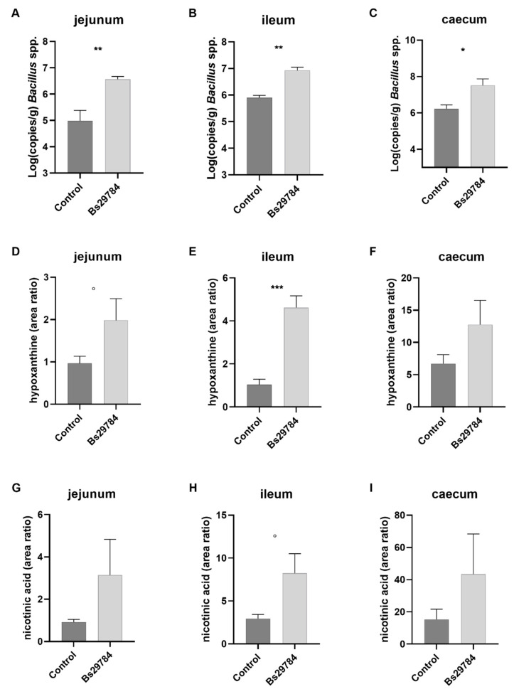
The total number of bacteria, as well as the number of Bacillus spp. in the jejunum, ileum and cecum were determined using qPCR. Supplementation of the diet with the probiotic B. subtilis strain Bs29784 did not introduce alterations in the total bacterial load (data not shown), but significantly increased the number of Bacillus spp. in the ileum (p = 0.005), jejunum (p = 0.008), and cecum (p = 0.014) (Figure 1A–C).
Figure 1.
Abundance of Bacillus spp. and metabolite concentrations in jejunum, ileum and cecum. The Bacillus load in the jejunum, ileum and cecum was measured via qPCR (A–C). The metabolites hypoxanthine (D–F) and nicotinic acid (G–I) are expressed as area ratio. ° p < 0.1, * p < 0.05, ** p < 0.01, *** p < 0.001.
To further assess whether this increase in Bacillus spp. was reflected in an increase in Bs29784 metabolites, the levels of hypoxanthine and nicotinic acid were determined. Overall, broilers fed a Bs29784-containing diet showed higher levels of hypoxanthine and nicotinic acid in the intestinal content. The increase in hypoxanthine was most pronounced in the ileum (p = 0.0003), but did not reach significance in the jejunum (p = 0.095) or cecum (p = 0.171) (Figure 1D–F). In-feed supplementation of Bs29784 tended to increase the level of nicotinic acid in the ileum (p = 0.051), as compared to birds fed the control diet, but had no effect on nicotinic acid levels in the jejunum (p = 0.223) or cecum (p = 0.306) (Figure 1G–I).
3.3. Effect of Bs29784 Supplementation in Broiler Feed on the Ileal and Cecal Microbial Diversity
The microbial complexity in the ileum and cecum was estimated by calculating the number of observed OTUs, the estimated OTU richness (Chao1) or the estimated community diversity (Shannon index) in each sample. In-feed supplementation of Bs29784 had no effect on the ileal microbial richness (observed OTUs or Chao1) (Table 2). However, addition of Bs2978 to the diet significantly reduced the ileal community diversity (Shannon index, p = 0.032). This is in contrast to the situation in the cecum, which had a tendency for higher microbial richness in birds fed the Bs29784-supplemented diet, as compared to the control diet (observed OTUs: p = 0.056, Chao1: p = 0.15), but no effect of Bs29784 on the microbial community diversity was observed (Table 2).
Table 2.
Taxonomic and functional alpha diversity of ileal and cecal microbial communities from broilers fed either a control or a Bs29784-supplemented feed.
| Control | Bs29784 | p-Value | |
|---|---|---|---|
| ILEUM | |||
| Taxonomic alpha diversity | |||
| nOTUs | 98.8 ± 29.95 | 90 ± 16.02 | 0.69 |
| Chao1 | 125.31 ± 49.39 | 107.59 ± 24.07 | 0.69 |
| Shannon | 1.72 ± 0.40 | 1.06 ± 0.43 | 0.032 * |
| Functional alpha diversity | |||
| nKOs | 4487 ± 257.13 | 4522.6 ± 145.87 | 1 |
| Chao1 | 4656.89 ± 375.39 | 4743.67 ± 298.32 | 1 |
| Shannon | 7.40 ± 0.23 | 7.16 ± 0.18 | 0.15 |
| CECUM | |||
| Taxonomic alpha diversity | |||
| nOTUs | 142.8 ± 5.45 | 181.2 ± 25.08 | 0.056 |
| Chao1 | 157.74 ± 7.13 | 196.50 ± 30.77 | 0.15 |
| Shannon | 2.91 ± 0.41 | 3.26 ± 0.58 | 0.42 |
| Functional alpha diversity | |||
| nKOs | 4228.4 ± 111.10 | 4205.0 ± 76.41 | 1 |
| Chao1 | 4554.97 ± 210.53 | 4414.80 ± 191.05 | 0.42 |
| Shannon | 7.71 ± 0.13 | 7.39 ± 0.14 | 0.016 * |
* Significant differences between the control and Bs29784 group (p < 0.05).
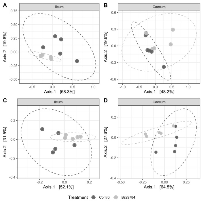
Bray–Curtis dissimilarity was used to investigate beta diversity between either the ileal or cecal microbiota from birds fed the control diet or the diet supplemented with B. subtilis strain 29874. Supplementation of Bs29784 to the broiler diet showed a significant clustering in the ileum, with 33.7% of the variation between the samples being explained by the Bs29784 supplementation to the feed (p = 0.028) (Figure 2A). However, no effect on the cecal microbial community composition was observed (diet explaining 17.4% of the variation, p = 0.15) (Figure 2B).
Figure 2.
PCoA plot of the taxonomic and functional microbial diversity from birds fed a control or Bs29784-supplemented diet. Principal coordinate analysis (PCoA) plots of bacterial taxonomic (OTU-level) (A,B) or functional (KO-level) (C,D) diversity calculated using the Bray–Curtis dissimilarity metric. Each dot represents an individual chicken microbiome. Significant separation of the microbial communities was observed in the ileum (p = 0.028) (A), but not the cecum (p = 0.153) (B). In both the ileum and cecum, significant grouping of the samples was observed based on the functional KO diversity (p = 0.024 and p = 0.029) (C,D).
3.4. Influence of Bs29784 on the Taxonomic Composition of the Ileal and Cecal Microbiome
The most abundant phyla in the ileum were Firmicutes (84.94% in control, 96.83% in Bs29784) and Proteobacteria (12.81% in control, 2.24% in Bs29784), with a minor portion belonging to the Verrucomicrobia (1.97% in control, 0.80% in Bs29784) and Actinobacteria (0.28% in control, 0.13% in Bs29784). Also in the cecum, the Firmicutes was the most prevalent phylum in both groups (48.16% in control, 68.37% in Bs29784), followed by the Proteobacteria (26.27% in control, 10.54% in Bs29784) and Verrucomicrobia (24.29% in control, 19.68% in Bs29784). The phylum Actinobacteria accounted for 1.28% and 1.41% of the cecal microbiome in birds fed the control or Bs29784-supplemented diet, respectively. Addition of Bs29784 to the broiler diet had no significant influence on either the ileal or cecal microbiome at phylum level.
In the ileum, the families Bacillaceae (<0.001% in control, 0.12% in Bs29784, padj = 0.06) and Enterococcaceae (45.25% in control, 82.47% in Bs29784, padj = 0.17) tended to be more abundant after probiotic supplementation, whereas both the family Leuconostocaceae (0.25% in control versus 0.0016% in Bs29784, padj = 0.06) and family Lactobacillaceae (24.45% in control and 2.51% in Bs29784, padj = 0.17) tended to be less abundant in the ileum of birds fed the Bs29784-supplemented diet. No significant effect of Bs29784 supplementation on the families in the cecum could be observed.
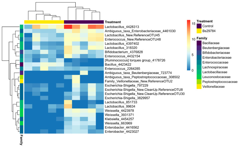
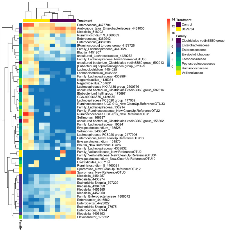
Differentially abundant genera and OTUs in the ileal or cecal microbiome from birds fed a Bs29784-supplemented diet as compared to the control diet were identified using DESeq2 (Table 3; Tables S2 and S3). Nine genera were differentially abundant between the ileal microbiota from birds fed either the control diet or the Bs29784 diet. Only the genus Bacillus was significantly increased in the ileal microbiota of birds fed the Bs29784-containing diet, a difference that could be fully attributed to a single OTU identified as Bacillus subtilis (OTU4423422, Figure 3, Table S2). The other significantly altered genera and OTUs in the ileal microbiome were all less abundant in Bs29784-fed birds, with multiple genera belonging to the Enterobacteriaceae family, including multiple OTUs belonging to genera Escherichia-Shigella and Enterobacter (Figure 3). Furthermore, addition of Bs29784 to the broiler feed resulted in a reduction of the genus Pediococcus and Weissella, as well as multiple OTUs belonging to the genus Lactobacillus in the ileal microbiome (Table 3, Figure 3). In the cecum, Bs29784 supplementation of the broiler feed significantly reduced the relative abundance of multiple genera belonging to the families Veillonellacaea and Enterobacteriaceae, with main OTUs belonging to the genus Klebsiella (Figure 4, Table S3). Additionally, an increase in members of the butyrate-producing families Ruminococcaceae and Lachnospiraceae was observed in the cecum of Bs29784-fed birds. Moreover, the genus Enterococcus, Clostridioides and a genus belonging to the Clostridiales vadinBB60 group were significantly increased in the cecum by Bs29784 supplementation of the feed (Table 3).
Table 3.
Differentially abundant genera in the ileal or cecal microbiota.
| Phylum | Class | Family | Genus | Mean Abundance (%) | Log2 Fold Change | Adjusted p-Value |
|
|---|---|---|---|---|---|---|---|
| Control | Bs29784 | ||||||
| ILEUM | |||||||
| Actinobacteria | Actinobacteria | Beutenbergiaceae | Ambiguous taxa Beutenbergiaceae | 0.046 | 0.000 | −23.36 | <0.001 |
| Firmicutes | Bacilli | Bacillaceae | Bacillus | 0.000 | 0.121 | 7.54 | <0.001 |
| Firmicutes | Bacilli | Lactobacillaceae | Pediococcus | 0.250 | 0.035 | −4.32 | 0.019 |
| Firmicutes | Bacilli | Leuconostocaceae | Weissella | 0.253 | 0.002 | −7.20 | <0.001 |
| Firmicutes | Clostridia | Peptostreptococcaceae | Ambiguous taxa Peptostreptococcaceae | 0.054 | 0.000 | −22.66 | <0.001 |
| Firmicutes | Negativicutes | Veillonellaceae | Family Veillonellaceae | 0.062 | 0.000 | −22.91 | <0.001 |
| Proteobacteria | Gammaproteobacteria | Enterobacteriaceae | Ambiguous taxa Enterobacteriaceae | 0.473 | 0.051 | −3.71 | 0.007 |
| Proteobacteria | Gammaproteobacteria | Enterobacteriaceae | Enterobacter | 0.045 | 0.002 | −6.32 | 0.001 |
| Proteobacteria | Gammaproteobacteria | Enterobacteriaceae | Klebsiella | 0.058 | 0.002 | −6.09 | 0.007 |
| CECUM | |||||||
| Firmicutes | Bacilli | Enterococcaceae | Enterococcus | 1.746 | 4.865 | 2.30 | 0.016 |
| Firmicutes | Clostridia | Clostridiales vadinBB60 group | uncultured bacterium_Clostridiales vadinBB60 group | 0.000 | 0.956 | 12.51 | <0.001 |
| Firmicutes | Clostridia | Lachnospiraceae | [Eubacterium] hallii group | 0.000 | 0.074 | 22.48 | <0.001 |
| Firmicutes | Clostridia | Lachnospiraceae | GCA-900066575 | 0.000 | 0.062 | 22.47 | <0.001 |
| Firmicutes | Clostridia | Lachnospiraceae | Lachnospiraceae FCS020 group | 0.004 | 0.219 | 7.32 | <0.001 |
| Firmicutes | Clostridia | Lachnospiraceae | Lachnospiraceae NK4A136 group | 0.000 | 0.556 | 25.64 | <0.001 |
| Firmicutes | Clostridia | Peptostreptococcaceae | Clostridioides | 0.000 | 0.066 | 23.25 | <0.001 |
| Firmicutes | Clostridia | Ruminococcaceae | Negativibacillus | 0.000 | 0.693 | 11.10 | <0.001 |
| Firmicutes | Clostridia | Ruminococcaceae | Ruminiclostridium 9 | 0.239 | 1.359 | 2.93 | 0.0461 |
| Firmicutes | Clostridia | Ruminococcaceae | Ruminococcaceae UCG-013 | 0.000 | 0.008 | 27.52 | <0.001 |
| Firmicutes | Negativicutes | Veillonellaceae | Family_Veillonellaceae | 1.272 | 0.000 | −27.55 | <0.001 |
| Firmicutes | Negativicutes | Veillonellaceae | Sporomusa | 3.657 | 0.000 | −28.07 | <0.001 |
| Proteobacteria | Gammaproteobacteria | Enterobacteriaceae | Ambiguous_taxa_Enterobacteriaceae | 5.518 | 0.758 | −2.48 | <0.001 |
| Proteobacteria | Gammaproteobacteria | Enterobacteriaceae | Enterobacter | 0.718 | 0.059 | −3.03 | 0.004 |
| Proteobacteria | Gammaproteobacteria | Enterobacteriaceae | Klebsiella | 3.221 | 0.745 | −2.33 | 0.006 |
Significant differences in genus level abundance in the ileal or cecal microbiota from birds fed the Bs29784-supplemented diet as compared to the control diet. The taxonomic classification and the log2 fold change (log2FC) (Bs29784/control) of the DESeq2-normalized abundance of each genus are shown. Positive values indicate an increase in abundance of the respective genus in the Bs29784 group, while negative values indicate a decrease.
Figure 3.
Differentially abundant OTUs in the ileal microbiome of birds fed either a control or Bs29784-supplemented diet. The abundance of the OTUs is shown as the log2 of the DESeq2-normalized counts. Each OTU is labelled with the genus information, or family information when no genus identification was possible, followed by the OTU number.
Figure 4.
Differentially abundant OTUs in the cecal microbiome of birds fed either a control or Bs29784-supplemented diet. The abundance of the OTUs is shown as the log2 of the DESeq2-normalized counts. Each OTU is labelled with the genus information, or family information when no genus identification was possible, followed by the OTU number.
3.5. Hypoxanthine and Nicotinic Acid Levels Are Associated with Specific Microbial Taxa in the Cecum
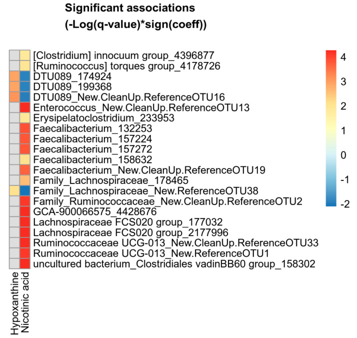
Associations between the hypoxanthine and nicotinic acid levels and microbial abundances in either the ileum or cecum were analyzed using multivariate association with linear models (MaAsLin2), while controlling for the type of diet (control diet or Bs29784-supplemented diet). In the ileum, no associations between metabolite levels and the abundance of specific microbial taxa were observed. In the cecum, the genus DTU089 (family Ruminoccocaceae) was significantly associated with the hypoxanthine levels (p = 0.001, q = 0.022) and inversely correlated with the nicotinic acid levels (p = 0.006, q = 0.099). These associations were also significant at the OTU level (Figure 5). Additionally, a similar association between metabolite levels and a single OTU belonging to the family Lachnospiraceae was observed (Figure 5). No other associations with hypoxanthine levels in the cecum could be observed. In contrast with the limited number of microbiome–hypoxanthine associations, the effect of nicotinic acid on the cecal microbiome was more pronounced. Nicotinic acid levels were positively associated with 17 OTUs, mainly ones belonging to the families Lachnospiraceae and Ruminococcaceae (Figure 5). Five out of seventeen OTUs (29.4%) that were associated with the cecal nicotinic acid levels belong to Faecalibacteria, and were mainly identified as F. prausnitzii (4/5 Faecalibacterium OTUs). These microbiome–nicotinic acid associations were also significant at the genus level, and even the family level, for both the family Ruminococcaceae (p = 0.012, q = 0.222) and family Clostridiales vadinBB60 group (p = 0.001, q = 0.024).
Figure 5.
Heatmap of microbial OTUs showing significant association with hypoxanthine or nicotinic acid levels in the cecum. Significant associations were identified using MaAsLin2 and are plotted as (−Log(q-value)*sign(coeff.)). Grey squares: no significant association.
3.6. In-Feed Bs29784 Supplementation Decreases the Abundance of Specific Microbial Metabolic Modules
To determine whether the Bs29784-induced alterations of the microbiota might have an effect on the microbial functions, the functional genes (KEGG orthologs) present in the ileal and cecal microbiome were in silico predicted and grouped into gut-specific metabolic modules (GMMs). In total, 5135 and 4674 KOs were identified in, respectively, the ileal and cecal microbiome. In-feed supplementation of Bs29784 had no effect on both the ileal and cecal functional richness (number of observed KOs or Chao1 richness estimator), but reduced the diversity of the functional genes (Shannon diversity, ileum: p = 0.15, cecum: p = 0.016) (Table 2). Beta-diversity analysis based on Bray–Curtis showed significant clustering in both the ileum and cecum with 28.0% and 33.8% of the variation between the samples being explained by the Bs29784 supplementation to the feed (ileum: p = 0.024, cecum: p = 0.029) (Figure 2C,D).
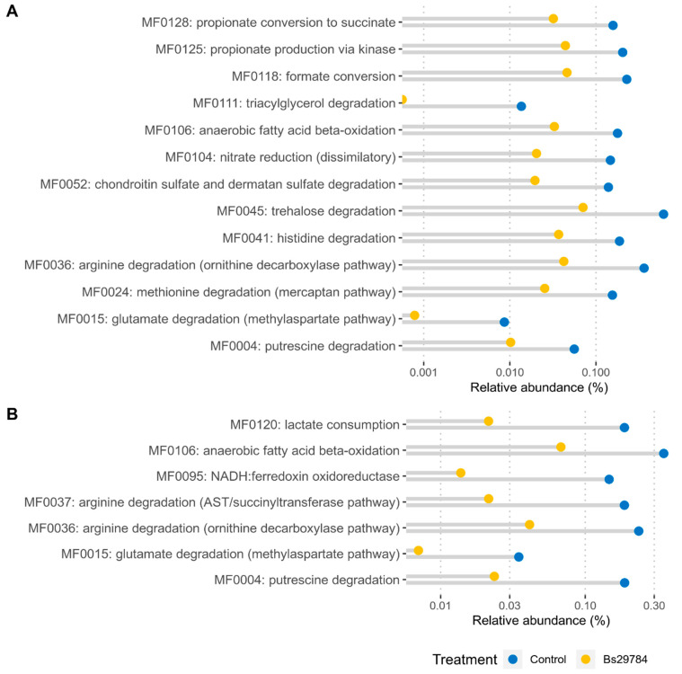
Based on the identified functional genes, 127 and 126 gut metabolic functional modules (GMM) could be constructed in, respectively, the ileum and cecum. None of the GMMs were significantly more abundant in either the ileum or cecum from birds receiving the Bs29784-supplemented feed. However, 13 GMMs were significantly less abundant in the ileum, whereas 7 GMMs were reduced in the cecum of Bs29784-fed birds (Tables S4 and S5). The affected GMMs can be classified in seven functional categories: amines and polyamines degradation (MF004), amino acid degradation (MF0015, MF0024, MF0036, MF0037 and MF0041), carbohydrate degradation (MF0045, MF0052), gas metabolism (MF0095), inorganic nutrient metabolism (MF0104), lipid degradation (MF0106, MF0111) and organic acid metabolism (MF0118, MF0120, MF0125, MF0128).
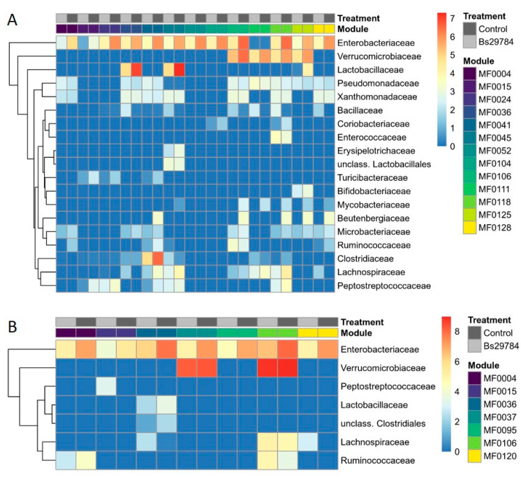
To further address the metagenomic potential of the ileal and cecal microbiota, the relative abundance of the GMMs of interest (Figure 6) as well as the microbial taxa putatively contributing to the selected pathways were identified (Figure 7, Tables S6 and S7). In the ileum, the majority of the changes in predicted metabolic modules could, at least partly, be attributed to members of the family Enterobacteriaceae (Figure 7A). Additionally, the genus Akkermansia within the family Verrucomicrobiaceae contributed for a large part to the observed reduction of a selection of GMMs (MF0106, MF0111, MF0118, MF0125), which are mainly involved in lipid degradation and organic acid metabolism (Figure 7A). In addition to the family Enterobacteriaceae, the Lactobacillaceae were main contributors to the arginine degradation (MF0036) and trehalose degradation (MF0045) modules, whereas the Clostridiaceae were in large part responsible for the histidine degradation (MF0041) module. Other bacterial families had only minor taxonomic contributions to the differences in metabolic modules encoded by the ileal microbiome from broilers fed a control or Bs29784-supplemented diet (Figure 7A, Table S6).
Figure 6.
Relative abundances of gut-specific metabolic modules (GMMs) in ileum (A) or cecum (B) of broilers with control and Bs29784-supplemented diets. Functional modules with a Log2FC > 2 and q-values < 0.1 are shown.
Figure 7.
Bacterial families responsible for each of the functional modules detected in ileum (A) or cecum (B) of control animals and broilers supplemented with Bs29784 in the feed. Metagenome contributions on the family level are sorted per functional module and per treatment (control: dark gray; Bs29784: light gray). The log2 of the module counts per family are shown on a blue–red scale. Only families that were present in at least 3 out of 5 samples from either treatment group were included. MF0004: putrescine degradation, MF0015: glutamate degradation, MF0024: methionine degradation, MF0036: arginine degradation (ornithine decarboxylase pathway), MF0037: arginine degradation (AST/succinyltransferase pathway), MF0041: histidine degradation, MF0045: trehalose degradation, MF0052: chondroitin sulfate and dermatan sulfate degradation, MF0095: NADH:ferredoxin oxidoreductase, MF00104: nitrate reduction, MF0106: anaerobic fatty acid beta-oxidataion, MF0111: triacylglycerol degradation, MF0118: formate conversion, MF0120: lactate consumption, MF0125: propionate production via kinase, MF0128: propionate conversion to succinate.
In the cecum, members of the family Enterobacteriaceae were contributing greatly to the observed differences in metabolic modules (Figure 7B). This effect of the Enterobacteriaceae is partially counteracted by a taxonomic increase of the families Ruminococcaceae and Lachnospiraceae, which specifically contribute to the modules encoding for arginine degradation (MF0036), anaerobic fatty acid beta-oxidation (MF0106) and lactate consumption (MF0120) (Figure 7B). Additionally, the genus Akkermansia (family Verrucomicrobiaceae) had a large share in the abundance of modules MF0106 and MF0037, but it did not influence the overall module abundance (Figure 7B).
4. Discussion
The Bacillus subtilis strain 29784 was previously shown to improve growth performance in broilers, turkeys and layer pullets [10,11,12], have a beneficial effect on the gut mucosal morphology in broilers [9] and increase the abundances of butyrate-producing bacteria in the ceca of both broilers and layer pullets [9,42]. Moreover, Bs29784 was shown to possess anti-inflammatory properties and enhance epithelial barrier integrity in vitro [13]. However, how Bs29784 modulates the microbiome and interacts with the host was largely unknown. In this study, we identified nicotinic acid and hypoxanthine as important metabolites that might contribute to the above-described host- and microbiome-modulating effects of Bs29784. Indeed, nicotinic acid and hypoxanthine were produced by Bs29784 in vitro and were also increased in the ileum of broilers fed a Bs29784-supplemented diet. Bacillus subtilis spores have been found to germinate in the small intestine of mice [43] and chickens [44]. The observed increase of hypoxanthine and nicotinic acid in the small intestine of broilers fed a Bs29784-supplemented diet indicates that the Bs29784 spores were germinating in the intestine and suggests that Bacillus-produced metabolites are able to actively contribute to the metabolite pool produced by the gastrointestinal microbiome.
In-feed supplementation of Bs29784 induces a shift in the cecal microbiome towards butyrate-producing bacteria, which can at least partly be explained by the metabolites produced by Bs29784. Although no changes were observed in the overall community structure, Bs29784 specifically decreased the abundance of multiple genera belonging to the families Veillonellaceae and Enterobacteriaceae, while increasing members of the families Clostridiales VadinBB60, Ruminococcaceae and Lachnospiraceae. This is in accordance with previous studies in both broilers and layers, where B. subtilis strain 29784 increased the cecal abundance of the butyrate-producing families Ruminococcaceae and Lachnospiraceae [9,42]. In this study, we showed that the abundance of various OTUs and genera belonging to the Clostridiales VadinBB60, Ruminococcaceae and Lachnospiraceae was significantly associated with nicotinic acid levels in the cecum. A similar association between nicotinic acid levels in the gut and the genus Faecalibacterium was previously observed in samples from inflammatory bowel disease (IBD) patients [45]. In both IBD patients and in our study, this association could mainly be attributed to Faecalibacterium prausnitzii. As F. prausnitzii is auxotroph for nicotinic acid, it has to acquire this nutrient form the environment [46,47], suggesting possible cross-feeding between B. subtilis strain 29784 and F. prausnitzii in the gut. Moreover, various members of the Ruminococcaceae and Lachnospiraceae lack the pathways for de novo synthesis of several other B-vitamins (mostly vitamin B1 (thiamin), B5 (pantothenate), B6 (pyridoxine) and B7 (biotin)), while these pathways were encoded in the genome of various B. subtilis strains [48]. Therefore, it might be that the observed association between nicotinic acid and these bacteria is caused by the production of other B vitamins by Bs29784. Indeed, we showed that Bs29784 is able to produce pantothenate in vitro. However, this vitamin was not further investigated in this study. Whether or not Bs29784 is able to produce other B-vitamins and steer the microbiome towards an anti-inflammatory community through cross-feeding remains to be elucidated.
Bs29784 addition to the broiler diet changes the microbial community structure in the ileum, thereby mainly reducing the abundance of various genera and OTUs belonging to the Lactobacillaceae and Enterobacteriaceae, while increasing the abundance of B. subtilis. This is in contrast to a previous study where in-feed supplementation of Bs29784 had no effect on the ileal microbiome in broilers [9]. This difference might be attributed to the age of the birds, where the aforementioned study used 42-day-old broilers, while our study aimed at studying the more dynamic microbiome of 13-day-old birds. Moreover, supplementation of B. subtilis strain 29784 in the feed of broilers reduced the abundance of several functional modules, which were mainly involved in amino acid degradation or organic acid metabolism. This effect on the microbial functional potential was less pronounced in the cecal microbiome and was in large part due to a reduction in Enterobacteriaceae. As no association was observed between hypoxanthine or nicotinic acid levels and the microbiome in the ileum, it is unclear how Bs29784 exerts its microbiome-modulating effect in the ileum. One possibility is that the observed microbiome effects are caused by the production of anti-microbial peptides by Bs29784 or through an indirect effect of Bs29784 on the host. Alternatively, it might be that the number of animals used in this study (n = 5 per group) did not yield enough statistical power to discover possible associations between the Bs29784-produced metabolites and the ileal microbiome.
In addition to the abovementioned effects on the microbiome, beneficial effects on intestinal health for both hypoxanthine and nicotinic acid were previously reported. Reduced faecal levels of hypoxanthine or nicotinic acid have both been linked with IBD [45,49,50]. Furthermore, both metabolites are able to ameliorate experimental colitis [51,52]. Additionally, nicotinic acid treatment promoted mucosal healing in patients with moderately active ulcerative colitis [51].
Hypoxanthine is a breakdown product of nucleic acids and can be taken up and incorporated by intestinal bacteria or the host via the nucleotide salvage pathway [53]. Additionally, hypoxanthine from the microbiota is salvaged for energy and nucleotide biosynthesis in intestinal epithelial cells, thereby supporting wound healing, mucus generation and intestinal barrier function [49,52,54]. Notably, hypoxanthine has also been shown to act as a substrate for the antimicrobial function of the enzyme xanthine oxidoreductase (XOR) which is located on the outer surface of epithelial cells [38,39]. XOR is responsible for the conversion of hypoxanthine to xanthine and from xanthine to uric acid. During both reactions, oxygen is reduced, generating hydrogen peroxide (H2O2) and reactive oxygen species (ROS) [55,56]. XOR-generated H2O2 has been shown to act as an effective antimicrobial agent against commensal microorganisms and anaerobes, although pathogenic bacteria could be more resistant [56]. Moreover, XOR-generated ROS have been hypothesized to initiate neutrophil infiltration in response to pro-inflammatory mediators [57]. These neutrophils can then help to combat infections. In chickens, XOR is mainly expressed in the intestine, liver and pancreas [58]. It is thus possible that hypoxanthine produced by Bs29784 contributes to intestinal health through enhancing epithelial barrier function and mucus production, while protecting the intestinal epithelial cells against microorganisms through H2O2 production. This could be one of the reasons a reduction in several genera of the Enterobacteriaceae, such as Enterobacter and Escherichia-Shigella, is seen in the ileum of broilers fed Bs29784-supplemented feed.
Nicotinic acid, or niacin (pyridine-3-carboxylic acid), is a form of vitamin B3, an essential nutrient for animals, including broilers. In humans and rodents, nicotinic acid is known to bind on the GPR109A receptor (aka HCA2 or HM74a in humans and NIACR1 in rodents), which is also one of the receptors for butyrate [59,60,61]. GPR109A has been shown to act as an anti-inflammatory mediator via the β-arrestin signaling pathway, protecting epithelial cells against inflammation and oxidative stress [61]. It is unclear whether nicotinic acid induces similar effects in birds, since an equivalent homologous receptor has not yet been identified. Nevertheless, nicotinic acid shows comparable effects on the regulation of the lipid transport apolipoproteins apoA and apoB in broilers as in humans which is mediated by GPR109A in the latter [62]. Furthermore, nicotinic acid is an important precursor for the coenzymes nicotinamide adenine dinucleotide (NAD) and nicotinamide adenine dinucleotide phosphate (NADP) that play an essential role in, among others, antioxidant protection [63,64]. This suggests that nicotinic acid, produced among others by Bs29784, may be taken up by the epithelial cells, protecting the cells from oxidative stress, while at the same time H2O2 is generated outside the cell by the action of the cell-surface xanthine oxidoreductase on hypoxanthine, also produced among others by Bs29784.
5. Conclusions
In conclusion, this study identified hypoxanthine and nicotinic acid as two important metabolites produced by B. subtilis strain 29784. The probiotic was shown to be metabolically active, producing these two metabolites in the intestine of broilers. These metabolites contribute, at least in part, to the interaction of Bs29784 with both the host and the microbiome, either through direct anti-inflammatory or anti-bacterial properties or by increasing the abundance of beneficial butyrate-producing bacteria in the cecum, potentially through cross-feeding.
Supplementary Materials
The following are available online at https://www.mdpi.com/article/10.3390/ani11051335/s1, Table S1: Metabolites detected in either blank LB medium or after 24 h growth of B. subtilis strain Bs29784 on LB medium, Table S2: Differentially abundant OTUs in the ileal microbiome of birds fed either the control or Bs29784-supplemented diet, Table S3: Differentially abundant OTUs in the cecal microbiome of birds fed either the control or Bs29784-supplemented diet, Table S4: Differentially abundant gut metabolic modules (GMM) in the ileal microbiome of birds fed either the control or Bs29784-supplemented diet, Table S5: Differentially abundant gut metabolic modules (GMM) in the cecal microbiome of birds fed either the control or Bs29784-supplemented diet, Table S6: Mean and SEM of the number of times a bacterial family contributes to a specific module in the ileum, Table S7: Mean and SEM of the number of times a bacterial family contributes to a specific module in the cecum.
Author Contributions
Conceptualization, P.C., L.R., E.D., R.D., F.V.I. and E.G.; formal analysis, P.C. and E.G.; funding acquisition, F.V.I.; investigation, P.C.; methodology, P.C., E.P., E.D.P. and L.V.; resources, F.V.I.; software, E.G.; supervision, R.D. and F.V.I.; writing—original draft, P.C., L.R., E.P., E.D., E.D.P., L.V., F.H., R.D., F.V.I. and E.G., All authors have read and agreed to the published version of the manuscript.
Funding
The MALDI-TOF mass spectrometer was financed by the Research Foundation Flanders (FWO) as Hercules project G0H2516N (AUGE/15/05). E.G. is supported by the Research Foundation Flanders (FWO) under grant number [12W8919N]. This research was funded by a Grant from Adisseo France SAS.
Institutional Review Board Statement
The study was undertaken following the guidelines of the ethics committee of the Faculty of Veterinary Medicine, Ghent University, in accordance with the EU Directive 2010/63/EU. Ethical review and approval were waived for this study because this study concerned a feed trial.
Informed Consent Statement
Not applicable.
Data Availability Statement
The raw sequencing data are available on NCBI SRA under the BioProject ID PRJNA716565. All other data are available from the corresponding author on reasonable request.
Conflicts of Interest
L.R., E.P. and E.D. are employees of Adisseo France SAS.
Footnotes
Publisher’s Note: MDPI stays neutral with regard to jurisdictional claims in published maps and institutional affiliations.
References
- 1.Higgins S.E., Erf G.F., Higgins J.P., Henderson S.N., Wolfenden A.D., Gaona-Ramirez G., Hargis B.M. Effect of probiotic treatment in broiler chicks on intestinal macrophage numbers and phagocytosis of Salmonella enteritidis by abdominal exudate cells. Poult. Sci. 2007;86:2315–2321. doi: 10.3382/ps.2007-00123. [DOI] [PubMed] [Google Scholar]
- 2.Li Y., Zhang H., Chen Y.P., Yang M.X., Zhang L.L., Lu Z.X., Zhou Y.M., Wang T. Bacillus amyloliquefaciens supplementation alleviates immunological stress and intestinal damage in lipopolysaccharide-challenged broilers. Anim. Feed Sci. Technol. 2015;208:119–131. doi: 10.1016/j.anifeedsci.2015.07.001. [DOI] [PubMed] [Google Scholar]
- 3.Fan Y., Zhao L., Ji C., Li X., Jia R., Xi L., Zhang J., Ma Q. Protective effects of Bacillus subtilis ANSB060 on serum biochemistry, histopathological changes and antioxidant enzyme activities of broilers fed moldy peanut meal naturally contaminated with aflatoxins. Toxins. 2015;7:3330–3343. doi: 10.3390/toxins7083330. [DOI] [PMC free article] [PubMed] [Google Scholar]
- 4.Aliakbarpour H.R., Chamani M., Rahimi G., Sadeghi A.A., Qujeq D. The Bacillus subtilis and lactic acid bacteria probiotics influences intestinal mucin gene expression, histomorphology and growth performance in broilers. Asian-Australas. J. Anim. Sci. 2012;25:1285–1293. doi: 10.5713/ajas.2012.12110. [DOI] [PMC free article] [PubMed] [Google Scholar]
- 5.Awad W.A., Ghareeb K., Abdel-Raheem S., Bohm J. Effects of dietary inclusion of probiotic and synbiotic on growth performance, organ weights, and intestinal histomorphology of broiler chickens. Poult. Sci. 2009;88:49–56. doi: 10.3382/ps.2008-00244. [DOI] [PubMed] [Google Scholar]
- 6.Bader J., Albin A., Stahl U. Spore-forming bacteria and their utilisation as probiotics. Benef. Microbes. 2012;3:67–75. doi: 10.3920/BM2011.0039. [DOI] [PubMed] [Google Scholar]
- 7.Hong H.A., Huang J.M., Khaneja R., Hiep L.V., Urdaci M.C., Cutting S.M. The safety of Bacillus subtilis and Bacillus indicus as food probiotics. J. Appl. Microbiol. 2008;105:510–520. doi: 10.1111/j.1365-2672.2008.03773.x. [DOI] [PubMed] [Google Scholar]
- 8.Rychen G., Aquilina G., Azimonti G., Bampidis V., Bastos M.D.L., Bories G., Chesson A., Cocconcelli P.S., Flachowsky G., Gropp J., et al. Safety and efficacy of Alterion NE® (Bacillus subtilis DSM 29784) as a feed additive for minor poultry species for fattening and reared for laying. EFSA J. 2018;16 doi: 10.2903/j.efsa.2018.5204. [DOI] [PMC free article] [PubMed] [Google Scholar]
- 9.Jacquier V., Nelson A., Jlali M., Rhayat L., Brinch K.S., Devillard E. Bacillus subtilis 29,784 induces a shift in broiler gut microbiome toward butyrate-producing bacteria and improves intestinal histomorphology and animal performance. Poult. Sci. 2019;98:2548–2554. doi: 10.3382/ps/pey602. [DOI] [PubMed] [Google Scholar]
- 10.Rhayat L., Jacquier V., Brinch K.S., Nielsen P., Nelson A., Geraert P.A., Devillard E. Bacillus subtilis strain specificity affects performance improvement in broilers. Poult. Sci. 2017;96:2274–2280. doi: 10.3382/ps/pex018. [DOI] [PubMed] [Google Scholar]
- 11.Neijat M., Shirley R.B., Welsher A., Barton J., Thiery P., Kiarie E. Growth performance, apparent retention of components, and excreta dry matter content in Shaver White pullets (5 to 16 week of age) in response to dietary supplementation of graded levels of a single strain Bacillus subtilis probiotic. Poult. Sci. 2019;98:3777–3786. doi: 10.3382/ps/pez080. [DOI] [PubMed] [Google Scholar]
- 12.Mohammadigheisar M., Shirley R.B., Barton J., Welsher A., Thiery P., Kiarie E. Growth performance and gastrointestinal responses in heavy Tom turkeys fed antibiotic free corn−soybean meal diets supplemented with multiple doses of a single strain Bacillus subtilis probiotic (DSM29784) Poult. Sci. 2019;98:5541–5550. doi: 10.3382/ps/pez305. [DOI] [PubMed] [Google Scholar]
- 13.Rhayat L., Maresca M., Nicoletti C., Perrier J., Brinch K.S., Christian S., Devillard E., Eckhardt E. Effect of Bacillus subtilis Strains on Intestinal Barrier Function and Inflammatory Response. Front. Immunol. 2019;10:1–10. doi: 10.3389/fimmu.2019.00564. [DOI] [PMC free article] [PubMed] [Google Scholar]
- 14.Brown A.C., Valiere A. Probiotics and medical nutrition therapy. Nutr. Clin. Care. 2004;7:56–68. [PMC free article] [PubMed] [Google Scholar]
- 15.Hamzehlou P., Sepahy A.A., Mehrabian S., Hosseini F. Production of vitamins B3, B6 and B9 by Lactobacillus isolated from traditional yogurt samples from 3 cities in Iran, winter 2016. Appl. Food Biotechnol. 2018;5:105–118. doi: 10.22037/afb.v%vi%i.18651. [DOI] [Google Scholar]
- 16.Lan Y., Verstegen M.W.A., Tamminga S., Williams B.A. The role of the commensal gut microbial community in broiler chickens. Worlds. Poult. Sci. J. 2005;61:95–104. doi: 10.1079/WPS200445. [DOI] [Google Scholar]
- 17.Dickinson D.N., La Duc M.T., Haskins W.E., Gornushkin I., Winefordner J.D., Powell D.H., Venkateswaran K. Species Differentiation of a Diverse Suite of Bacillus Spores by Mass Spectrometry-Based Protein Profiling. Appl. Environ. Microbiol. 2004;70:475–482. doi: 10.1128/AEM.70.1.475-482.2004. [DOI] [PMC free article] [PubMed] [Google Scholar]
- 18.Weisburg W.G., Barns S.M., Pelletier D.A., Lane D.J. 16S ribosomal DNA amplification for phylogenetic study. J. Bacteriol. 1991;173:697–703. doi: 10.1128/JB.173.2.697-703.1991. [DOI] [PMC free article] [PubMed] [Google Scholar]
- 19.Rombouts C., Hemeryck L.Y., Van Hecke T., De Smet S., De Vos W.H., Vanhaecke L. Untargeted metabolomics of colonic digests reveals kynurenine pathway metabolites, dityrosine and 3-dehydroxycarnitine as red versus white meat discriminating metabolites. Sci. Rep. 2017;7:1–13. doi: 10.1038/srep42514. [DOI] [PMC free article] [PubMed] [Google Scholar]
- 20.Vanden Bussche J., Marzorati M., Laukens D., Vanhaecke L. Validated High Resolution Mass Spectrometry-Based Approach for Metabolomic Fingerprinting of the Human Gut Phenotype. Anal. Chem. 2015;87:10927–10934. doi: 10.1021/acs.analchem.5b02688. [DOI] [PubMed] [Google Scholar]
- 21.Kamleh M.A., Ebbels T.M.D., Spagou K., Masson P., Want E.J. Optimizing the use of quality control samples for signal drift correction in large-scale urine metabolic profiling studies. Anal. Chem. 2012;84:2670–2677. doi: 10.1021/ac202733q. [DOI] [PubMed] [Google Scholar]
- 22.Wang L., Meeus I., Rombouts C., Van Meulebroek L., Vanhaecke L., Smagghe G. Metabolomics-based biomarker discovery for bee health monitoring: A proof of concept study concerning nutritional stress in Bombus terrestris. Sci. Rep. 2019;9 doi: 10.1038/s41598-019-47896-w. [DOI] [PMC free article] [PubMed] [Google Scholar]
- 23.Griffiths R.I., Whiteley A.S., O’Donnell A.G., Bailey M.J. Rapid method for coextraction of DNA and RNA from natural environments for analysis of ribosomal DNA- and rRNA-based microbial community composition. Appl. Environ. Microbiol. 2000;66:5488–5491. doi: 10.1128/AEM.66.12.5488-5491.2000. [DOI] [PMC free article] [PubMed] [Google Scholar]
- 24.Aguirre M., Vuorenmaa J., Valkonen E., Kettunen H., Callens C., Haesebrouck F. In—Feed resin acids reduce matrix metalloproteinase activity in the ileal mucosa of healthy broilers without inducing major effects on the gut microbiota. Vet. Res. 2019:1–14. doi: 10.1186/s13567-019-0633-3. [DOI] [PMC free article] [PubMed] [Google Scholar]
- 25.Han G.Q., Xiang Z.T., Yu B., Chen D.W., Qi H.W., Mao X.B., Chen H., Mao Q., Huang Z.Q. Effects of different starch sources on Bacillus spp. in intestinal tract and expression of intestinal development related genes of weanling piglets. Mol. Biol. Rep. 2012;39:1869–1876. doi: 10.1007/s11033-011-0932-x. [DOI] [PMC free article] [PubMed] [Google Scholar]
- 26.Hopkins M.J., Macfarlane G.T., Furrie E., Fite A., Macfarlane S. Characterisation of intestinal bacteria in infant stools using real-time PCR and northern hybridisation analyses. FEMS Microbiol. Ecol. 2005;54:77–85. doi: 10.1016/j.femsec.2005.03.001. [DOI] [PubMed] [Google Scholar]
- 27.Klindworth A., Pruesse E., Schweer T., Peplies J., Quast C., Horn M., Glockner F.O. Evaluation of general 16S ribosomal RNA gene PCR primers for classical and next-generation sequencing-based diversity studies. Nucleic Acids Res. 2013;41:e1. doi: 10.1093/nar/gks808. [DOI] [PMC free article] [PubMed] [Google Scholar]
- 28.Bolger A.M., Lohse M., Usadel B. Trimmomatic: A flexible trimmer for Illumina sequence data. Bioinformatics. 2014;30:2114–2120. doi: 10.1093/bioinformatics/btu170. [DOI] [PMC free article] [PubMed] [Google Scholar]
- 29.Masella A.P., Bartram A.K., Truszkowski J.M., Brown D.G., Neufeld J.D. PANDAseq: PAired-eND Assembler for Illumina sequences. BMC Bioinform. 2012;13:31. doi: 10.1186/1471-2105-13-31. [DOI] [PMC free article] [PubMed] [Google Scholar]
- 30.Edgar R.C., Haas B.J., Clemente J.C., Quince C., Knight R. UCHIME improves sensitivity and speed of chimera detection. Bioinformatics. 2011;27:2194–2200. doi: 10.1093/bioinformatics/btr381. [DOI] [PMC free article] [PubMed] [Google Scholar]
- 31.Edgar R.C. Search and clustering orders of magnitude faster than BLAST. Bioinformatics. 2010;26:2460–2461. doi: 10.1093/bioinformatics/btq461. [DOI] [PubMed] [Google Scholar]
- 32.Quast C., Pruesse E., Yilmaz P., Gerken J., Schweer T., Yarza P., Peplies J., Glockner F.O. The SILVA ribosomal RNA gene database project: Improved data processing and web-based tools. Nucleic Acids Res. 2013;41:D590–D596. doi: 10.1093/nar/gks1219. [DOI] [PMC free article] [PubMed] [Google Scholar]
- 33.Caporaso J.G., Kuczynski J., Stombaugh J., Bittinger K., Bushman F.D., Costello E.K., Fierer N., Pena A.G., Goodrich J.K., Gordon J.I., et al. QIIME allows analysis of high-throughput community sequencing data. Nat. Methods. 2010;7:335–336. doi: 10.1038/nmeth.f.303. [DOI] [PMC free article] [PubMed] [Google Scholar]
- 34.Bokulich N.A., Subramanian S., Faith J.J., Gevers D., Gordon J.I., Knight R., Mills D.A., Caporaso J.G. Quality-filtering vastly improves diversity estimates from Illumina amplicon sequencing. Nat. Methods. 2013;10:57–59. doi: 10.1038/nmeth.2276. [DOI] [PMC free article] [PubMed] [Google Scholar]
- 35.Langille M.G., Zaneveld J., Caporaso J.G., McDonald D., Knights D., Reyes J.A., Clemente J.C., Burkepile D.E., Vega Thurber R.L., Knight R., et al. Predictive functional profiling of microbial communities using 16S rRNA marker gene sequences. Nat. Biotechnol. 2013;31:814–821. doi: 10.1038/nbt.2676. [DOI] [PMC free article] [PubMed] [Google Scholar]
- 36.Darzi Y., Falony G., Vieira-Silva S., Raes J. Towards biome-specific analysis of meta-omics data. ISME J. 2016;10:1025–1028. doi: 10.1038/ismej.2015.188. [DOI] [PMC free article] [PubMed] [Google Scholar]
- 37.Vieira-Silva S., Falony G., Darzi Y., Lima-Mendez G., Garcia Yunta R., Okuda S., Vandeputte D., Valles-Colomer M., Hildebrand F., Chaffron S., et al. Species-function relationships shape ecological properties of the human gut microbiome. Nat. Microbiol. 2016;1:1–8. doi: 10.1038/nmicrobiol.2016.88. [DOI] [PubMed] [Google Scholar]
- 38.McMurdie P.J., Holmes S. phyloseq: An R package for reproducible interactive analysis and graphics of microbiome census data. PLoS ONE. 2013;8:e61217. doi: 10.1371/journal.pone.0061217. [DOI] [PMC free article] [PubMed] [Google Scholar]
- 39.Dixon P. Computer program review VEGAN, a package of R functions for community ecology. J. Veg. Sci. 2003;14:927–930. doi: 10.1111/j.1654-1103.2003.tb02228.x. [DOI] [Google Scholar]
- 40.Love M.I., Huber W., Anders S. Moderated estimation of fold change and dispersion for RNA-seq data with DESeq2. Genome Biol. 2014;15:550. doi: 10.1186/s13059-014-0550-8. [DOI] [PMC free article] [PubMed] [Google Scholar]
- 41.Morgan X.C., Tickle T.L., Sokol H., Gevers D., Devaney K.L., Ward D.V., Reyes J.A., Shah S.A., LeLeiko N., Snapper S.B., et al. Dysfunction of the intestinal microbiome in inflammatory bowel disease and treatment. Genome Biol. 2012;13:R79. doi: 10.1186/gb-2012-13-9-r79. [DOI] [PMC free article] [PubMed] [Google Scholar]
- 42.Neijat M., Habtewold J., Shirley R.B., Welsher A., Barton J., Thiery P., Kiarie E. Bacillus subtilis Strain DSM 29784 Modulates the Cecal Microbiome, Concentration of Short-Chain Fatty Acids, and Apparent Retention of Dietary Components in Shaver White Chickens during Grower, Developer, and Laying Phases. Appl. Environ. Microbiol. 2019;85:1–20. doi: 10.1128/AEM.00402-19. [DOI] [PMC free article] [PubMed] [Google Scholar]
- 43.Casula G., Cutting S.M. Bacillus probiotics: Spore germination in the gastrointestinal tract. Appl. Environ. Microbiol. 2002;68:2344–2352. doi: 10.1128/AEM.68.5.2344-2352.2002. [DOI] [PMC free article] [PubMed] [Google Scholar]
- 44.Cartman S.T., La Ragione R.M., Woodward M.J. Bacillus subtilis spores germinate in the chicken gastrointestinal tract. Appl. Environ. Microbiol. 2008;74:5254–5258. doi: 10.1128/AEM.00580-08. [DOI] [PMC free article] [PubMed] [Google Scholar]
- 45.Santoru M.L., Piras C., Murgia A., Palmas V., Camboni T., Liggi S., Ibba I., Lai M.A., Orrù S., Blois S., et al. Cross sectional evaluation of the gut-microbiome metabolome axis in an Italian cohort of IBD patients. Sci. Rep. 2017;7 doi: 10.1038/s41598-017-10034-5. [DOI] [PMC free article] [PubMed] [Google Scholar]
- 46.Soto-Martin E.C., Warnke I., Farquharson F.M., Christodoulou M., Horgan G., Derrien M., Faurie J.M., Flint H.J., Duncan S.H., Louis P. Vitamin biosynthesis by human gut butyrate-producing bacteria and cross-feeding in synthetic microbial communities. MBio. 2020;11:1–18. doi: 10.1128/mBio.00886-20. [DOI] [PMC free article] [PubMed] [Google Scholar]
- 47.Magnúsdóttir S., Ravcheev D., de Crécy-Lagard V., Thiele I. Systematic genome assessment of B-vitamin biosynthesis suggests co-operation among gut microbes. Front. Genet. 2015;6:148. doi: 10.3389/fgene.2015.00148. [DOI] [PMC free article] [PubMed] [Google Scholar]
- 48.Rodionov D.A., Arzamasov A.A., Khoroshkin M.S., Iablokov S.N., Leyn S.A., Peterson S.N., Novichkov P.S., Osterman A.L. Micronutrient requirements and sharing capabilities of the human gut microbiome. Front. Microbiol. 2019;10:1316. doi: 10.3389/fmicb.2019.01316. [DOI] [PMC free article] [PubMed] [Google Scholar]
- 49.Scott Lee J., Wang R.X., Alexeev E.E., Lanis J.M., Battista K.D., Glover L.E., Colgan S.P. Hypoxanthine is a checkpoint stress metabolite in colonic epithelial energy modulation and barrier function. J. Biol. Chem. 2018;293:6039–6051. doi: 10.1074/jbc.RA117.000269. [DOI] [PMC free article] [PubMed] [Google Scholar]
- 50.Santoru M.L., Piras C., Murgia F., Spada M., Tronci L., Leoni V.P., Serreli G., Deiana M., Atzori L. Modulatory effect of nicotinic acid on the metabolism of Caco-2 cells exposed to IL-1β and LPS. Metabolites. 2020;10:204. doi: 10.3390/metabo10050204. [DOI] [PMC free article] [PubMed] [Google Scholar]
- 51.Li J., Kong D., Wang Q., Wu W., Tang Y., Bai T., Guo L., Wei L., Zhang Q., Yu Y., et al. Niacin ameliorates ulcerative colitis via prostaglandin D 2 -mediated D prostanoid receptor 1 activation. EMBO Mol. Med. 2017;9:571–588. doi: 10.15252/emmm.201606987. [DOI] [PMC free article] [PubMed] [Google Scholar]
- 52.Lee J.S., Wang R.X., Goldberg M.S., Clifford G.P., Kao D.J., Colgan S.P. Microbiota-Sourced Purines Support Wound Healing and Mucous Barrier Function. iScience. 2020;23:101226. doi: 10.1016/j.isci.2020.101226. [DOI] [PMC free article] [PubMed] [Google Scholar]
- 53.Yutaka K., Toshiya I.I., Tohru K. Absorption and metabolism of purines by the small intestine of the chicken. Comp. Biochem. Physiol. Part A Physiol. 1991;99:235–240. doi: 10.1016/0300-9629(91)90265-E. [DOI] [PubMed] [Google Scholar]
- 54.Lee J.S., Wang R.X., Alexeev E.E., Colgan S.P. Intestinal Inflammation as a Dysbiosis of Energy Procurement: New Insights into an Old Topic. Gut Microbes. 2021;13:1–20. doi: 10.1080/19490976.2021.1880241. [DOI] [PMC free article] [PubMed] [Google Scholar]
- 55.Galbusera C., Orth P., Fedida D., Spector T. Superoxide radical production by allopurinol and xanthine oxidase. Biochem. Pharmacol. 2006;71:1747–1752. doi: 10.1016/j.bcp.2006.02.008. [DOI] [PubMed] [Google Scholar]
- 56.Crane J.K., Naeher T.M., Broome J.E., Boedeker E.C. Role of host xanthine oxidase in infection due to enteropathogenic and shiga-toxigenic Escherichia coli. Infect. Immun. 2013;81:1129–1139. doi: 10.1128/IAI.01124-12. [DOI] [PMC free article] [PubMed] [Google Scholar]
- 57.Martin H.M., Hancock J.T., Salisbury V., Harrison R. Role of xanthine oxidoreductase as an antimicrobial agent. Infect. Immun. 2004;72:4933–4939. doi: 10.1128/IAI.72.9.4933-4939.2004. [DOI] [PMC free article] [PubMed] [Google Scholar]
- 58.Carro M.D., Falkenstein E., Blemings K.P., Klandorf H. Determination of xanthine oxidoreductase activity in broilers: Effect of pH and temperature of the assay and distribution in tissues. Poult. Sci. 2009;88:2406–2414. doi: 10.3382/ps.2009-00278. [DOI] [PubMed] [Google Scholar]
- 59.Graff E.C., Fang H., Wanders D., Judd R.L. Anti-inflammatory effects of the hydroxycarboxylic acid receptor 2. Metabolism. 2016;65:102–113. doi: 10.1016/j.metabol.2015.10.001. [DOI] [PubMed] [Google Scholar]
- 60.Chen G., Ran X., Li B., Li Y., He D., Huang B., Fu S., Liu J., Wang W. Sodium Butyrate Inhibits Inflammation and Maintains Epithelium Barrier Integrity in a TNBS-induced Inflammatory Bowel Disease Mice Model. EBioMedicine. 2018;30:317–325. doi: 10.1016/j.ebiom.2018.03.030. [DOI] [PMC free article] [PubMed] [Google Scholar]
- 61.Singh N., Gurav A., Sivaprakasam S., Brady E., Padia R., Shi H., Thangaraju M., Prasad P.D., Manicassamy S., Munn D.H., et al. Activation of Gpr109a, receptor for niacin and the commensal metabolite butyrate, suppresses colonic inflammation and carcinogenesis. Immunity. 2014;40:128–139. doi: 10.1016/j.immuni.2013.12.007. [DOI] [PMC free article] [PubMed] [Google Scholar]
- 62.Jiang R.R., Zhao G.P., Zhao J.P., Chen J.L., Zheng M.Q., Liu R.R., Wen J. Influence of dietary nicotinic acid supplementation on lipid metabolism and related gene expression in two distinct broiler breeds of female chickens. J. Anim. Physiol. Anim. Nutr. 2014;98:822–829. doi: 10.1111/jpn.12138. [DOI] [PubMed] [Google Scholar]
- 63.Ilkhani F., Hosseini B., Saedisomeolia A. Niacin and Oxidative Stress: A Mini-Review. J. Nutr. Med. Diet Care. 2016;2:14. doi: 10.23937/2572-3278.1510014. [DOI] [Google Scholar]
- 64.Perumal S.S., Shanthi P., Sachdanandam P. Augmented efficacy of tamoxifen in rat breast tumorigenesis when gavaged along with riboflavin, niacin, and CoQ10: Effects on lipid peroxidation and antioxidants in mitochondria. Chem. Biol. Interact. 2005;152:49–58. doi: 10.1016/j.cbi.2005.01.007. [DOI] [PubMed] [Google Scholar]
Associated Data
This section collects any data citations, data availability statements, or supplementary materials included in this article.
Supplementary Materials
Data Availability Statement
The raw sequencing data are available on NCBI SRA under the BioProject ID PRJNA716565. All other data are available from the corresponding author on reasonable request.


