Abstract
The filoviruses Zaire ebolavirus (EBOV), Marburg marburgvirus (MARV), and Sudan ebolavirus (SUDV) are some of the most lethal infectious agents known. To date, the Zaire ebolavirus vaccine (ERVEBO®) is the only United States Food and Drug Administration (FDA) approved vaccine available for any species of filovirus. However, the ERVEBO® vaccine requires cold-chain storage not to exceed −60°C. Such cold-chain requirements are difficult to maintain in low- and middle-income countries where filovirus outbreaks originate. To improve the thermostability of filovirus vaccines in order to potentially relax or eliminate these cold-chain requirements, monovalent subunit vaccines consisting of glycoproteins from EBOV, MARV, and SUDV were stabilized within amorphous disaccharide glasses through lyophilization. Lyophilized formulations and liquid controls were incubated for up to 12 weeks at 50°C to accelerate degradation. To identify a stability-indicating assay appropriate for monitoring protein degradation and immunogenicity loss during these accelerated stability studies, filovirus glycoprotein secondary, tertiary, and quaternary structures and vaccine immunogenicity were measured. Size-exclusion chromatography was the most sensitive indicator of glycoprotein stability in the various formulations for all three filovirus immunogens. Degradation of the test vaccines during accelerated stability studies was reflected in changes in quaternary structure, which were discernable with size-exclusion chromatography. Filovirus glycoproteins in glassy lyophilized formulations retained secondary, tertiary, and quaternary protein structure over the incubation period, whereas the proteins within liquid controls both aggregated to form higher molecular weight species and dissociated from their native quaternary structure to form a variety of structurally-perturbed lower molecular weight species.
Keywords: Lyophilization, Glycoproteins, Vaccines, Formulation, Stability, Immunogenicity, Biopharmaceutical characterization
1. Introduction
Zaire ebolavirus (EBOV), Marburg marburgvirus (MARV), and Sudan ebolavirus (SUDV) are the most prevalent species of filoviruses, each causing severe disease often including hemorrhagic fevers in humans. They are among the most lethal infectious agents known, with up to 89% fatality rates in human cases1,2. Despite increasing epidemic activity that demonstrates a clear need for multi-species filovirus vaccines, the recently approved EBOV vaccine ERVEBO® developed by Merck & Co., Inc.3 is the only licensed vaccine for a filovirus4, and no vaccines are currently available for MARV or SUDV.
All vaccines lose potency over time, and in most cases, this loss of potency depends directly on temperature5. To minimize temperature-dependent potency losses, vaccines must be manufactured, transported, and stored under carefully regulated, continuously-controlled temperature conditions known as the “cold-chain”6. The typical temperature range for shipping and storage of vaccines in cold-chain temperatures is 2 to 8 °C, although certain vaccines require lower temperatures7–9. Cold-chain storage, especially in the −60 to −80°C range, is difficult to maintain in low- and middle-income countries (LMIC) that often do not have the requisite reliable access to electricity and healthcare infrastructure5. For example, ERVEBO® (rVSV-ZEBOV), which has been used since August 201810 in an attempt to control the ongoing outbreak of EBOV in the North Kivu region of the Democratic Republic of Congo, requires storage as a frozen solid at −60 to −80°C11. After thawing, the liquid formulation of rVSV-ZEBOV is stable at 25°C for only one day. The vaccine is not stable at temperatures higher than 25°C11 and unused vaccine doses must be discarded. Thus, delivery of the vaccine to patients in remote villages requires complicated refrigeration and secondary containment. Since filoviruses are endemic in hot, tropical LMICs, development of thermostable vaccines that can withstand higher temperatures for longer periods of time is essential.
There are several ways to stabilize protein subunit vaccines against temperature-dependent degradation, but the most commonly used method is lyophilization5. Lyophilization of protein subunit vaccine antigens from solutions that contain judiciously chosen excipients (e.g., non-reducing disaccharides) results in formulations wherein the antigens are embedded in a glassy excipient matrix. These glassy matrices restrict molecular motions of embedded antigens12, inhibiting many common protein degradation pathways, extending vaccine shelf life, and potentially allowing for storage at higher temperatures.
Previous work by our group showed that lyophilization could be used to stabilize an EBOV glycoprotein (EBOV-GP) subunit vaccine against degradation for up to 12 weeks at temperatures as high as 40°C while still retaining immunogenicity13. Biophysical characterization of EBOV-GP vaccines showed that the assembly state of EBOV-GP changed during high temperature storage of liquid formulations, whereas in lyophilized formulations the assembly state of EBOV-GP was unaffected. These lyophilized formulations contained trehalose, a disaccharide dessicoprotectant that forms glassy solids during freeze-drying. Relatively high amounts of trehalose were used in the formulation to form an isotonic solution (9.5% w/v = 280 mM) that minimizes the time during the lyophilization process that the antigen spends in a highly cryoconcentrated state before reaching the glass transition temperature. Additionally, ionic strength of the formulation was controlled with ammonium acetate, which partially volatilizes during lyophilization. Previous studies we conducted revealed that, independent of its starting concentration, about two-thirds of the ammonium acetate volatilizes from these formulations during lyophilization. This reduces the potentially detrimental effects that cryoconcentrated salts may exert on antigens and adjuvants, especially during reconstitution of lyophilized formulations where pH shifts14 and colloidal instabilities may foster aggregation. It should be noted that the acidity of ammonium (pKa = 9.25) balances the basicity of acetate (pKb = 9.25) so that ammonium acetate produces a neutral pH solution, but it is not considered a buffer at neutral pH15.
Although EBOV vaccines have been the main focus of filovirus vaccine research, vaccines are also needed for MARV and SUDV due to their epidemic potential. To improve thermostability and reduce cold-chain cost requirements, we tested whether glycoprotein subunits of MARV and SUDV (hereafter denoted as MARV-GP and SUDV-GP) could also be stabilized within glassy solids using lyophilization. In this study, our focus was developing stability-indicating physical and chemical assays that could be used in accelerated stability studies conducted at elevated temperatures (25°C, 40°C, or 50°C) to follow degradation of filovirus glycoprotein antigens over time. Analytical methods tested included far-UV circular dichroism (CD), near-UV CD, intrinsic fluorescence quenching, size-exclusion high performance liquid chromatography (SE-HPLC), and gel electrophoresis. From these assays, information can be drawn about the secondary through quaternary structure and protein molecular weight. In addition, thermally stressed antigens were tested for immunogenicity in mice by assessing humoral responses.
2. Materials and methods
2.1. Materials
Recombinant viral glycoproteins EBOV-GP, MARV-GP, and SUDV-GP were produced at the University of Hawaiʻi at Mānoa as previously described16. Ammonium acetate was purchased from J.T. Baker (Avantor, Radnor, PA) and trehalose was generously donated from Pfanstiehl, Inc. (Waukegan, IL). FIOLAX® 3 mL alumino-borosilicate glass vials were obtained from Schott (Lebanon, PA), and 13 mm two-leg butyl rubber stoppers and 13 mm aluminum seals were purchased from DWK Life Sciences, LLC (Millville, NJ). Coomassie Brilliant Blue G-250 was purchased from Amresco (Solon, OH). Mini-Protean® TGX™ Gels, 4x Laemmli Sample Buffer, and Precision Plus Protein™ All Blue Standards were purchased from Bio-Rad Laboratories (Hercules, CA). The following materials were purchased from Sigma Aldrich (St. Louis, MO): 10x phosphate buffered saline (PBS), DL-dithiothreitol (DTT), sodium dodecyl sulfate (SDS), sodium sulfate, sodium phosphate dibasic, and acrylamide. Materials from Thermo Fisher Scientific (Waltham, MA) included polysorbate 20, tris(hydroxymethyl)aminomethane, glycine, and sodium phosphate monobasic.
2.2. EBOV-GP vaccine formulations
EBOV-GP stock solution was stored at −80°C at a concentration of 0.56 mg/mL in 10 mM PBS at pH 7.4. After thawing at room temperature until no ice could be visually detected, stock EBOV-GP was dialyzed overnight with three buffer exchanges into 10 mM ammonium acetate at pH 7. After dialysis, the EBOV-GP in ammonium acetate solution was centrifuged at 50,000 × g for 1 hour to remove large particulates. The supernatant was diluted with 10 mM ammonium acetate and sufficient trehalose to obtain a final concentration of 0.1 mg/mL EBOV-GP with 9.5% (w/v) trehalose in 10 mM ammonium acetate at pH 7. The formulation was filter sterilized (0.22-micron PES membrane) and 1.2 mL aliquots were dispensed into autoclaved 3 mL alumino-borosilicate glass vials and stoppered with rubber stoppers. For vaccine formulations that were not lyophilized (denoted as EBOV-GP LIQ), the vials were sealed with aluminum caps. Unincubated samples were stored at 5°C, whereas incubated samples were stored at either 25°C or 40°C for up to 12 weeks.
Liquid controls also included EBOV-GP diluted in 10 mM PBS at pH 7.4 to a final antigen concentration of 0.1 mg/mL (denoted as EBOV-GP PBS). The formulation was also filter sterilized and 1.2 mL was aliquoted into autoclaved glass vials, stoppered, sealed, and subjected to the same incubation conditions as above.
2.3. MARV-GP vaccine formulations
A similar study was conducted using MARV-GP. Here, nine different formulation conditions were investigated by varying the parameters of salt concentration, surfactant concentration, and pH to further explore stability-defining parameters. MARV-GP stock was stored at 0.68 mg/mL in 10 mM PBS at pH 7.4 at −80°C. Similar to EBOV-GP formulations, the stock MARV-GP solution was dialyzed overnight with three buffer exchanges into 10 mM ammonium acetate at pH 5.5, 6.5, 7, or 7.5. Dialyzed MARV-GP was diluted to 0.1 mg/mL with additions of trehalose, polysorbate-20 (PS-20), and ammonium acetate at proper pH values to reach the final concentrations for the nine formulation groups shown in Table 1. Aliquoting and lyophilization procedures were the same as above. After reconstitution of these lyophilized formulations, pH values were measured and were found to be within 0.1 pH units of the target values.
Table 1.
Summarized excipient conditions for MARV-GP vaccine formulations. For PS-20, the critical micelle concentration (CMC) is 0.06 mg/mL.
| Group # | Trehalose conc. | Surfactant level (PS-20) | Salt level (ammonium acetate) | pH |
|---|---|---|---|---|
| MARV-GP LYO Salt #1 | 9.5% (w/v) | --- | 25 mM | 7 |
| MARV-GP LYO Salt #2 | 9.5% (w/v) | --- | 50 mM | 7 |
| MARV-GP LYO Salt #3 | 9.5% (w/v) | --- | 100 mM | 7 |
| MARV-GP LYO Surf #1 | 9.5% (w/v) | ½× CMC = 0.03 mg/mL | 10 mM | 7 |
| MARV-GP LYO Surf #2 | 9.5% (w/v) | 2× CMC = 0.12 mg/mL | 10 mM | 7 |
| MARV-GP LYO Surf #3 | 9.5% (w/v) | 4× CMC = 0.24 mg/mL | 10 mM | 7 |
| MARV-GP LYO pH #1 | 9.5% (w/v) | --- | 10 mM | 5.5 |
| MARV-GP LYO pH #2 | 9.5% (w/v) | --- | 10 mM | 6.5 |
| MARV-GP LYO pH #3 | 9.5% (w/v) | --- | 10 mM | 7.5 |
To initially screen candidate MARV-GP formulations, a short but high-temperature incubation study was conducted on all lyophilized MARV-GP formulations for 2 weeks at 50°C. After incubation, lyophilized samples were subjected to biophysical characterization and immunogenicity testing in mice and were compared to unincubated samples.
After the initial incubation study, a longer, 12-week incubation study was conducted. Only one lyophilized formulation was tested, “MARV-GP LYO pH #2”, based on results from the short incubation study and its similarity to the lyophilized formulation that were previously shown to confer stability for EBOV-GP13. The lyophilized MARV-GP formulation was also compared to a liquid formulation in PBS (0.1 mg/mL antigen in 10 mM PBS) and a liquid formulation containing ammonium acetate (the same formulation that was lyophilized to form sample “MARV-GP LYO pH #2” listed in Table 1). Samples were incubated for 12 weeks at both 25°C and 40°C. All samples underwent biophysical characterization. Liquid formulations in PBS and lyophilized formulations were also tested for immunogenicity in mice.
2.4. SUDV-GP vaccine formulations
Based on results from studies with the other glycoproteins, formulations tested were similar to the standard EBOV-GP formulation (0.1 mg/mL SUDV-GP in 10 mM ammonium acetate, pH 7 with 9.5% (w/v) trehalose) as well as to the same formulations with increased salt levels of 50 mM and 100 mM ammonium acetate. These formulations were compared to liquid vaccine formulations of SUDV-GP in PBS. Formulations were incubated for 0, 4, 8, and 12 weeks at 25°C and 40°C, after which time biophysical characterization was done. Immunogenicity of the various SUDV-GP vaccines were measured in mice.
2.5. Lyophilization
Samples of filovirus vaccine formulations were lyophilized in an FTS Systems LyoStar lyophilizer (Warminster, PA) using a previously described protocol13. Vaccine formulations were formulated with each component at 1.2× concentration to allow for lyophilization of 1 mL solutions that could be reconstituted to 1.2 mL to obtain a final concentration of 0.1 mg/mL glycoprotein and 9.5% (w/v) trehalose. Autoclaved 3 mL alumino-borosilicate glass vials were filled with 1 mL of 1.2× concentrated formulation with 13mm rubber butyl stoppers inserted halfway to allow for air flow. Sample vials were placed on lyophilizer shelves that were pre-cooled to −10°C. Sample vials were surrounded by unstoppered “dummy” vials containing 1 mL water to minimize variations in heat transfer due to radiative heat transfer from the lyophilizer walls and door. During the freezing stage of the lyophilization cycle, the shelf temperature was decreased at a rate of 0.5°C/min to −40°C. To ensure that the vial contents were completely frozen, the temperature was held at −40°C for one hour. Primary drying began by decreasing the pressure to 60 mTorr and increasing the temperature to −20°C at a rate of 1°C/min, where it was then held for 20 hours. Next, during secondary drying, the shelf temperature was first increased to 0°C at 0.2°C/min, then increased to 30°C at 0.5°C/min, and finally held at 30°C for 5 hours while maintaining pressure at 60 mTorr. After the cycle was compete, the lyophilizer was back-filled with filtered nitrogen until pressure returned to atmospheric pressure, after which time the vials were fully stoppered automatically by moving the lyophilizer shelves. The vials were removed from the lyophilizer and immediately secured with aluminum caps. Lyophilized samples were either kept at 5°C or incubated at elevated temperatures as described above. Prior to use for analysis or administration in mice, lyophilized vaccine formulations were reconstituted with sterile, deionized (MilliQ®) water or HyClone HyPure water for injection (GE Healthcare Life Sciences, Chicago, IL) to 1.2 mL total volume.
2.6. Size-exclusion high performance liquid chromatography (SE-HPLC)
SE-HPLC using an Agilent 1100 series system (Santa Clara, CA) was used to monitor changes in the assembly state of the glycoproteins before and after incubation. Samples were first centrifuged at 10,000 × g for 5 min to sediment any particulates. The supernatant was passed through a TSKgel guard column and a TSKgel G3000SWXL column (TOSOH Biosciences, Montgomeryville, PA) with a mobile phase containing 100 mM sodium sulfate, 50 mM sodium phosphate monobasic, 50mM sodium phosphate dibasic, and 0.05% (w/v) sodium azide, pH 6.7 at a flow rate of 0.6 mL/min. UV absorbance of the eluent was measured at 280 nm.
Peak areas were quantified using OriginPro, Version 2019b (Northampton, MA). For EBOV-GP chromatograms, the area under the curve from 9 minutes to 12.3 minutes was used to quantify the high molecular weight (HMW) species, the area under the curve from 12.3 to 13.9 minutes was used to quantify the expanded monomeric species, and areas from 13.9 to 17.3 minutes was used to quantify the monomer folding variants, all with a baseline at 0 mAU. Total soluble protein retention (percent) was determined by adding the areas from HMW, monomer, and monomer folding variants peak areas and dividing by the total peak area in samples that were unincubated. For MARV-GP samples, total soluble protein retention was calculated as the area under the curves from 9 to 14 minutes elution time, divided by the total area in unincubated samples. For SUDV-GP chromatograms, HMW peak areas were defined as any peaks eluted between 8 and 12.2 minutes, monomer peak area was defined as peaks between 12.2 and 14.2 minutes eluted, and monomer folding variants were between 14.2 and 18 minutes. Again, total area was calculated as the sum of HMW, monomer, and monomer folding variants peak areas with normalization completed by dividing values by areas measured on unincubated antigen samples.
2.7. Size-exclusion chromatography with multi-angle light scattering (SEC-MALS)
To separate and then determine the molecular weights of glycoprotein species, unincubated protein samples were analyzed using SEC-MALS. SEC-MALS was performed using an ÄKTApurifier™ system (GE Healthcare Life Sciences, Marlborough, MA) with an in-line Wyatt Dawn Heleos II 18-angle light scattering detector (Santa Barbara, CA) and a Wyatt Optilab rEX refractive index detector. The Dawn Heleos II module was also equipped with an integrated QELS detector for simultaneous dynamic light scattering measurements. Samples used were stock solutions of EBOV-GP, MARV-GP, and SUDV-GP stored in PBS to allow for sufficient loading to obtain accurate molecular weights for each protein peak that eluted from the column. Before injection, samples were filtered through a 0.1 μm centrifugal filter (MilliporeSigma, Burlington, MA). The supernatant was passed through a TSKgel guard column and a TSKgel G3000SWXL column with the same mobile phase as used in SE-HPLC experiments. The system was operated at a flow rate of 0.5 mL/min.
2.8. Intrinsic fluorescence quenching
Tertiary structure changes were observed via intrinsic fluorescence quenching of tryptophan residues using acrylamide. Samples were measured on a Photon Technology International Fluorimeter (Birmingham, NJ) with a 3-mm path length Hellma cuvette (Mülheim, Germany). As a control, unfolded EBOV-GP was prepared by diluting stock EBOV-GP with a concentrated urea solution to obtain a final sample at an antigen concentration of 0.1 mg/mL in 8 M urea. The unfolded control was rotated end-over-end overnight and compared to EBOV-GP samples before and after incubation. To measure fluorescence signals from tryptophan residues, samples were excited at 295 nm and the emission spectra were measured between 315 and 350 nm. Samples were quenched with 2 μL aliquots of an acrylamide stock solution (3.25 M in 10 mM PBS) until a total concentration of 0.4 M acrylamide in the sample was reached. Quenching experiments were performed in triplicate for each sample. Data were plotted on a Stern-Volmer plot17, where the Stern-Volmer constant was obtained from the initial slope.
2.9. Circular dichroism
Secondary and tertiary protein structure were measured via near- and far-UV circular dichroism (CD) using an Applied Photophysics ChirascanPlus (Surrey, United Kingdom). Far-UV spectra were collected from 200 to 260 nm in a 10 × 0.5 mm Hellma quartz cuvette whereas near-UV spectra were collected from 240 to 360 nm in a 10 × 10 mm Hellma quartz cuvette. For data processing, spectra were analyzed using OriginPro after blank subtraction with the appropriate buffer.
Second derivative spectra for near-UV CD data were calculated and smoothed with a 5th order Savitzy-Golay polynomial. The derivative spectra were spline interpolated with 10 points to every measured value to give 0.1-nm resolution. From these data, the first intersection with the x-axis was calculated. For far-UV spectra, spectral minima were analyzed by first interpolating data using a cubic spline to 0.1-nm resolution and then analyzing the minima from the interpolated spectra.
2.10. SDS-PAGE
Antigen samples were characterized using SDS-PAGE and Coomassie staining. Reduced (containing 100 mM DTT) and non-reduced samples were analyzed on a Mini-Protean® TGX™ Gel with a Tris-glycine running buffer. Staining was completed using a protocol developed by Studier18.
2.11. Immunogenicity testing in Swiss-Webster mice and microsphere immunoassay (MIA) characterization of antibody responses
Each vaccination group consisted of five female and five male seven- to eight-week old Swiss-Webster mice purchased from Taconic (Rensselaer, NY) or bred at University of Hawaiʻi from stocks purchased from Taconic. Mice were acclimatized for one week before the start of the study. Immediately before injection into mice, 10 mg/ml of CoVaccine HT™, a microemulsion adjuvant from Protherics Medicines Development Ltd. (London, United Kingdom)19, was added to most of the reconstituted lyophilized vaccines and liquid PBS vaccines; samples without added CoVaccine HT™ are denoted as “w/o CoV”.
Mice were immunized intramuscularly three times with 10 μg EBOV-GP, SUDV-GP, or MARV-GP containing 1 mg CoVaccine HT™ three weeks apart (Days 0, 21, and 42). Serum samples were collected by tail vein bleed on Days 14 and 35, and mice were euthanized and bled by cardiac puncture on Day 56. Antibody responses were measured using a Luminex®-based (Austin, TX) microsphere immunoassay (MIA) as previously described20,21, but with the following modifications:
i) The median fluorescence intensity (MFI) readouts of the experimental samples were converted to antibody concentrations using purified antibody standards. The antibody standards were prepared from high titer mouse antiserum to each of the filovirus GPs. In brief, purified IgG was isolated from the mouse antisera by protein A affinity chromatography. The purified IgG was then subjected to immunoaffinity chromatography (IAC) using a solid phase matrix (NHS-Sepharose; GE Healthcare, Piscataway, NJ) coupled to purified homologous GP. After loading the column with the IgG and washing with PBS containing 0.05% Tween 20 (polysorbate 20), the antigen-specific antibody was eluted from the IAC column with 20 mM glycine buffer, pH 2.5, and then neutralized with PBS, pH 7.2. The purified GP-specific IgG was quantified by measuring the absorbance of the solution at 280 nm.
ii) The purified IgG was diluted to concentrations in the range of 7.8 ng/mL to 4000 ng/mL and analyzed in the MIA assay. The resulting MFI values were analyzed using a sigmoidal dose-response, variable slope computer model (GraphPad Prism, San Diego, CA), with antibody concentrations transformed to log10 values. The resulting curves yielded r2 values > 0.99 with well-defined top, bottom, and EC50 values.
iii) The experimental samples were assayed at two dilutions (1:8,000 and 1:40,000) along with the antibody standards and the experimental sample IgG concentrations were interpolated from the standard curves (using the same computer program). The interpolated values were multiplied by the dilution factor (8,000 or 40,000) and finally plotted as antibody concentrations (ng/mL) for the experimental samples.
Mouse experiments were approved by the University of Hawaiʻi Institutional Animal Care and Use Committee (IACUC), and conducted in strict accordance with local, state, federal, and institutional policies established by the National Institutes of Health and the University of Hawaiʻi IACUC. The University of Hawaiʻi John A. Burns School of Medicine (JABSOM) Laboratory Animal Facility is accredited by the American Association for Accreditation of Laboratory Animal Care (AAALAC). All animal experiments were conducted in consultation with veterinary and animal care staff at the University of Hawaiʻi.
2.12. Statistical Analysis
Significant differences in IgG antibody concentrations between multiple groups of animals were determined using an ANOVA analysis with multiple comparisons (GraphPad Prism, San Diego, CA). For comparison of results between two sets of biophysical data, determination of significant differences used unpaired t tests. P-values < 0.05 were considered significant.
3. Results
3.1. Stability-indicating assays for EBOV-GP vaccine formulations
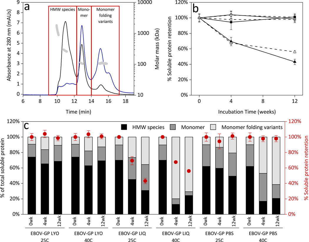
EBOV-GP vaccine formulations were tested with various biophysical characterization methods before and after incubation for up to 12 weeks at both 25°C and 40°C. EBOV-GP naturally forms a homotrimer on the surface of the viral membrane, and each monomer unit consists of a GP1 and GP2 subunit linked via a disulfide bridge22. Figure 1 shows the size-exclusion chromatograms for the lyophilized, liquid in ammonium acetate, and liquid in PBS formulations. The sample chromatograms in Figure 1a reflect the peaks identified as high molecular weight (HMW) species, monomer, and monomer folding variants. The molecular weights of the species eluting in each of the peaks were identified using SEC-MALS, with molecular weights shown as gray markers on the right axis in Figure 1a. It was determined that the peak eluting at 11 minutes was an EBOV-GP trimer (with an average molecular weight of 273.1 ± 9.5 kDa) and the peak eluting at 13 minutes was expanded monomer (107.6 ± 2.8 kDa). The peaks that eluted later than the monomer were determined to be folding variants of the EBOV-GP monomer because they had the same molecular weight (106.8 ± 1.7 kDa) as the monomer but had longer retention times in size exclusion chromatography due to their more compact size. The earlier-eluting monomer species had a hydrodynamic radius of 2.6 ± 0.3 nm compared to a hydrodynamic radius of 1.3 ± 0.1 nm for the monomeric folding variants. This is in good agreement with the characterization of an expanded conformations of intrinsically-disordered proteins being 1.5–2.0 times larger than corresponding globular proteins23. Peaks eluting earlier than 11 minutes were associated with species having molecular weights > 877 kDa, and were considered to be mixtures of oligomers larger than a trimer and were therefore classified as HMW species along with the trimer peak.
Figure 1.
(a) Representative size-exclusion chromatogram of unincubated EBOV-GP liquid in PBS formulation (black) and 4-week incubated liquid in PBS at 40°C (blue). Peak classifications were made using SEC-MALS molecular weight data (gray markers, right axis). High molecular weight (HMW) species were defined peaks that eluted between 9 and 12.3 minutes, monomer was defined as peaks eluting between 12.3 and 13.9 minutes, and monomer folding variants were defined as the peaks eluting between 13.9 and 17.3 minutes. (b) Area under the entire chromatogram was calculated and normalized to areas of each unincubated formulation to represent protein retention over time, where solid markers represent incubation at 25°C and open markers represent incubation at 40°C for lyophilized (circles), liquid in ammonium acetate with trehalose (triangles), and liquid in PBS (squares) formulations. Error bars are standard deviation from the average of three replicates. (c) Total soluble protein area was divided into HMW species, monomer, and monomer folding variants and is shown as the percent of the total area (bars, left y-axis) compared to soluble protein retention (red markers, right y-axis).
Total area under the chromatogram was calculated and normalized to that for the respective unincubated formulation, shown in Figure 1b. The amount of soluble protein in both the lyophilized and liquid in PBS formulations was unchanged after incubation for 12 weeks at both temperatures, whereas a loss of soluble protein due to aggregation was evident in the liquid in ammonium acetate formulation. The total area under the curve was broken down into HMW species, monomer, and monomer folding variants (Figure 1c). Lyophilized formulations maintained a high fraction of HMW species regardless of incubation temperature, but increasing fractions of monomer folding variants were seen in both liquid formulations, especially after incubation at 40°C.
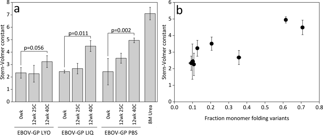
Changes in tertiary structure of EBOV-GP were observed via intrinsic fluorescence spectroscopy and quenching of tryptophan residues, of which EBOV-GP has 1322. As the protein unfolds, these hydrophobic tryptophan residues become more exposed to solvent and the Stern-Volmer constant (KSV) increases. Figure 2a shows the unfolded control had a KSV significantly higher than any of the vaccine formulations. All formulations had similar KSV values at the 0-week time point, indicating that formulation and lyophilization did not noticeably alter the tertiary structure. After incubation at 40°C for 12 weeks, samples of EBOV-GP in liquid formulations containing ammonium acetate and in PBS exhibited higher KSV values than those in unincubated samples. KSV values measured in the various formulations were positively correlated with the fraction of monomer folding variants shown in Figure 2b, with a coefficient of determination of 0.76.
Figure 2.
(a) Stern-Volmer constants for freshly prepared and incubated EBOV-GP vaccines for lyophilized (EBOV-GP LYO), liquid in ammonium acetate (EBOV-GP LIQ), and liquid in PBS (EBOV-GP PBS) compared to unfolded control in 8M urea. (b) Comparison of the Stern-Volmer constant with monomer folding variant area from size-exclusion chromatograms shows a positive correlation between degree of unfolding and the fraction of the total soluble protein present as monomeric folding variants. Error bars are standard deviation from the average of three replicates.
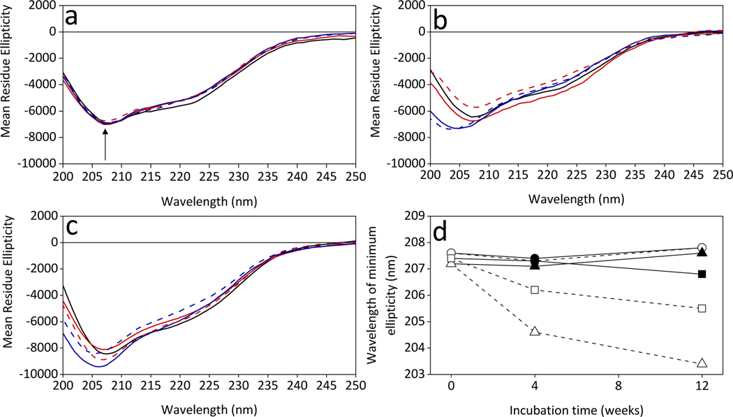
EBOV-GP secondary structure was monitored using far-UV CD on the lyophilized (Figure 3a), liquid in ammonium acetate with trehalose (Figure 3b), and liquid in PBS (Figure 3c) formulations. The spectra show a main minimum at 208 nm and a smaller minimum at 222 nm, which both correspond to alpha-helical structures24. The larger minimum at 208 nm was observed to shift to lower wavelengths over the incubation period (Figure 3d) for both liquid formulations; the shift was most apparent after incubation at 40°C. Comparing the wavelength of minimum ellipticity to relative amounts of monomer folding variants showed that lower wavelengths correlated to higher amounts of monomer folding variants (R2 = 0.82, data not shown).
Figure 3.
Far-UV circular dichroism spectra of EBOV-GP (a) lyophilized formulations, (b) liquid in ammonium acetate formulations, and (c) liquid in phosphate buffered saline formulations that were freshly prepared (black solid line), incubated for 4 weeks (red solid line for 25°C and blue solid line for 40°C), or incubated for 12 weeks (red dashed line for 25°C and blue dashed line for 40°C). Non-incubated spectral shapes consisted of an overall peak minimum around 208 nm, indicated by an arrow shown in (a). Changes in overall peak minimum were monitored over the incubation period, shown in (d) where solid markers represent incubation at 25°C and open markers represent incubation at 40°C for lyophilized (circles), liquid in ammonium acetate with trehalose (triangles), and liquid in PBS (squares) formulations.
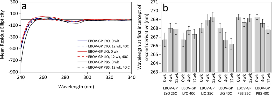
Tertiary structure was also probed using near-UV CD, which provides structural insight into the environment surrounding aromatic amino acids25. As seen in Figure 4a, changes in near-UV CD spectra were minor, even after incubation for 12 weeks at 40°C. In order to quantitatively determine any minor changes, second derivatives of the spectra (shown in Supplementary Figure 1) in the 240–270 nm range were computed, and the wavelength where the second derivative spectrum first intersected the x-axis (approximately 267 nm) was recorded. These wavelengths (Figure 4b) were compared in all the samples. Slight differences were observed after incubation in all three formulations.
Figure 4.
(a) EBOV-GP near-UV circular dichroism spectra for unincubated and incubated for 12 weeks at 40°C samples in lyophilized (EBOV-GP LYO), liquid in ammonium acetate with trehalose (EBOV-GP LIQ), and liquid in PBS (EBOV-GP PBS) formulations. (b) Changes in near-UV CD were assessed by monitoring the wavelength corresponding to the first intercept in the second derivative of each spectrum (referring to an inflection point in the CD spectrum) over the incubation period. Error bars represent the standard deviation from n = 8 replicates.
Figure 5 shows the SDS-PAGE of the EBOV-GP vaccines with Coomassie staining. Under non-reducing conditions, two bands were present in the 50–75 kDa range. Under reducing conditions, the apparent molecular weight of the protein in the two bands was each reduced by 20 kDa, while an additional band appeared at 20 kDa, likely assignable to the 20 kDa GP2 portion of the EBOV-GP after reducing the disulfide bond. For the EBOV-GP vaccines formulated in liquid solutions containing ammonium acetate and trehalose (Figure 5b), there are bands present at ~250 kDa under both non-reducing and reducing conditions in the 40°C samples, indicating aggregation that is unperturbed by DTT and SDS treatment. Liquid in PBS samples (Figure 5c and 5d) showed changes in band intensity over the incubation period. In general, the lyophilized samples (Figure 5a) were the most stable after incubation in terms of band pattern and intensity.
Figure 5.
Analysis of (a) lyophilized, (b) liquid in ammonium acetate with trehalose, and (c) liquid in PBS EBOV-GP formulations before and after incubation by SDS-PAGE using Coomassie staining. Lanes for other samples that were used for comparison purposes were excised from these pictures. The unincubated sample for liquid in PBS formulation was reanalyzed, shown in (d) because the initial analysis showed anomalous band patterns compared to the incubated samples.
3.2. Immunogenicity of lyophilized and liquid in PBS EBOV-GP vaccine formulations
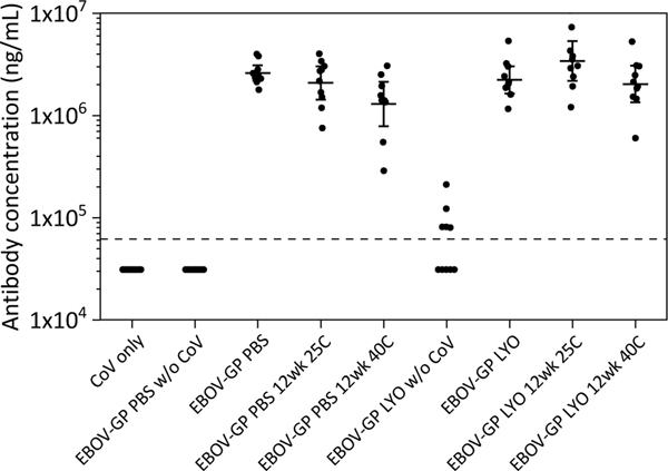
Antibody responses against EBOV-GP were determined by the MIA assay and are shown in Figure 6 for EBOV-GP formulated in PBS and in lyophilized formulations. Samples without adjuvant yielded negligible immune responses. All adjuvanted formulations elicited similar antibody levels, and incubation for 12 weeks at 25°C or 40°C did not affect the total antigen specific antibody response (including both neutralizing (data not shown) and non-neutralizing GP-reactive antibody response), with geometric mean titers (GMT) between 1.3 and 3.4 mg/mL. Statistical significance between each group is shown in Supplementary Table 1.
Figure 6.
Mouse immunogenicity of EBOV-GP PBS and EBOV-GP LYO formulations before and after incubation. Data are shown as geometric mean titers (GMT) +/− 95% confidence interval from serum collected post dose three at Day 56. Each dot represents the antibody concentration measured from serum of one individual mouse. Sera were diluted 1:8000 before being analyzed using a microsphere immunoassay. The dotted line represents the limit of quantification (LOQ); any values measured below this limit are shown here as half the LOQ.
3.3. Size exclusion chromatography and immunogenicity analysis of MARV-GP and SUDV-GP vaccines after accelerated stability studies
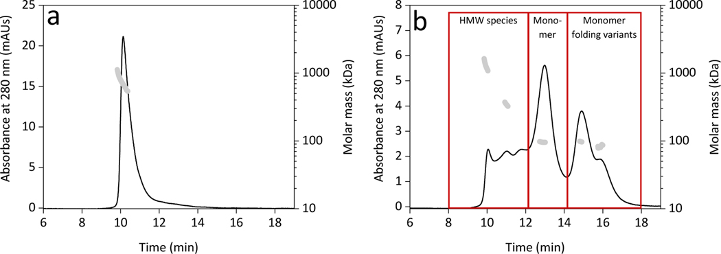
Because SE-HPLC proved to be the most sensitive stability-indicating assay of temperature-dependent degradation of EBOV-GP vaccines, similar studies were conducted to assess thermostability of MARV-GP and SUDV-GP vaccines under similar accelerated stability conditions. In Figure, representative chromatograms are shown for MARV-GP and SUDV-GP with peak molecular weights from SEC-MALS analysis overlaid and shown as gray markers. Unlike EBOV-GP, for MARV-GP (Figure 7a) only one species was present in the chromatogram, indicating MARV-GP assembly was not prone to dissociation. This species was identified as an oligomer that was around 760 kDa (Table 2). SUDV-GP (Figure 7b), however, was similar to EBOV-GP in that there were multiple species present, including monomeric folding variants that have the same molecular weight as the monomer but elute at different times. Peak molecular weights for both MARV-GP and SUDV-GP are shown in Table 2.
Figure 7.
Representative size-exclusion chromatograms of unincubated liquid in PBS formulations for (a) MARV-GP and (b) SUDV-GP. Peak classifications were made using SEC-MALS molecular weight data (gray markers, right axis). No distribution of oligomerization states was seen in MARV-GP samples, so only total peak area was used for area analyses. For SUDV-GP, high molecular weight (HMW) species were defined as any peak eluted between 8 and 12.2 minutes, monomer was defined as the peak between 12.2 and 14.2 minutes, and monomer folding variants were those peaks eluted between 14.2 and 18 minutes, shown in boxes in (b).
Table 2.
Molecular weight (MW) determinations from SEC-MALS peaks for unincubated MARV-GP and SUDV-GP, both stored as liquid in PBS. Classifications were assigned based on relative MW, with monomer size known to be around 100 kDa. MWs shown are the average ± standard deviation from n = 3 replicates.
| Antigen | Oligomers MW (kDa) | Trimer MW (kDa) | Monomer MW (kDa) | Folding variants MW (kDa) |
|---|---|---|---|---|
| MARV-GP PBS | 763.1 ± 45.7 | --- | --- | --- |
| SUDV-GP PBS | 1232.1 ± 131.4 | 346.0 ± 52.2 | 95.8 ± 7.3 | 93.8 ± 12.6 |
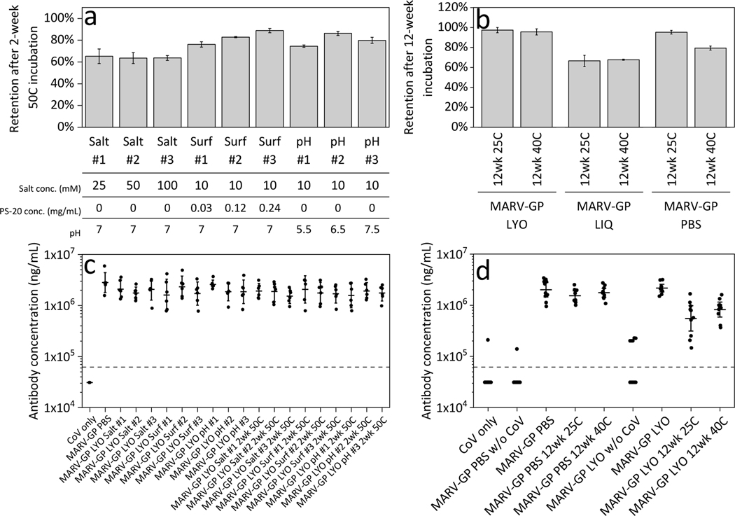
Over the incubation period, the MARV-GP chromatograms did not show evidence of redistribution of assembly states. However, due to aggregation during storage at elevated temperatures, the total amount of soluble protein decreased over time. Figure 8a and 8b show the relative amount of soluble protein remaining over time, defined as the total area in the chromatograms for the incubated samples normalized to the corresponding area for samples that were not incubated at elevated temperatures. Values of relative amounts of soluble protein less than 100% indicate that aggregates formed that were retained by the guard column. Figure 8a shows that increasing the amount of salt did not affect the level of soluble protein following 2-week incubation at 50°C. All three salt formulations had a similar 60% retention of soluble protein after 2 weeks. Non-ionic surfactants such as polysorbate-20 are often added to protein pharmaceuticals to inhibit aggregation26,27. An increase in retention of soluble protein after 2-week incubation at 50°C is seen with increasing surfactant concentration, suggesting that the surfactant inhibits aggregation. By varying pH, we observed that aggregation was lower at pH 6.5 than at pH 5.5 or 7.5. Figure 8c shows the results of the immunogenicity testing of lyophilized samples, both unincubated and incubated at 50°C for 2 weeks. Total antibody responses to all adjuvanted samples were statistically indistinguishable, with GMTs between 1.5 mg/mL and 2.8 mg/mL.
Figure 8.
MARV-GP accelerated stability studies results for size-exclusion chromatography and vaccine immunogenicity for the (a, c) 2-week 50°C incubation study and (b, d) 12-week incubation study. (a) Area under the entire chromatogram was calculated and normalized to areas of each unincubated formulation to represent retention of total protein after 50°C incubation. (b) Area under the entire chromatogram, normalized to unincubated areas, was calculated to signify protein retention after 12 weeks of incubation for the MARV-GP LYO pH #2 formulation. Liquid in ammonium acetate at the same conditions and liquid in PBS were also incubated and analyzed for comparison. Error bars are standard deviation from the average of three replicates in both (a) and (b). (c) Mouse immunogenicity of MARV-GP lyophilized formulations at 0 weeks incubation and 2 weeks at 50°C incubation shown as GMTs +/−95% confidence interval. (d) Mouse immunogenicity of MARV-GP LYO pH #2 formulation compared to MARV-GP liquid in PBS. Each dot represents the measured antibody concentration from serum of one individual mouse in both (c) and (d). Sera were diluted 1:8000 for analysis. The dotted line in (c) and (d) represents the LOQ, with values below this limit shown as half the LOQ.
The 12-week incubation study was completed on MARV-GP formulation group pH #2 (See Table 1 for conditions) and compared in the liquid and lyophilized form as well as to liquid in PBS. In Figure 8b, total protein retention was close to 100% in the lyophilized formulation incubated for 12 weeks at 25°C and 40°C. Liquid samples containing ammonium acetate lost 40% of total protein by the end of the incubation. Liquid formulations of the MARV-GP in PBS exhibited high soluble protein retention values after incubation at 25°C, but incubation at 40°C lowered soluble protein retention to 80%. Prior to incubation, liquid formulations of MARV-GP liquid in PBS and lyophilized MARV-GP formulations elicited similar antibody levels in mice (Figure 8d). For liquid formulations in PBS, incubation did not affect the immunogenicity, but lyophilized formulations showed a significantly lower antibody response after incubation for 12 weeks at both 25°C and 40°C (p < 0.0001 for both temperatures). All pairwise comparisons from ANOVA analysis are shown in Supplementary Table 2.
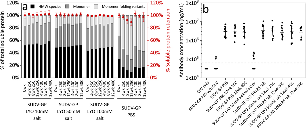
Similar to chromatograms from EBOV-GP studies, the size-exclusion chromatograms for SUDV-GP formulations showed multiple peaks that redistributed after incubation in liquid vaccine formulations. Although little aggregation was observed in the liquid formulations incubated at 40°C, and in all three lyophilized formulations at both incubation temperatures (Figure 9a), incubation of liquid vaccine formulations in PBS resulted in a large increase in the percentage of monomer folding variants. The unincubated liquid sample had 30% monomer folding variants, but by the end of the 12 weeks of incubation, the amounts of folding variants increased to 70% for the 25°C incubation and 60% for the 40°C incubation. Because there was a loss of total protein via aggregation observed in the PBS samples that were incubated at 25°C, the amount of folding variants on a weight basis was not statistically different (p = 0.11) between the two liquid in PBS samples after 12 weeks of incubation. Lyophilized formulations maintained an assembly state consisting of around 50% HMW species after incubation at both temperatures at all three salt levels.
Figure 9.
SUDV-GP accelerated stability study results for size-exclusion chromatography and vaccine immunogenicity. (a) Area under the entire chromatogram was calculated and normalized to areas of each unincubated formulation to represent protein retention over time, shown in red markers on the right y-axis. Error bars are standard deviation from the average of three replicates. Total soluble protein area was divided into HMW, monomer, and monomer folding variants and is shown as the percent of the total area as bars on the left y-axis. (b) Mouse immunogenicity of SUDV-GP lyophilized formulations with varying salt levels compared to liquid in PBS formulations shown as GMTs +/− 95% confidence interval. Each dot represents the antibody concentration from serum of one individual mouse. The dotted line represents the LOQ, with values below this limit shown as half the LOQ.
In spite of the large changes in assembly state that were observed when SUDV-GP liquid vaccines were incubated at 40°C for up to 12 weeks, all of the tested SUDV-GP vaccines that contained both antigen and CoVaccine HT™ produced statistically indistinguishable total antibody responses. Immunization with these formulations yielded antibody levels at least an order of magnitude above the limit of quantification in all mice, with GMTs consistently around 3.0 mg/mL. Thus, incubation at 40 °C for up to 12 weeks appeared to have no impact on immunogenicity of SUDV-GP in either liquid or lyophilized formulations. All pairwise analyses are shown in Supplementary Table 3.
4. Discussion
Lyophilized EBOV-GP formulations were more stable during storage than their liquid counterparts. Although this increased stability was reflected to a degree in each of the biophysical characterization techniques that we employed, it was most evident in the size-exclusion chromatography results. The soluble trimeric form of the protein, in addition to dissociating into monomers, can also oligomerize to form higher order assembly states. Additionally, SEC analysis revealed peaks that eluted later than the expanded monomer that had the same molecular weight, indicating that a variety of monomeric folding states were present. Over time, the relative amounts of these more compact monomeric variants increased when liquid EBOV-GP formulations were stored. Storing the EBOV-GP as a lyophilized product not only reduced refolding but also inhibited dissociation of HMW species so that a high percentage of HMW species was maintained. These higher molecular weight species, which consist of trimer and larger oligomers, appear to generate more potent virus-specific responses (data not shown) because they preserve the structural integrity and represent the native oligomerization state of the glycoproteins. Dissociated monomers and monomer folding variants allow for additional epitopes to be exposed (or necessary epitopes to be buried) that could lead to the rise of more non-specific or non-neutralizing antibodies. Size-exclusion chromatography revealed that EBOV-GP has a complicated assembly structure that is prone to redistribution of species. EBOV-GP liquid in PBS formulations had increased amounts of monomer and monomeric folding variants after 12 weeks of incubation, which allows for the potential of those formulations to elicit antibodies to non-exposed regions of the native trimeric EBOV-GP. In addition to detailing the distribution of different protein quaternary structures in a vaccine sample, SE-HPLC also allows the determination of the total amount of soluble protein. Large particles and soluble aggregates are retained by the guard column prior to injection on the analytical column. Because the same amount of protein was loaded each time (0.01 mg), aggregation can be quantified using a mass balance referencing the total amount of soluble protein in unincubated control samples. Due to this ability to accurately report on total amount of soluble protein as well as the distribution of protein species, SE-HPLC is a powerful tool that was determined to be the most sensitive assay to indicate stability in the three filovirus GP subunit vaccines.
Changes to the tertiary structure of EBOV-GP were observed via intrinsic fluorescence quenching and near-UV CD. After incubation, the EBOV-GP vaccines were still sufficiently folded to sequester tryptophan residues. Because the monomeric folding variants exhibit smaller hydrodynamic radii than that of the expanded monomer, it might have been expected that tryptophan residues in the more compact conformation would be less solvent exposed. Interestingly, it was observed that higher Stern-Volmer constants correlated positively with increased fractions of the folding variants species. These tertiary structure changes were also verified with near-UV CD. However, unlike SE-HPLC, where peaks can easily be identified, the ability of near-UV CD to give significant structural insights that correspond to certain parts of the spectrum is limited25 so conclusions have to be made from changes to reference material, in this case unincubated samples. In order to quantify these differences, second derivative spectra were calculated and it was found that tracking the wavelength of the first intercept of the derivative spectra was the best indicator of these changes. After incubation for 12 weeks at 40°C, there was not a statistically significant difference in the wavelength of the first intercept from second derivative near-UV CD spectra for the lyophilized formulation, but there were statistically significant differences in the wavelength for both liquid formulations (p < 0.0001 for both).
Far-UV CD was used to see differences in secondary structure between EBOV-GP formulations. The shift to lower wavelengths of spectral minima seen in both liquid EBOV-GP formulations that had increased amounts of monomer folding variants suggests that the changes in tertiary structure also affected the secondary structure. There were also differences seen in the intensity of the mean residue ellipticity, which should be the same when corrected for concentration. Greenfield24 states that these differences are often due to the protein not being in a monomeric state or due to the protein aggregating. Therefore, because EBOV-GP has a complicated assembly structure that is prone to both aggregating and forming oligomers, far-UV CD is not as sensitive when there are dramatic differences in tertiary and quaternary structure.
Due to inclusion of SDS in sample buffer for analysis of EBOV-GP using gel electrophoresis, oligomers are broken down into monomers. Therefore, higher order structural information cannot be gathered. We observed a decrease in band intensity after incubation in liquid EBOV-GP vaccines, which could indicate loss of soluble protein by aggregation. SDS-PAGE proved insensitive to changes in quaternary structure, which was determined to be one of the main mechanisms of degradation.
We hoped that our stability-indicating assay would help us to predict the conformational long-term stability of EBOV-GP in a vaccine formulation. Even though there was a similar immune response to all EBOV-GP vaccines tested, we did see evidence of degradation (via multimer redistribution and monomer refolding) from the stability-indicating assays that did not affect total immunogenicity in liquid in PBS EBOV-GP samples. It is important to note that immunogenicity data such as antigen-binding IgG concentration as presented herein may not correlate with vaccine efficacy (protection against disease), particularly in the case of filoviruses as correlates of protection have not yet been established. Moreover, the total antigen response may include both neutralizing and non-neutralizing antibody responses. However, it is possible that the immunizing dose used in this study was sufficiently high so that even though the EBOV-GP liquid in PBS vaccine that was incubated for 12 weeks at 40°C consisted 60% of monomer folding variants, there was still a sufficient amount of HMW species remaining to yield a maximum humoral response. The fact that incubated EBOV-GP subunit vaccines were as immunogenic as fresh, refrigerated material is significant. The marketed vaccine for Zaire ebolavirus (ERVEBO®) cannot experience temperatures higher than 25°C11, while the vaccines tested here could withstand temperatures up to 40°C.
For the MARV-GP and SUDV-GP thermostability studies, it was similarly concluded that SE-HPLC was the best stability-indicating assay. Therefore, only the SE-HPLC results were shown here. MARV-GP had a robust assembly state that did not dissociate upon thermal stress, but significant levels of aggregation were inferred from the decreased soluble protein retention observed in incubated liquid in PBS samples. Despite this aggregation, immunogenicity results suggested that this physical degradation did not have a significant impact on the antibody response in mice, with the same caveats as listed above for EBOV-GP. SUDV-GP study results showed that like EBOV-GP, the assembly state was highly labile, leading to significant redistribution of species after high temperature incubation. Similarly, the immunogenicity of the SUDV-GP antigen was unaffected by lyophilization and incubation.
Although lyophilized EBOV-GP and SUDV-GP retained their immunogenicity after 12 weeks of incubation at 25 or 40 °C, lyophilized MARV-GP vaccines generated antibody responses that were lower than those for the unincubated material. These results may be anomalous; immunogenicity losses were not observed in the lyophilized MARV-GP samples incubated at 50 °C for two weeks, and samples incubated for 12 weeks at 25 °C gave somewhat lower responses than those incubated at 40 °C.
Because immunogenicity data were converted into antibody concentrations, we can directly compare the antibody responses across different studies. The SUDV-GP vaccine groups containing adjuvant showed higher antibody levels, with all samples producing antibody concentrations above 2.6 mg/mL. In contrast, the MARV-GP 12-week study adjuvanted vaccine samples were less immunogenic, producing antibody concentrations below 2.2 mg/mL. The adjuvanted EBOV-GP vaccine groups elicited intermediate antibody concentrations between 1.3 and 3.4 mg/mL.
Antibody responses between unincubated liquid in PBS and lyophilized formulations with CoVaccine HT showed no significant differences. However, in all three 12-week accelerated stability studies, unadjuvanted liquid in PBS groups had lower GMTs than the corresponding unadjuvanted lyophilized groups. In addition, groups given lyophilized vaccines without CoVaccine had more individual mice with total antibody responses above the limit of quantification compared to liquid in PBS vaccines. This indicates that even without the addition of an adjuvant, the lyophilized glycoprotein samples were more immunogenic.
5. Conclusions
In the search for stability-indicating assays that can help predict long-term vaccine stability, we concluded that size-exclusion chromatography was the best method for all three filovirus glycoproteins tested. The mechanism of degradation of the glycoproteins appeared to be mainly through aggregation and changes in quaternary structure. Size-exclusion chromatography can distinguish both losses of protein via aggregation and changes in quaternary structure by analyzing distributions of peak areas and evolution of total peak area over time. Even though immunogenicity testing showed nearly equivalent total antibody responses regardless of formulation or incubation, physical degradation was observed in the stability-indicating assays. Because Ebola and Marburg viruses are endemic in Africa, it is important to have vaccines that can withstand high temperature storage that align with maximum ambient temperatures in the region. It is imperative to develop assays that are sensitive enough to detect whether heat-induced degradation has occurred. These assays enable the continued evaluation of stability after high temperature storage to determine thermostability of vaccine candidates. Our findings suggest that size-exclusion chromatography may be the best method to test new filovirus vaccine formulations and determine if degradation has occurred after long-term storage.
We do not expect our current formulation to provide cross-protection between the three filoviruses. Therefore, we hope to build on our work here by developing a trivalent vaccine that can induce a high level of protective antibodies against all three viruses. By studying glycoproteins from each virus individually, we have identified pathways of degradation and corresponding stability-indicating assays that may further be used in determining stability of trivalent candidate vaccines.
Supplementary Material
6. Acknowledgements
Funding for this project was provided by the National Institute for Allergy and Infection Diseases under grants R01AI119185 and R01AI132323 (PI: Dr. Axel Lehrer) and sponsored by Soligenix, Inc. We would like to thank our collaborators at Hawaii Biotech, Inc. for generating the Drosophila expression cell line used to produce the SUDV-GP supporting this study. We further want to thank Protherics Medicines Development (London, UK) for the gift of CoVaccine HT™ for the conduct of this study. We also thank the Biochemistry Shared Instruments Facility at the University of Colorado Boulder for the use of their SEC-MALS, Photon Technology International Fluorimeter, and Applied Photophysics ChirascanPlus CD instruments.
References
- 1.Rouquet P, Froment JM, Bermejo M, et al. Wild animal mortality monitoring and human ebola outbreaks, Gabon and Republic of Congo, 2001–2003. Emerg Infect Dis. 2005. doi: 10.3201/eid1102.040533 [DOI] [PMC free article] [PubMed] [Google Scholar]
- 2.CDC. Years of Ebola Virus Disease Outbreaks. https://www.cdc.gov/vhf/ebola/history/chronology.html. Published 2019. Accessed November 7, 2019.
- 3.U.S. Food and Drug Administration. First FDA-approved vaccine for the prevention of Ebola virus disease, marking a critical milestone in public health preparedness and response. https://www.fda.gov/news-events/press-announcements/first-fda-approved-vaccine-prevention-ebola-virus-disease-marking-critical-milestone-public-health. Accessed January 16, 2020.
- 4.CDC. Prevention | Ebola (Ebola Virus Disease) | CDC. https://www.cdc.gov/vhf/ebola/prevention/index.html. Accessed August 15, 2019. [Google Scholar]
- 5.Chen D, Kristensen D. Opportunities and challenges of developing thermostable vaccines Opportunities and challenges of developing thermostable vaccines. 2014;(January):547–557. doi: 10.1586/erv.09.20 [DOI] [PubMed] [Google Scholar]
- 6.Brandau DT, Jones LS, Wiethoff CM, Rexroad J, Middaugh CR. Thermal stability of vaccines. J Pharm Sci. 2003;92(2):218–231. doi: 10.1002/jps.10296 [DOI] [PubMed] [Google Scholar]
- 7.PATH. Summary of Stability Data for Licensed Vaccines. Seattle, WA; 2012. https://path.azureedge.net/media/documents/TS_vaccine_stability_table.pdf. [Google Scholar]
- 8.American Academy of Pediatrics. Vaccine Storage Chart. https://www.aap.org/en-us/advocacy-and-policy/aap-health-initiatives/immunizations/Practice-Management/Pages/vaccine-storage-chart.aspx. Accessed August 5, 2019.
- 9.Kumru OS, Joshi SB, Smith DE, Middaugh CR, Prusik T, Volkin DB. Vaccine instability in the cold chain: Mechanisms, analysis and formulation strategies. Biologicals. 2014;42(5):237–259. doi: 10.1016/j.biologicals.2014.05.007 [DOI] [PubMed] [Google Scholar]
- 10.World Heath Organization. At 1-year mark, we mourn the lives lost to Ebola, and call for solidarity. https://www.who.int/news-room/detail/31-07-2019-at-1-year-mark-we-mourn-the-lives-lost-and-call-for-solidarity. Published 2019. Accessed August 5, 2019.
- 11.Arnemo M, Viksmoen Watle SS, Schoultz KM, et al. Stability of a Vesicular Stomatitis Virus-Vectored Ebola Vaccine. J Infect Dis. 2016;213(6):930–933. doi: 10.1093/infdis/jiv532 [DOI] [PubMed] [Google Scholar]
- 12.Wang B, Tchessalov S, Cicerone MT, Warne NW, Pikal MJ. Impact of sucrose level on storage stability of proteins in freeze-dried solids: II. Correlation of aggregation rate with protein structure and molecular mobility. J Pharm Sci. 2009;98(9):3145–3166. doi: 10.1002/jps.21622 [DOI] [PubMed] [Google Scholar]
- 13.Chisholm CF, Kang TJ, Dong M, et al. Thermostable Ebola virus vaccine formulations lyophilized in the presence of aluminum hydroxide. Eur J Pharm Biopharm. 2019;136(2019):213–220. doi: 10.1016/j.ejpb.2019.01.019 [DOI] [PMC free article] [PubMed] [Google Scholar]
- 14.Nail SL, Akers MJ. Development and Manufacture of Protein Pharmaceuticals.; 2002. [Google Scholar]
- 15.Konermann L. Addressing a Common Misconception: Ammonium Acetate as Neutral pH “Buffer” for Native Electrospray Mass Spectrometry. J Am Soc Mass Spectrom. 2017;28(9):1827–1835. doi: 10.1007/s13361-017-1739-3 [DOI] [PubMed] [Google Scholar]
- 16.Lehrer AT, Wong TAS, Lieberman MM, et al. Recombinant proteins of Zaire ebolavirus induce potent humoral and cellular immune responses and protect against live virus infection in mice. Vaccine. 2018;36(22):3090–3100. doi: 10.1016/j.vaccine.2017.01.068 [DOI] [PMC free article] [PubMed] [Google Scholar]
- 17.Lakowicz Joseph R.. Principles of Fluorescence Spectroscopy. Vol 390. Third. Springer US; 2006. doi: 10.1007/978-0-387-46312-4 [DOI] [Google Scholar]
- 18.Studier FW. Protein production by auto-induction in high density shaking cultures. Protein Expr Purif. 2005;41(1):207–234. doi: 10.1016/j.pep.2005.01.016 [DOI] [PubMed] [Google Scholar]
- 19.Hilgers LAT, Blom AG. Sucrose fatty acid sulphate esters as novel vaccine adjuvant. Vaccine. 2006;24(SUPPL. 2):S81–S82. doi: 10.1016/j.vaccine.2005.01.133 [DOI] [PubMed] [Google Scholar]
- 20.To A, Medina LO, Mfuh KO, et al. Recombinant Zika Virus Subunits Are Immunogenic and Efficacious in Mice. mSphere. 2018;3(1):1–16. doi: 10.1128/msphere.00576-17 [DOI] [PMC free article] [PubMed] [Google Scholar]
- 21.Haun BK, Kamara V, Dweh AS, et al. Serological evidence of Ebola virus exposure in dogs from affected communities in Liberia: A preliminary report. PLoS Negl Trop Dis. 2019;13(7):1–11. doi: 10.1371/journal.pntd.0007614 [DOI] [PMC free article] [PubMed] [Google Scholar]
- 22.Lee JE, Fusco ML, Hessell AJ, Oswald WB, Burton DR, Saphire EO. Structure of the Ebola virus glycoprotein bound to an antibody from a human survivor. Nature. 2008;454(7201):177–182. doi: 10.1038/nature07082 [DOI] [PMC free article] [PubMed] [Google Scholar]
- 23.Tompa P.Structure and Function of Intrinsically Disordered Proteins. CRC Press; 2010. [Google Scholar]
- 24.Greenfield NJ. Using circular dichroism spectra to estimate protein secondary structure. Nat Protoc. 2006;1(6):2876–2890. doi: 10.1038/nprot.2006.202. [DOI] [PMC free article] [PubMed] [Google Scholar]
- 25.Kelly SM, Jess TJ, Price NC. How to study proteins by circular dichroism. Biochim Biophys Acta - Proteins Proteomics. 2005;1751(2):119–139. doi: 10.1016/j.bbapap.2005.06.005 [DOI] [PubMed] [Google Scholar]
- 26.Chi EY, Krishnan S, Randolph TW, Carpenter JF. Physical stability of proteins in aqueous solution: Mechanism and driving forces in nonnative protein aggregation. Pharm Res. 2003;20(9):1325–1336. doi: 10.1023/A:1025771421906 [DOI] [PubMed] [Google Scholar]
- 27.Randolph TW, Jones LS. Surfactant-Protein Interactions. In: Carpenter JF, Manning MC, eds. Rational Design of Stable Protein Formulations: Theory and Practice. New York: Kluwer Academic/Plenum Publishers; 2002:159–175. [Google Scholar]
Associated Data
This section collects any data citations, data availability statements, or supplementary materials included in this article.


