Correction to: Scientific Reports 10.1038/srep44847, published online 24 March 2017
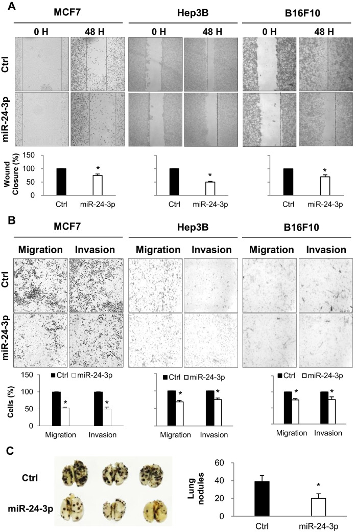
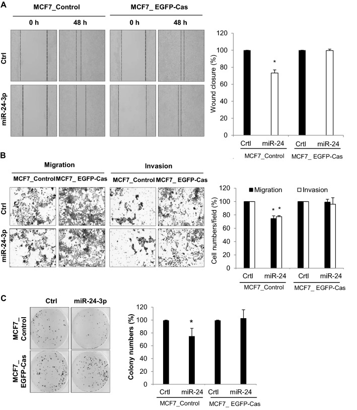
This article contains errors in MCF7 cell images of Figures 2 and 5. The correct Figures 2 and 5 appear below as Figures 1 and 2.
Figure 1.
(A) Analysis of wound closure after miRNA transfection. MCF7, Hep3B, and B16F10 cells were transfected with either miR-24-3p mimic or control miRNAs and cultured until they reached confluency. After wounds were created, the cell migration distance was analyzed 48 h later. (B) Migration and invasion assay. After the transfection of miRNAs, cells were cultured in transwell with or without matrigel, and migrated cells were stained and analyzed by counting cells from three different fields. (C) After transfection of miR-24-3p and control miRNA, B16F10 cells (3 × 10⁵ cells/mouse) were injected into the tail vein of C57BL6 mice (n = 5). 17 days later, mice were sacrificed and the lungs were isolated. The number of B16F10 colonies present on the surface of each set of lungs was determined by visual inspection. Data are representative from three independent experiments. *p < 0.05.
Figure 2.
(A,B) MCF7_Control and MCF7_EGFP-Cas cells were transfected with either miR-24-3p mimic or control miRNAs and cell migration/invasion was analyzed. After the cells reached confluency, wound closure was determined by measuring cell migration distance (A). Cells were cultured in transwell with or without matrigel, and migrated cells were stained and analyzed by counting cells from three different fields (B). (C) 1 × 10³ of miRNA-transfected cells were cultured for 3 weeks. The colonies were stained with crystal violet and counted in 3 randomly selected visual fields. The images were representative of three independent experiments and graphs indicate the mean ± SEM of three independent experiments. *p < 0.05.
Contributor Information
Wook Kim, Email: wookkim21@ajou.ac.kr.
Eun Kyung Lee, Email: leeek@catholic.ac.kr.


