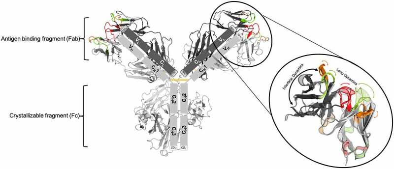
Figure 1.

Structure of an IgG1 antibody and schematic illustration of the unique modular anatomy. the arms of the Y-shaped structure allow the antibody to carry out two functions, on the one hand antigen-binding and on the other hand biological activity mediation. the arms of the antibody are known as antigen-binding fragments (Fabs). The Fab is composed of a constant and a variable domain of each of the heavy and the light chain. the variable domains shape the antigen binding site (paratope) at the amino-terminal end of the antibody. the variable fragment (Fv) is highlighted in the picture. the CDR 1 loops are colored in green, the CDR 2 loops are depicted in orange and the CDR 3 loops are shown in red. The close up to the Fv also indicates the high flexibility of the CDR loops and the relative VH-VL interface and shows that the antibody binding site exists as ensembles of paratope states. the tail region of the antibody, also known as Fc region, is responsible for the communication with the immune system and interacts with the cell surface receptors, called Fc receptors
