Abstract
Microbes colonize the gastrointestinal tract are considered as highest complex ecosystem because of having diverse bacterial species and 150 times more genes as compared to the human genome. Imbalance or dysbiosis in gut bacteria can cause dysregulation in gut homeostasis that subsequently activates the immune system, which leads to the development of inflammatory bowel disease (IBD). Neuromediators, including both neurotransmitters and neuropeptides, may contribute to the development of aberrant immune response. They are emerging as a regulator of inflammatory processes and play a key role in various autoimmune and inflammatory diseases. Neuromediators may influence immune cell’s function via the receptors present on these cells. The cytokines secreted by the immune cells, in turn, regulate the neuronal functions by binding with their receptors present on sensory neurons. This bidirectional communication of the enteric nervous system and the enteric immune system is involved in regulating the magnitude of inflammatory pathways. Alterations in gut bacteria influence the level of neuromediators in the colon, which may affect the gastrointestinal inflammation in a disease condition. Changed neuromediators concentration via dysbiosis in gut microbiota is one of the novel approaches to understand the pathogenesis of IBD. In this article, we reviewed the existing knowledge on the role of neuromediators governing the pathogenesis of IBD, focusing on the reciprocal relationship among the gut microbiota, neuromediators, and host immunity. Understanding the neuromediators and host-microbiota interactions would give a better insight in to the disease pathophysiology and help in developing the new therapeutic approaches for the disease.
Keywords: Inflammation, Gut microbiota, Inflammatory bowel disease, Enteric nervous system, Neuroimmunomodulation, Neuromediator
Core Tip: Dysbiosis in gut bacteria is a well-established factor, and the abnormality in the enteric nervous system is an emerging aspect that influences the gut inflammation. Both of them contribute to inflammatory bowel disease (IBD) pathogenesis by modulating the host immune response. Through this review, we linked the two pathological mechanisms and explained how neuroimmunomodulation by gut bacteria play a crucial role in IBD. We elaborated all the known neuromediators produced by gut bacteria and the role of each neuromediator as well as the respective gut bacteria in inflammatory signaling pathways especially in IBD.
INTRODUCTION
The gastrointestinal tract (GIT) is equipped with the most extensive immune system, and the largest network of neurons outside the central nervous system (CNS) called the enteric nervous system (ENS). Sometimes, ENS also referred to as “brain in gut” because it does not require any intermediate input from the brain for its functioning. The structure of ENS is organised into two Plexi, myenteric plexus and submucosal (Meissner’s) plexus. Myenteric plexus is located between the longitudinal and circular muscle of muscularis propria and regulates the intestinal motility. Submucosal plexus is located in the submucosa of the intestine and regulates secretion, absorption, and blood flow[1]. Neurons of these Plexi releases various neurotransmitters that regulate the secretory and motor functions of GIT. During inflammatory bowel disease (IBD), there are morphological, histological, and immunohistochemical abnormalities in the ENS which causes neuronal hyperplasia, necrosis, ganglion, and axonal degeneration, alteration in synthesis and release of neurotransmitters. It leads to a defect in the secretory and motor functions of GIT[2].
The neurotransmitters and neuropeptides released from ENS can alter various immune cell functions. Immune cells residing in colon express various receptors for neurotransmitters, and once neurotransmitter binds to these receptors, there would be an initiation of signal transduction pathways of cytokine production[3]. These cytokines, in turn, bind to their specific receptors, expressed on sensory nerve fibers to trigger neuronal response, thus establishing a bidirectional communication. This bidirectional cross-talk between ENS and the enteric immune system is crucial to maintain visceral homeostasis. This cross-talk regulates the magnitude of inflammatory response via the production of cytokines, disruption of epithelial tight junctions, neutrophil recruitment, phagocytosis, modification in lymphocyte differentiation, and ultimately cell death ensues[4].
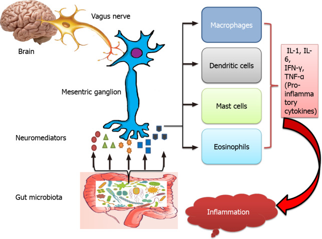
During the early postnatal life, ENS undergoes extensive development in parallel to the colonisation of gut microbiota and maturation of mucosal immune system in GIT. In germ-free mice, structural and functional abnormalities of the ENS have been observed, which suggests the role of gut microbiota in ENS development. Microbiota interacts with the nervous system through modulation of neurotransmitters production. Indeed, bacteria have been found to have the capability to produce a range of significant neurotransmitters in the gut. Therefore, gut microbiota fine-tunes the interaction between enteric nervous and immune system by altering the level of neuromediators (Figure 1).
Figure 1.
Modulation of cross-talk between the enteric nervous system and the enteric immune system via gut bacteria. Gut microbiota and vagus nerve stimulate mesenteric ganglion (enteric neuron) to produce neuromediators. Neuromediators act on various immune cells and influence their ability to release pro-inflammatory cytokines. During inflammatory bowel disease, dysbiosis in gut microbiota and abnormality in the enteric nervous system affect the level of neuromediators that results in overproduction of pro-inflammatory cytokines and promote inflammation. IL: Interleukin; TNF-α: Tumour necrosis factor-α; IFN-γ: Interferon-γ.
A more thorough understanding of the interactions among neuromediators, inflammation, and neuromediators producing gut microbiota is required to ensure the effectiveness of neuromediators as a treatment option for IBD. Herein, we review the current knowledge of the role of neuromediators and bacteria that produce neuromediators which might be a potential option in the treatment of IBD.
NEUROMEDIATORS AND IBD
A variety of neuropeptides and neurotransmitters are known to involve in the pathogenesis of IBD. Neuropeptides such as substance P (SP), neurotensin (NT), vasoactive intestinal peptide (VIP), neuropeptide Y (NPY), corticotrophin-releasing hormone (CRH), galanin (GAL) and calcitonin gene-related peptides (CGRP) and neurotransmitters like serotonin, nitric oxide (NO), acetylcholine, noradrenaline (NA) and γ-aminobutyric acid (GABA) regulates inflammatory processes by employing immunomodulatory pathways. Role of each of these neuromediators are briefly summarized in Table 1.
Table 1.
List of neuromediators and their role in gut inflammation
|
Neuromediator
|
Distribution
|
Binding receptor
|
Function
|
| SP | Neurons and inflammatory cells like lymphocytes, macrophages, and dendritic cells | NK-1R | Exerts pro-inflammatory effects in epithelial and immune cells and contributes to inflammatory diseases. In murine model of colitis, it plays regulatory role |
| NT | Nervous system and intestine | NTR1 | Recognized as an immunomodulator. By interacting with immune cells, it enhances the chemotaxis and induces the cytokine release to modulate the immune response. In IBD, it exerts its pro-inflammatory effects by promoting the expression of miR-210 in intestinal epithelial cells |
| NPY | Central and peripheral nervous system and immune cells | Out of five receptors of NPY, NPYY1 is known to play a crucial role in immunomodulation | Regulates various immune cell functions such as T helper cell differentiation, neutrophil chemotaxis, natural killer cell activity, and granulocyte oxidative burst and NO production. In the gut, NPY is known to exert pro-inflammatory effects |
| VIP | Neuronal and lymphoid cells | VIPR1 and VIPR2 | Identified as an anti-inflammatory molecule. administration of VIP nanomedicine in the form of VIP-SSM are capable of alleviating the symptoms of DSS- induced mice model of colitis |
| GAL | Vasculature, immune cells and colonic epithelial cells | GAL (1-3) receptor | Exerts anti-inflammatory effects in TNBS induced colitis model by reducing the expression and activity of iNOS |
| CRH | Immune cells | CRH-R1 and CRH-R2 | It acts as a pro-inflammatory peptide. The expression pattern of CRH 1 and CRH 2 varies in ulcerative colitis. Inhibition of CRH1 and overexpression of CRH2 may have the therapeutic potential in IBD |
| CGRP | Sensory nerves projecting to the lymphoid organs, airways, and pulmonary neuroendocrine cells | CGRP receptors | CGRP negatively regulates innate immune responses and thus has potential anti-inflammatory effects. Its expression reduced in the colon of an animal model of colitis |
| NA | Nerves innervating the peripheral lymphoid organs | Adrenergic α and β receptors | immunomodulatory effect of NA is administered via cAMP. Activation of NA receptors that stimulate cAMP resulting in a shift toward Th2 responses which are anti-inflammatory and neuroprotective whereas decreased cAMP stimulates Th1 responses resulting in cell destruction and inflammation |
| Acetylcholine | Central and peripheral nervous system, immune cells, keratinocytes, endothelial cells, urothelial cells of the urinary bladder, airways and epithelial cells of the placenta | Nicotinic and muscarinic receptors | Muscarinic receptors mediate pro-inflammatory responses and nicotinic receptors enhance anti-inflammatory responses. Treatment of UC via nicotine suggests the role of the cholinergic pathway in colonic inflammation |
| NO | Neuron synapses and immune cells | NO does not act via receptors. its specificity for target cell depends on its concentration, its activity and response, and territory of target cells | NO is oxidised to reactive nitrogen oxide species which mediate most of the immunological effects. It regulates the growth, functional activity, and death of immune cells. It acts as a biomarker for monitoring disease activity due to its increased serum concentration during the active phase of both UC and CD and reduced concentration during the inactive phase of the disease |
| Serotonin or 5-HT | Central nervous system and EC cells of GIT | 5-HT receptor | It promotes activation of lymphocytes and secretion of pro-inflammatory cytokines. It activates the signalling molecules of the NF-kB pathway during gut inflammation |
| GABA | Nervous system and immune system | GABA- AR and GABA-BR | GABA has several effects on immune cells, including modulation of cytokine secretion, regulation of cell proliferation, and migration. Activation of GABA-A receptor aggravates DSS induced mice model of colitis |
SP: Substance P; GABA: γ-aminobutyric acid; 5-HT: 5-hydroxytryptamine; EC: Enterochromaffin; NF-kB: Nuclear factor kB; NO: Nitric oxide; NA: Noradrenaline; CD: Crohn’s disease; UC: Ulcerative colitis; cAMP: Cyclic adenosine monophosphate; CGRP: Calcitonin gene-related peptide; IBD: Inflammatory bowel disease; CRH: Corticotropin-releasing hormone; iNOS: Inducible nitric oxide synthase; GAL: Galanin; TNBS: 2,4,6-trinitrobenzenesulfonic acid; VIP: Vasoactive intestinal peptide; SSM: Sterically stabilised micelles; NPY: Neuropeptide Y; NK-1R: Neurokinin-1 receptor; NTR1: Neurotensi receptor 1; DSS: Dextran sodium sulfate.
SP
SP is released from neurons and also from inflammatory cells like lymphocytes, macrophages, and dendritic cells. It acts by binding to the neurokinin-1 receptor (NK-1R). It plays a vital role in the amplification of inflammatory response by inducing the release of cytokines, reactive oxygen species, and stimulates leukocyte recruitment. Increased level of SP has been observed in the colon of IBD patients and, in the synovial fluid and serum of rheumatoid arthritis (RA) patients. Also, the enhanced expression of NK-1R was reported in the colon of IBD and synoviocytes of RA patients. SP has pro-inflammatory effects in epithelial and immune cells and contributes to many inflammatory diseases, including sarcoidosis, asthma, chronic bronchitis, RA, and IBD[5]. However, in murine models of colitis, SP plays a regulatory action[6]. In a recent study, SP was observed as an accelerator for healing the dextran sodium sulfate (DSS)-induced damaged intestine via inhibiting inflammatory responses through the modulation of cytokine expression[7].
NT
NT is a tridecapeptide, a pro-inflammatory neuropeptide widely distributed in the nervous system and intestine. It binds to NT receptor 1 (NTR1) which is a high-affinity receptor and expressed in neurons, immune cells, colonic epithelial cells and colon cancer cell lines. It regulates various peripheral processes including gut motility, intestinal epithelial cell proliferation, secretion, and vascular smooth muscle activity, but recently it is recognized as an immunomodulator. NT interacts with leukocytes, dendritic cells and peritoneal mast cells, inducing the release of cytokines and enhancing chemotaxis in order to modulate the immune response. The elevated level of NT and increased expression of NTR1 have been reported in the colonic mucosa of the experimental model of colitis and ulcerative colitis (UC) patients. NT is implicated in various acute and chronic inflammatory diseases, including lung and intestinal inflammation[8-11]. In IBD, NT exerts its pro-inflammatory effects by promoting the expression of miR-210 in intestinal epithelial cells[12].
NPY
NPY is a peptide of 36 amino acids and produced abundantly by the central and peripheral nervous system and also by immune cells. Neuronal functions of NPY include modulation of blood pressure, nociception, anxiety, and appetite. It also has diverse effects on innate and adaptive immunity, including immune cell migration, cytokine release from macrophages and T helper cells, and antibody production. Out of five receptors of NPY, NPYY1 is known to play a crucial role in immunomodulation. To modulate inflammation, NPY regulates various immune cell functions such as T helper cell differentiation, neutrophil chemotaxis, natural killer cell activity, and granulocyte oxidative burst and NO production. In the gut, NPY is known to exert pro-inflammatory effects. Several clinical studies reported the role of NPY in immune or inflammatory disorders such as arthritis, asthma, and IBD[4,13-15].
VIP
VIP is a 28 amino acid neuropeptide, produced by neuronal cells and lymphoid cells. It controls the homeostasis of the immune system by carrying out a wide range of immunological functions. Recently, it has been identified as an anti-inflammatory molecule. It is reported to inhibit pro-inflammatory cytokines and chemokines production from macrophages, dendritic cells, and microglial cells. Furthermore, VIP reduces the expression of costimulatory molecules on antigen-presenting cells, resulting in the promotion of Th2 type responses and reduction in Th1 type responses. VIP has been considered as a promising target for the treatment of autoimmune as well as acute and chronic inflammatory diseases such as multiple sclerosis, RA, Crohn’s disease (CD), septic shock, or autoimmune diabetes[4,16-18]. Recombinant VIP analogue protects the intestinal mucosal barrier function effectively in rats. This analogue of VIP ameliorates 2,4,6-trinitrobenzenesulfonic acid (TNBS)-induced colonic injury and inflammation through downregulating the expression of tumour necrosis factor-α and upregulating the interleukin (IL)-10 expression[15,19]. Though the administration of VIP shown anti-inflammatory effect but its therapeutic use is restricted due to its rapid degradation and continuous infusion. Recently, the administration of VIP nanomedicine in the form of sterically stabilized micelles has been observed to overcome the barriers and are capable of alleviating the symptoms of DSS-induced mice model of colitis[20].
GAL
GAL is a 30 amino acid long sensory neuropeptide known to attenuate neurogenic inflammation. Among the receptors (GAL1-3), GAL-3 is most abundantly expressed on the vasculature, and immune cells and GAL-1 is the only receptor expressed in colonic epithelial cells. Various studies indicate the role of GAL-3 in inflammatory disease conditions. GAL-1 has multiple recognition sites for nuclear factor kB (NF-kB), and its expression is increased in colonic tissues of IBD patients. NF-kB is a significant player in IBD; thus, specific antagonists of GAL-1 may be used in the treatment of IBD. Administration of GAL in the TNBS-induced colitis model exerts anti-inflammatory effects by reducing the expression and activity of inducible NO synthase (iNOS)[21]. GAL may act as an immunomodulatory peptide because of its ability to sensitize natural killer cells and polymorphonuclear neutrophils towards pro-inflammatory cytokines. In neutrophil-dominated autoimmune arthritis, activation of GAL-3 can be considered as a substantial anti-inflammatory pathway. In multiple sclerosis, GAL-2 agonist has been reported to be a promising therapeutic target[22-26].
CRH
CRH is 41 amino acid neuropeptide, produced by various immune cells to regulate immune/inflammatory responses. This locally produced CRH in the peripheral organs, also called peripheral CRH. Peripheral CRH is expressed in various inflamed sites where it acts as a pro-inflammatory peptide. It is also found in the testes, adrenal medulla, ovaries, GIT, cardiovascular system, spinal cord, pancreas, lung, endometrium, and placenta. It has also shown pro-inflammatory effects in the female reproductive system. CRH exerts its biological effects by CRH-Receptor R1 and CRH-R2. CRH and CRH-Rs are known to be expressed in several components of the immune system and regulates various inflammatory phenomena. Due to its pro-inflammatory properties, the antagonist of CRH has been proposed as a potential therapeutic target in the treatment of allergic conditions (asthma, eczema, urticaria) and also in the treatment of lower gastrointestinal inflammatory diseases (chronic inflammatory bowel syndromes, irritable bowel disease, and UC)[23,27]. The expression pattern of CRH-1 and CRH-2 is found to be altered in UC. Based on their differential expression, their therapeutic role is advocated in IBD. Inhibition of CRH-1 and overexpression of CRH-2 may have the therapeutic potential[28]. Activation of CRH-1 signaling upregulates the production of vascular endothelial growth factor-A via cyclic adenosine monophosphate (cAMP) response-element binding protein (CREB) transcriptional activity, which results in inflammatory angiogenesis in the gut. Therefore by targeting CREB inactivation, symptoms of colitis may be ameliorated[29]. CRH is also reported to enhance gut permeability by activating mast cells that worsen the IBD pathogenesis. Thus blocking CRH receptors with appropriate antagonists can inhibit mast cell activation and may be considered as a promising therapeutic target for chronic gastrointestinal inflammatory diseases, including IBD[30,31].
CGRP
CGRP is a 37 amino acid peptide that is expressed by sensory nerves projecting to the lymphoid organs, airways, and by pulmonary neuroendocrine cells. Peripheral CGRP is a vasodilator and responsible for acute neurogenic inflammation. It upregulates the expression of IL-10 and inhibits activation of NF-kB by acting on innate immune cells. It also inhibits the production of pro-inflammatory cytokines and presentation of antigens to T cells by directly acting on dendritic cells and macrophages. CGRP negatively regulates innate immune responses and thus has potential anti-inflammatory effects. Available pieces of evidence suggest CGRP contributes to limiting tissue damage in liver inflammation, joint inflammation, and also in chronic obstructive pulmonary disease. Decreased level of CGRP was observed in the colon of an animal model of colitis which suggests its role in intestinal inflammation[32-36].
NA
NA is a primary neurotransmitter of the sympathetic nervous system, released from nerves innervating the peripheral lymphoid organs. Some evidence suggests that the immunomodulatory effect of NA is administered via cAMP. NA influences immune response directly by alteration in expression of adrenergic β receptors on macrophages or indirectly by alteration in level of endogenous NA. Activation of α2 adrenoceptors located on sympathetic nerve terminals results in decreased extracellular NA concentration by a negative feedback effect. Activation of NA receptors that stimulate adenylate cyclase to produce cAMP resulting in a shift toward Th2 responses which are anti-inflammatory and neuroprotective whereas decreased cAMP stimulates Th1 responses resulting in cell destruction and inflammation[3,37]. The use of the α2-adrenoceptor antagonist might be a novel therapeutic approach for the management of colitis[38].
ACETYLCHOLINE
Previously it was thought that acetylcholine is synthesised by only neurons of the parasympathetic and sympathetic nervous system, but now it is established that acetylcholine is also synthesized by immune cells, keratinocytes, endothelial cells, urothelial cells of the urinary bladder, airways and epithelial cells of the placenta. Acetylcholine released from these cells has been reported to modulate local inflammatory processes. Muscarinic and nicotinic are the two receptor subtypes of acetylcholine. T-cells express both subtypes and activation of each subtype exhibit differential effect. Muscarinic receptors mediate pro-inflammatory responses and nicotinic receptors enhance anti-inflammatory responses. Acetylcholine binds to α7 nicotinic receptors thus inhibits the release of pro-inflammatory cytokines from macrophages, and it is referred to as “cholinergic anti-inflammatory pathway”. Acetylcholinesterase is an enzyme that catabolizes acetylcholine; thus, inhibitors of acetylcholinesterase may be considered for attenuating inflammation. In the murine model of sepsis, levels of pro-inflammatory cytokines can be brought down by injecting acetylcholinesterase inhibitors intraperitoneally. Reduced level of acetylcholine has been observed in multiple sclerosis, which is characterized by heightened inflammation. In mice, lacking the α7 subunit of the nicotinic acetylcholine receptor (α7nAChR-/-), the severity of colitis was found to be enhanced[39]. Treatment of UC via nicotine also suggests the role of the cholinergic pathway in colonic inflammation. Acetylcholine is well evident to play an essential role in acute or chronic inflammation or autoimmune diseases, including RA[40,41].
NO
NO is a major non-adrenergic non-cholinergic potent neurotransmitter at the neuron synapses. It is involved in the regulation of apoptosis. NO is a gaseous signaling molecule, synthesized by many cells that are involved in immunity and inflammation. However, low levels of NO gives an anti-inflammatory effect and maintain homeostasis but overproduction of NO induces inflammation and causes tissue destruction. The key enzyme involved in NO synthesis is iNOS-2. At high concentrations, NO is oxidized to reactive nitrogen oxide species which mediate most of the immunological effects. NO does not act via receptors, its specificity for target cell depends on its concentration, its activity and response, and territory of target cells. In the cardiovascular system, it induces vasodilation. It also regulates the growth, functional activity, and death of various cells including T lymphocytes, atrial premature complexes, neutrophils, mast cells, NK cells, and most importantly macrophages, which release NO in high concentration. Available information suggests that it contributes to the pathogenesis of inflammatory diseases of joint, gut and lungs[37,42,43]. NO may act as a biomarker for monitoring disease activity due to its increased serum concentration during the active phase of both UC and CD and reduced concentration during the inactive phase of the disease[44].
SEROTONIN OR 5-HYDROXYTRYPTAMINE
Five-hydroxytryptamine (5-HT) is a monoamine neurotransmitter and hormone which is traditionally recognized by its functions in the CNS where it is known to regulate sleep, appetite, mood, body temperature, metabolism, and sexuality. The majority of 5-HT is localized to the intestine and tryptophan hydroxylase (TPH1) enzyme catalysis the synthesis of serotonin in enterochromaffin (EC) cells of GIT. EC cells produce 5-HT more than all neuronal and other sources combined. 5-HT is reported to promote activation of lymphocytes and secretion of pro-inflammatory cytokines[45]. 5-HT is considered a potent immunomodulator and it can affect various immune cells including dendritic cells, macrophages, lymphocytes, enteric epithelial cells, and endothelial cells through 5-HT receptors and also via a process of serotonylation. During intestinal inflammation, 5-HT is known to mediate activation of signaling molecules of the NF-kB pathway[46]. Upregulated TPH1 and downregulated serotonin transporter (5-HT) expression leads to increased 5-HT availability resulting in enhanced 5-HT signalling, which is associated with inflammation in CD[47]. The role of 5-HT is not only limited to intestinal inflammation, but the alteration in its levels has also been observed in patients with RA and allergic airway inflammation[37,48].
GABA
GABA is an amino acid that is synthesised by decarboxylation of the glutamate with the help of enzyme glutamic acid decarboxylase. It is a classical neurotransmitter and best studied in CNS where it acts as an inhibitory neurotransmitter. Recently it has been found that the immune system is capable of synthesising GABA. GABA has several effects on immune cells, including modulation of cytokine secretion, regulation of cell proliferation, and migration. It can regulate immune responses in various autoimmune and inflammatory diseases such as multiple sclerosis, RA, psoriasis, and type 1 diabetes[49]. Reduced GABAergic signaling is reported to contribute in the pathogenesis of IBD[50]. However, a recent study demonstrated the aggravation of DSS-induced colitis through activation of GABA-A receptor[51].
NEUROMEDIATORS PRODUCING GUT MICROBIOTA AND IBD
Several commensal gut bacteria have emerged as the producers of a variety of neuromediators. These neuromediators are the result of the metabolism of indigestible fibres by gut bacteria. Many bacteria genera are recognised to produce different neuromediators. Bacillus family is reported to contribute to the synthesis of dopamine, various species of Bacteroides, Parabacteroides, Lactobacillus and Bifidobacteria are known to produce GABA. Similarly, serotonin is produced by Enterococcus, Streptococcus, and Escherichia families. Some species of Lactobacilli are involved in acetylcholine synthesis. Some species of Bacillus and Escherichia also produce noradrenaline[52].
Bacillus
Despite the low abundance of Bacillus species in the human gut, it has many beneficial effects, including probiotic features in GIT. Administration of Bacillus subtilis in DSS-induced mice model of colitis attenuated the gut inflammation and dysbiosis of gut microbiota[53]. It balances the pro and anti-inflammatory cytokines during disease conditions. It has also shown it’s protective effects in IBD patients[54]. Bacillus is reported to produce bioactive metabolites, including neurotransmitters, that further affect the host inflammatory responses[55].
Bacteroidetes
Bacteroidetes is one of the most dominant genera of gut microbiota. It is comprised of Bacteroides, Parabacteroides, and Alistipes. In IBD patients, a low abundance of Bacteroidetes has been observed. Bacteroidetes confer protection against colitis by expressing polysaccharide A, which can induce the growth of regulatory T cell[56]. Various species of Bacteroidetes including Bacteroides fragilis, Bacteroides vulgatus, Bacteroides ovatus, Bacteroides thetaiotamicron, Parabacteriodes, Alistipes indistinctus, Alistipes finegoldiiand, Alistipes putredinis are evident to produce GABA[57,58]. Administration of these species in LPS induced intestinal epithelial cells and animal model of colitis ameliorated colonic inflammation[59,60]. Significant reduction in the severity of gut inflammation in DSS induced mice model of colitis have been observed after oral administration of Parabacteroides distasonis[61].
Bifidobacterium
Bifidobacteria is considered the early colonisers of human GIT. The beneficial effects of this genus are very well established[62]. It is widely used in the preparation of probiotics and reported to exert anti-inflammatory effects. Many species such as Bifidobacterium dentium, Bifidobacterium breve, Bifidobacterium bifidum are found to produce GABA[58]. These species, together with some other species like Bifidobacterium longum, Bifidobacterium adolescentis are known to confer beneficial effects to IBD patients by inhibiting the NF-kB activation, blocking pro-inflammatory cytokines expression and ultimately attenuating the inflammation[63,64].
Enterococcus
Enterococcus primarily resides in the small and large intestine of human GIT. The strains of Enterococcus represent approximately 1% of human faecal flora. Enterococcus faecalis and Enterococcus faecium are the two dominant species found in the human gut[65]. Enterococcus is comprised of both commensals as well as nosocomial pathogens. However, commensals have shown several beneficial effects including antimicrobial properties, by releasing bacteriocins and genetically they are very distinct from pathogenic but still, they are not considered safe due to its pathogenic strains. Enterococcus is found to be actively involved in the biosynthesis of serotonin[66]. Increased abundance of Enterococcus faecalis has been observed in IBD patients where it contributes toward pathogenesis[67]. In IL-10 knockout mice, Enterococcus faecalis can also induce IBD[68]. Daily administration of probiotic strain of Enterococcus faecium in combination with Lactobacillus helveticus 416 and Bifidobacterium longum ATCC 15707 is known to relieve the symptoms in DSS-induced colitis in rats[69].
Escherichia
Escherichia coli is the regular inhabitant of human GIT. It is the most diverse member of gut microbiota which can act like commensal, probiotic, and pathogenic as well. Increased abundance of Escherichia is evident in several mouse models of colitis[70]. A newly identified pro-inflammatory strain of Escherichia coli (E. coli), adherent-invasive E. coli is detected in UC, CD and colorectal cancer. It is highly prevalent and associated with CD pathogenesis as compared to UC[71,72]. E. coli Nissle 1917 (EcN) is reported to produce serotonin and also enhance its bioavailability by interacting with the host. Clinical trials demonstrated the beneficial role of EcN in maintaining the UC in remission phase[73-75]. Serotonin signalling was reported to be altered in IBD patients[76]. Some strains of E. coli are found to exacerbate the gut inflammation, which suggested the strain-specific effects of E. coli[77].
Lactobacillus
Despite having a low abundance, this genus is well known for its probiotic effects[74]. The population of Lactobacillus is either positively or negatively associated with many diseases, including IBD[78]. Significant reduction in the Lactobacillus population has been observed in UC patients, and there are reports suggested the improvement in clinical symptoms of UC patients after consuming food containing Lactobacillus. It showed a beneficial effect in intestinal inflammation by modulating Treg cells which maintain intestinal homeostasis by secreting anti-inflammatory cytokines[79]. Various species of Lactobacillus like Lactobacillus acidophilus, Lactobacillus plantarum, Lactobacillus brevis, Lactobacillus reuteri and Lactobacillus rhamnosus are reported to produce GABA[57].
Additionally, acetylcholine is also produced by various strains of Lactobacillus, especially Lactobacillus plantarum[80]. In a recent study, the effect of dietary probiotics is investigated in IBD induced murine model where Lactobacillus rhamnosus is observed as a significant producer of IL-10 and interferon-γ[81]. Group of animal studies, human trials, and in vitro studies revealed that these species of Lactobacillus are involved in controlling inflammation either by inhibiting the NF-κB induced release of pro-inflammatory cytokines or by maintaining the intestinal barrier integrity[82-88].
Streptococcus
Streptococcus is a luminal microbial genus, dominant in the distal oesophagus, duodenum, and jejunum. The most common species are Streptococcus salivarius, Streptococcus thermophilus, and Streptococcus parasanguinis[89]. Streptococcus species, including Streptococcus thermophilus is reported to produce serotonin[90]. Increased abundance of streptococcus has been observed in IBD patients that indicated the involvement of this genus in the severity of IBD. Streptococcus bovis is found to be associated with colon cancer and IBD. Streptococcus is known to interact with immune cells and modulate the secretion of pro-inflammatory cytokines that could initiate the inflammatory response in different organs[91]. Recently, immunoglobulin enriched streptococcus is reported in IBD patients that implicate a prominent role of oropharyngeal bacteria in IBD pathogenesis by triggering host immune response[92].
SIGNIFICANCE OF NEUROIMMUNOMODULATION BY GUT BACTERIA
Gut bacteria have been known to be crucial for human health. It deliberate number of benefits to the host, including digestion of indigestible carbohydrates that leads to the production of short-chain fatty acids (SCFA) and prevent the colonisation of pathogenic bacteria by producing antimicrobial peptides. SCFAs are involved in various functions like protection from epithelial injury, synthesise vitamins (vitamin B12, vitamin K and folic acid) and essential amino acids, regulate fat metabolism, boost intestinal angiogenesis, cause intestinal motility and promote proper development of immune system[93-95]. Studies conducted in IBD patients and mice models have indicated the central role of gut bacteria in the gut inflammation[96]. The new research in the field opens up new avenues to understand the IBD pathogenesis. Through numerous mechanisms, bacteria execute their part in disease pathogenesis. The revelation of secretion of neuromediators from gut microbes introduced a new area for research and a unique way of looking at the pathophysiology of IBD.
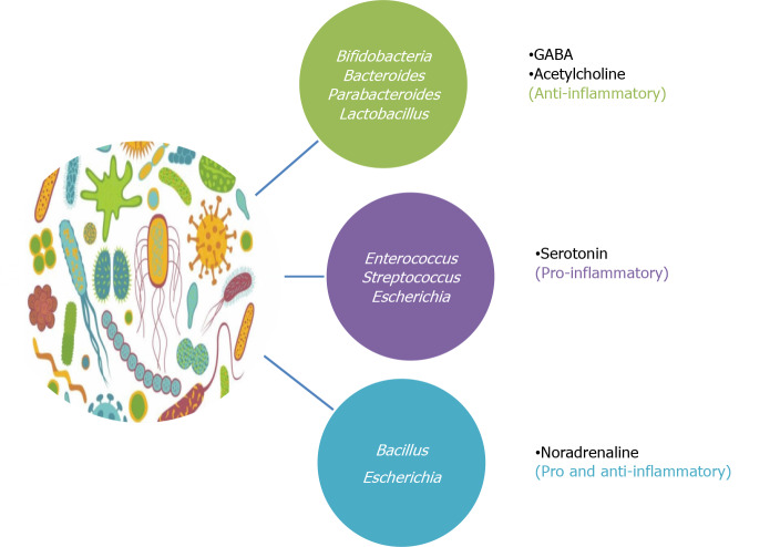
Neuromediators, apart from their classical neuronal functions, are currently being recognised as a pillar in maintaining the gut homeostasis. There are different sources of neuromediators in GIT, including enteric neurons, gut microbiota, immune cells and gut epithelial cells. Out of all the sources, microbial content is the only factor which can be extrinsically varied. Altering the neuromediators via gut bacteria can affect the gut physiology, signalling and immune cells secretions and function in GIT. The available literature on the signalling pathways of a variety of neuromediators and their respective gut bacteria in IBD indicated that the neuromediators released by bacteria being used as probiotic are having anti-inflammatory properties and bacteria which were reported to increase disease severity produce neuromediators with pro-inflammatory properties. For instance, GABA and acetylcholine are the anti-inflammatory neuromediators, produced by those bacteria which are very well established to attenuate gut inflammation in both DSS-induced mice model of colitis and IBD patients. Serotonin which is a pro-inflammatory neuromediator is produced by bacteria that are involved in the severity of IBD. Besides, noradrenaline, having both anti and pro-inflammatory properties, produced by two different types of bacteria, one having the beneficial role and other having the debatable role in IBD (Figure 2). This interrelation suggests that bacteria impart their effects in gut inflammation through releasing neuromediators as one of the mechanism.
Figure 2.
Inter-relation of diverse gut microbiota and their respective neuromediators with gut inflammation. Bacteria used as probiotics in inflammatory bowel disease (IBD) (green box) produces anti-inflammatory neuromediators (γ-aminobutyric acid, acetylcholine), bacteria having a detrimental role in IBD (purple box) releases pro-inflammatory neuromediator (serotonin) and bacteria having a debatable role in IBD (blue box) secrete neuromediator (noradrenaline) having both pro and anti-inflammatory properties. GABA: γ-aminobutyric acid.
CONCLUSION
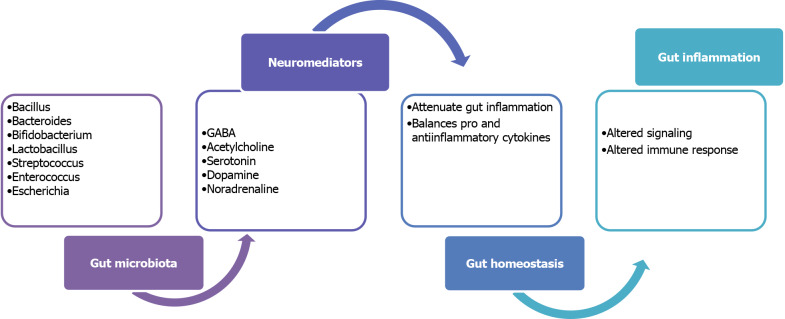
Neuromediators are emerging as essential players in IBD pathogenesis. These are influenced by the complex interaction of gut microbiota, host immunity, and intestinal epithelium. During gut inflammation or IBD, dysbiosis in gut microbiota and alteration in neuromediators complicate the mechanism of gut homeostasis resulting in perturbed equilibrium (Figure 3). In-depth mechanism of neuroimmunomodulation due to gut bacteria needs to be explored more, to settle the gut homeostasis during disease. These neuromediators may prove to be a great tool to clinicians in treating inflammatory diseases. Through this review, we summarized various neuromediators produced by different gut microbiota and their significance as an immunomodulatory entity in the colon. Using gut bacteria that can produce neuromediators having anti-inflammatory properties for treating IBD patients may be a novel therapeutic approach and also the fertile area for future research.
Figure 3.
Role of neuromediators producing gut microbiota during gut inflammation. Gut microbiota produces various neuromediators that attenuate the gut inflammation by balancing the pro and anti-inflammatory cytokines to maintain gut homeostasis. During inflammation, dysbiosis in gut microbiota leads to alteration in respective neuromediators which may lead to altered the host immune response. GABA: γ-aminobutyric acid.
Footnotes
Conflict-of-interest statement: Authors declare no conflict of interests for this article.
Manuscript source: Invited manuscript
Peer-review started: December 14, 2020
First decision: February 14, 2021
Article in press: April 20, 2021
Specialty type: Gastroenterology and hepatology
Country/Territory of origin: India
Peer-review report’s scientific quality classification
Grade A (Excellent): 0
Grade B (Very good): B
Grade C (Good): 0
Grade D (Fair): 0
Grade E (Poor): 0
P-Reviewer: Iizuka M S-Editor: Fan JR L-Editor: A P-Editor: Yuan YY
Contributor Information
Surbhi Aggarwal, Department of Biochemical Engineering and Biotechnology, Indian Institute of Technology, Delhi 110016, India; School of Life Sciences, Jawaharlal Nehru University, Delhi 110067, India. aggarwalsurbhi28@gmail.com.
Raju Ranjha, School of Life Sciences, Jawaharlal Nehru University, Delhi 110067, India; Field Unit Raipur, ICMR-National Institute of Malaria Research, Raipur 492015, Chhattisgarh, India.
Jaishree Paul, School of Life Sciences, Jawaharlal Nehru University, Delhi 110067, India.
References
- 1.Brookes SJ. Classes of enteric nerve cells in the guinea-pig small intestine. Anat Rec. 2001;262:58–70. doi: 10.1002/1097-0185(20010101)262:1<58::AID-AR1011>3.0.CO;2-V. [DOI] [PubMed] [Google Scholar]
- 2.Vasina V, Barbara G, Talamonti L, Stanghellini V, Corinaldesi R, Tonini M, De Ponti F, De Giorgio R. Enteric neuroplasticity evoked by inflammation. Auton Neurosci. 2006;126-127:264–272. doi: 10.1016/j.autneu.2006.02.025. [DOI] [PubMed] [Google Scholar]
- 3.Szelényi J, Vizi ES. The catecholamine cytokine balance: interaction between the brain and the immune system. Ann N Y Acad Sci. 2007;1113:311–324. doi: 10.1196/annals.1391.026. [DOI] [PubMed] [Google Scholar]
- 4.Chandrasekharan B, Nezami BG, Srinivasan S. Emerging neuropeptide targets in inflammation: NPY and VIP. Am J Physiol Gastrointest Liver Physiol. 2013;304:G949–G957. doi: 10.1152/ajpgi.00493.2012. [DOI] [PMC free article] [PubMed] [Google Scholar]
- 5.O’Connor TM, O’Connell J, O’Brien DI, Goode T, Bredin CP, Shanahan F. The role of substance P in inflammatory disease. J Cell Physiol. 2004;201:167–180. doi: 10.1002/jcp.20061. [DOI] [PubMed] [Google Scholar]
- 6.Weinstock JV. Substance P and the regulation of inflammation in infections and inflammatory bowel disease. Acta Physiol (Oxf) 2015;213:453–461. doi: 10.1111/apha.12428. [DOI] [PubMed] [Google Scholar]
- 7.Hong HS, Hwang DY, Park JH, Kim S, Seo EJ, Son Y. Substance-P alleviates dextran sulfate sodium-induced intestinal damage by suppressing inflammation through enrichment of M2 macrophages and regulatory T cells. Cytokine. 2017;90:21–30. doi: 10.1016/j.cyto.2016.10.002. [DOI] [PubMed] [Google Scholar]
- 8.Brun P, Mastrotto C, Beggiao E, Stefani A, Barzon L, Sturniolo GC, Palù G, Castagliuolo I. Neuropeptide neurotensin stimulates intestinal wound healing following chronic intestinal inflammation. Am J Physiol Gastrointest Liver Physiol. 2005;288:G621–G629. doi: 10.1152/ajpgi.00140.2004. [DOI] [PubMed] [Google Scholar]
- 9.Law IK, Pothoulakis C. MicroRNA-133α regulates neurotensin-associated colonic inflammation in colonic epithelial cells and experimental colitis. RNA Dis. 2015;2 doi: 10.14800/rd.472. [DOI] [PMC free article] [PubMed] [Google Scholar]
- 10.Moura LI, Silva L, Leal EC, Tellechea A, Cruz MT, Carvalho E. Neurotensin modulates the migratory and inflammatory response of macrophages under hyperglycemic conditions. Biomed Res Int. 2013;2013:941764. doi: 10.1155/2013/941764. [DOI] [PMC free article] [PubMed] [Google Scholar]
- 11.Seufferlein T, Rozengurt E. Galanin, neurotensin, and phorbol esters rapidly stimulate activation of mitogen-activated protein kinase in small cell lung cancer cells. Cancer Res. 1996;56:5758–5764. [PubMed] [Google Scholar]
- 12.Bakirtzi K, Law IK, Xue X, Iliopoulos D, Shah YM, Pothoulakis C. Neurotensin Promotes the Development of Colitis and Intestinal Angiogenesis via Hif-1α-miR-210 Signaling. J Immunol. 2016;196:4311–4321. doi: 10.4049/jimmunol.1501443. [DOI] [PMC free article] [PubMed] [Google Scholar]
- 13.El-Salhy M, Hausken T. The role of the neuropeptide Y (NPY) family in the pathophysiology of inflammatory bowel disease (IBD) Neuropeptides. 2016;55:137–144. doi: 10.1016/j.npep.2015.09.005. [DOI] [PubMed] [Google Scholar]
- 14.Wheway J, Herzog H, Mackay F. NPY and receptors in immune and inflammatory diseases. Curr Top Med Chem. 2007;7:1743–1752. doi: 10.2174/156802607782341046. [DOI] [PubMed] [Google Scholar]
- 15.El-Salhy M, Solomon T, Hausken T, Gilja OH, Hatlebakk JG. Gastrointestinal neuroendocrine peptides/amines in inflammatory bowel disease. World J Gastroenterol. 2017;23:5068–5085. doi: 10.3748/wjg.v23.i28.5068. [DOI] [PMC free article] [PubMed] [Google Scholar]
- 16.Abad C, Gomariz R, Waschek J, Leceta J, Martinez C, Juarranz Y, Arranz A. VIP in inflammatory bowel disease: state of the art. Endocr Metab Immune Disord Drug Targets. 2012;12:316–322. doi: 10.2174/187153012803832576. [DOI] [PubMed] [Google Scholar]
- 17.Delgado M, Abad C, Martinez C, Juarranz MG, Arranz A, Gomariz RP, Leceta J. Vasoactive intestinal peptide in the immune system: potential therapeutic role in inflammatory and autoimmune diseases. J Mol Med (Berl) 2002;80:16–24. doi: 10.1007/s00109-001-0291-5. [DOI] [PubMed] [Google Scholar]
- 18.Gonzalez-Rey E, Delgado M. Role of vasoactive intestinal peptide in inflammation and autoimmunity. Curr Opin Investig Drugs. 2005;6:1116–1123. [PubMed] [Google Scholar]
- 19.Xu CL, Guo Y, Qiao L, Ma L, Cheng YY. Recombinant expressed vasoactive intestinal peptide analogue ameliorates TNBS-induced colitis in rats. World J Gastroenterol. 2018;24:706–715. doi: 10.3748/wjg.v24.i6.706. [DOI] [PMC free article] [PubMed] [Google Scholar]
- 20.Jayawardena D, Anbazhagan AN, Guzman G, Dudeja PK, Onyuksel H. Vasoactive Intestinal Peptide Nanomedicine for the Management of Inflammatory Bowel Disease. Mol Pharm. 2017;14:3698–3708. doi: 10.1021/acs.molpharmaceut.7b00452. [DOI] [PMC free article] [PubMed] [Google Scholar]
- 21.Talero E, Sánchez-Fidalgo S, Calvo JR, Motilva V. Chronic administration of galanin attenuates the TNBS-induced colitis in rats. Regul Pept. 2007;141:96–104. doi: 10.1016/j.regpep.2006.12.029. [DOI] [PubMed] [Google Scholar]
- 22.Botz B, Kemény Á, Brunner SM, Locker F, Csepregi J, Mócsai A, Pintér E, McDougall JJ, Kofler B, Helyes Z. Lack of Galanin 3 Receptor Aggravates Murine Autoimmune Arthritis. J Mol Neurosci. 2016;59:260–269. doi: 10.1007/s12031-016-0732-9. [DOI] [PMC free article] [PubMed] [Google Scholar]
- 23.Gross KJ, Pothoulakis C. Role of neuropeptides in inflammatory bowel disease. Inflamm Bowel Dis. 2007;13:918–932. doi: 10.1002/ibd.20129. [DOI] [PubMed] [Google Scholar]
- 24.Kofler B, Brunner S, Koller A, Wiesmayr S, Locker F, Lang R, Botz B, Kemény À, Helyes Z. Contribution of the galanin system to inflammation. Springerplus. 2015;4:L57. doi: 10.1186/2193-1801-4-S1-L57. [DOI] [PMC free article] [PubMed] [Google Scholar]
- 25.Lang R, Kofler B. The galanin peptide family in inflammation. Neuropeptides. 2011;45:1–8. doi: 10.1016/j.npep.2010.10.005. [DOI] [PubMed] [Google Scholar]
- 26.Wraith DC, Pope R, Butzkueven H, Holder H, Vanderplank P, Lowrey P, Day MJ, Gundlach AL, Kilpatrick TJ, Scolding N, Wynick D. A role for galanin in human and experimental inflammatory demyelination. Proc Natl Acad Sci USA. 2009;106:15466–15471. doi: 10.1073/pnas.0903360106. [DOI] [PMC free article] [PubMed] [Google Scholar]
- 27.Nezi M, Mastorakos G, Mouslech Z. Corticotropin Releasing Hormone And The Immune/Inflammatory Response. 2015 Jul 30. In: Feingold KR, Anawalt B, Boyce A, Chrousos G, de Herder WW, Dungan K, Grossman A, Hershman JM, Hofland J, Kaltsas G, Koch C, Kopp P, Korbonits M, McLachlan R, Morley JE, New M, Purnell J, Singer F, Stratakis CA, Trence DL, Wilson DP, editors. Endotext [Internet]. South Dartmouth (MA): MDText.com, Inc., 2000. [Google Scholar]
- 28.Chatzaki E, Anton PA, Million M, Lambropoulou M, Constantinidis T, Kolios G, Taché Y, Grigoriadis DE. Corticotropin-releasing factor receptor subtype 2 in human colonic mucosa: down-regulation in ulcerative colitis. World J Gastroenterol. 2013;19:1416–1423. doi: 10.3748/wjg.v19.i9.1416. [DOI] [PMC free article] [PubMed] [Google Scholar]
- 29.Rhee SH, Ma EL, Lee Y, Taché Y, Pothoulakis C, Im E. Corticotropin Releasing Hormone and Urocortin 3 Stimulate Vascular Endothelial Growth Factor Expression through the cAMP/CREB Pathway. J Biol Chem. 2015;290:26194–26203. doi: 10.1074/jbc.M115.678979. [DOI] [PMC free article] [PubMed] [Google Scholar]
- 30.González-Moret R, Cebolla A, Cortés X, Baños RM, Navarrete J, de la Rubia JE, Lisón JF, Soria JM. The effect of a mindfulness-based therapy on different biomarkers among patients with inflammatory bowel disease: a randomised controlled trial. Sci Rep. 2020;10:6071. doi: 10.1038/s41598-020-63168-4. [DOI] [PMC free article] [PubMed] [Google Scholar]
- 31.Overman EL, Rivier JE, Moeser AJ. CRF induces intestinal epithelial barrier injury via the release of mast cell proteases and TNF-α. PLoS One. 2012;7:e39935. doi: 10.1371/journal.pone.0039935. [DOI] [PMC free article] [PubMed] [Google Scholar]
- 32.Eysselein VE, Reinshagen M, Patel A, Davis W, Nast C, Sternini C. Calcitonin gene-related peptide in inflammatory bowel disease and experimentally induced colitis. Ann N Y Acad Sci. 1992;657:319–327. doi: 10.1111/j.1749-6632.1992.tb22779.x. [DOI] [PubMed] [Google Scholar]
- 33.Holzmann B. Antiinflammatory activities of CGRP modulating innate immune responses in health and disease. Curr Protein Pept Sci. 2013;14:268–274. doi: 10.2174/13892037113149990046. [DOI] [PubMed] [Google Scholar]
- 34.Kroeger I, Erhardt A, Abt D, Fischer M, Biburger M, Rau T, Neuhuber WL, Tiegs G. The neuropeptide calcitonin gene-related peptide (CGRP) prevents inflammatory liver injury in mice. J Hepatol. 2009;51:342–353. doi: 10.1016/j.jhep.2009.03.022. [DOI] [PubMed] [Google Scholar]
- 35.Springer J, Geppetti P, Fischer A, Groneberg DA. Calcitonin gene-related peptide as inflammatory mediator. Pulm Pharmacol Ther. 2003;16:121–130. doi: 10.1016/S1094-5539(03)00049-X. [DOI] [PubMed] [Google Scholar]
- 36.Walsh DA, Mapp PI, Kelly S. Calcitonin gene-related peptide in the joint: contributions to pain and inflammation. Br J Clin Pharmacol. 2015;80:965–978. doi: 10.1111/bcp.12669. [DOI] [PMC free article] [PubMed] [Google Scholar]
- 37.Skobowiat C. Contribution of Neuropeptides and Neurotransmitters in colitis. Vet Sci Tech. 2011 [Google Scholar]
- 38.Bai A, Lu N, Guo Y, Chen J, Liu Z. Modulation of inflammatory response via alpha2-adrenoceptor blockade in acute murine colitis. Clin Exp Immunol. 2009;156:353–362. doi: 10.1111/j.1365-2249.2009.03894.x. [DOI] [PMC free article] [PubMed] [Google Scholar]
- 39.Ghia JE, Blennerhassett P, Deng Y, Verdu EF, Khan WI, Collins SM. Reactivation of inflammatory bowel disease in a mouse model of depression. Gastroenterology. 2009;136:2280–2288.e1-4. doi: 10.1053/j.gastro.2009.02.069. [DOI] [PubMed] [Google Scholar]
- 40.Reale M, de Angelis F, di Nicola M, Capello E, di Ioia M, Luca Gd, Lugaresi A, Tata AM. Relation between pro-inflammatory cytokines and acetylcholine levels in relapsing-remitting multiple sclerosis patients. Int J Mol Sci. 2012;13:12656–12664. doi: 10.3390/ijms131012656. [DOI] [PMC free article] [PubMed] [Google Scholar]
- 41.Rosas-Ballina M, Tracey KJ. Cholinergic control of inflammation. J Intern Med. 2009;265:663–679. doi: 10.1111/j.1365-2796.2009.02098.x. [DOI] [PMC free article] [PubMed] [Google Scholar]
- 42.Coleman JW. Nitric oxide in immunity and inflammation. Int Immunopharmacol. 2001;1:1397–1406. doi: 10.1016/s1567-5769(01)00086-8. [DOI] [PubMed] [Google Scholar]
- 43.Sharma JN, Al-Omran A, Parvathy SS. Role of nitric oxide in inflammatory diseases. Inflammopharmacology. 2007;15:252–259. doi: 10.1007/s10787-007-0013-x. [DOI] [PubMed] [Google Scholar]
- 44.Avdagić N, Zaćiragić A, Babić N, Hukić M, Seremet M, Lepara O, Nakaš-Ićindić E. Nitric oxide as a potential biomarker in inflammatory bowel disease. Bosn J Basic Med Sci. 2013;13:5–9. doi: 10.17305/bjbms.2013.2402. [DOI] [PMC free article] [PubMed] [Google Scholar]
- 45.Bertrand PP, Barajas-Espinosa A, Neshat S, Bertrand RL, Lomax AE. Analysis of real-time serotonin (5-HT) availability during experimental colitis in mouse. Am J Physiol Gastrointest Liver Physiol. 2010;298:G446–G455. doi: 10.1152/ajpgi.00318.2009. [DOI] [PubMed] [Google Scholar]
- 46.Ghia JE, Li N, Wang H, Collins M, Deng Y, El-Sharkawy RT, Côté F, Mallet J, Khan WI. Serotonin has a key role in pathogenesis of experimental colitis. Gastroenterology. 2009;137:1649–1660. doi: 10.1053/j.gastro.2009.08.041. [DOI] [PubMed] [Google Scholar]
- 47.Shajib MS, Chauhan U, Adeeb S, Chetty Y, Armstrong D, Halder SLS, Marshall JK, Khan WI. Characterization of Serotonin Signaling Components in Patients with Inflammatory Bowel Disease. J Can Assoc Gastroenterol. 2019;2:132–140. doi: 10.1093/jcag/gwy039. [DOI] [PMC free article] [PubMed] [Google Scholar]
- 48.Shajib MS, Khan WI. The role of serotonin and its receptors in activation of immune responses and inflammation. Acta Physiol (Oxf) 2015;213:561–574. doi: 10.1111/apha.12430. [DOI] [PubMed] [Google Scholar]
- 49.Jin Z, Mendu SK, Birnir B. GABA is an effective immunomodulatory molecule. Amino Acids. 2013;45:87–94. doi: 10.1007/s00726-011-1193-7. [DOI] [PMC free article] [PubMed] [Google Scholar]
- 50.Aggarwal S, Ahuja V, Paul J. Attenuated GABAergic Signaling in Intestinal Epithelium Contributes to Pathogenesis of Ulcerative Colitis. Dig Dis Sci. 2017;62:2768–2779. doi: 10.1007/s10620-017-4662-3. [DOI] [PubMed] [Google Scholar]
- 51.Ma X, Sun Q, Sun X, Chen D, Wei C, Yu X, Liu C, Li Y, Li J. Activation of GABAA Receptors in Colon Epithelium Exacerbates Acute Colitis. Front Immunol. 2018;9:987. doi: 10.3389/fimmu.2018.00987. [DOI] [PMC free article] [PubMed] [Google Scholar]
- 52.Sarkar A, Lehto SM, Harty S, Dinan TG, Cryan JF, Burnet PWJ. Psychobiotics and the Manipulation of Bacteria-Gut-Brain Signals. Trends Neurosci. 2016;39:763–781. doi: 10.1016/j.tins.2016.09.002. [DOI] [PMC free article] [PubMed] [Google Scholar]
- 53.Li Y, Zhang T, Guo C, Geng M, Gai S, Qi W, Li Z, Song Y, Luo X, Wang N. Bacillus subtilis RZ001 improves intestinal integrity and alleviates colitis by inhibiting the Notch signalling pathway and activating ATOH-1. Pathog Dis. 2020;78 doi: 10.1093/femspd/ftaa016. [DOI] [PubMed] [Google Scholar]
- 54.Zhang HL, Li WS, Xu DN, Zheng WW, Liu Y, Chen J, Qiu ZB, Dorfman RG, Zhang J, Liu J. Mucosa-reparing and microbiota-balancing therapeutic effect of Bacillus subtilis alleviates dextrate sulfate sodium-induced ulcerative colitis in mice. Exp Ther Med. 2016;12:2554–2562. doi: 10.3892/etm.2016.3686. [DOI] [PMC free article] [PubMed] [Google Scholar]
- 55.Ilinskaya ON, Ulyanova VV, Yarullina DR, Gataullin IG. Secretome of Intestinal Bacilli: A Natural Guard against Pathologies. Front Microbiol. 2017;8:1666. doi: 10.3389/fmicb.2017.01666. [DOI] [PMC free article] [PubMed] [Google Scholar]
- 56.Zhou Y, Zhi F. Lower Level of Bacteroides in the Gut Microbiota Is Associated with Inflammatory Bowel Disease: A Meta-Analysis. Biomed Res Int. 2016;2016:5828959. doi: 10.1155/2016/5828959. [DOI] [PMC free article] [PubMed] [Google Scholar]
- 57.Pokusaeva K, Johnson C, Luk B, Uribe G, Fu Y, Oezguen N, Matsunami RK, Lugo M, Major A, Mori-Akiyama Y, Hollister EB, Dann SM, Shi XZ, Engler DA, Savidge T, Versalovic J. GABA-producing Bifidobacterium dentium modulates visceral sensitivity in the intestine. Neurogastroenterol Motil. 2017;29 doi: 10.1111/nmo.12904. [DOI] [PMC free article] [PubMed] [Google Scholar]
- 58.Strandwitz P, Kim KH, Terekhova D, Liu JK, Sharma A, Levering J, McDonald D, Dietrich D, Ramadhar TR, Lekbua A, Mroue N, Liston C, Stewart EJ, Dubin MJ, Zengler K, Knight R, Gilbert JA, Clardy J, Lewis K. GABA-modulating bacteria of the human gut microbiota. Nat Microbiol. 2019;4:396–403. doi: 10.1038/s41564-018-0307-3. [DOI] [PMC free article] [PubMed] [Google Scholar]
- 59.Delday M, Mulder I, Logan ET, Grant G. Bacteroides thetaiotaomicron Ameliorates Colon Inflammation in Preclinical Models of Crohn’s Disease. Inflamm Bowel Dis. 2019;25:85–96. doi: 10.1093/ibd/izy281. [DOI] [PMC free article] [PubMed] [Google Scholar]
- 60.Tan H, Zhao J, Zhang H, Zhai Q, Chen W. Novel strains of Bacteroides fragilis and Bacteroides ovatus alleviate the LPS-induced inflammation in mice. Appl Microbiol Biotechnol. 2019;103:2353–2365. doi: 10.1007/s00253-019-09617-1. [DOI] [PubMed] [Google Scholar]
- 61.Kverka M, Zakostelska Z, Klimesova K, Sokol D, Hudcovic T, Hrncir T, Rossmann P, Mrazek J, Kopecny J, Verdu EF, Tlaskalova-Hogenova H. Oral administration of Parabacteroides distasonis antigens attenuates experimental murine colitis through modulation of immunity and microbiota composition. Clin Exp Immunol. 2011;163:250–259. doi: 10.1111/j.1365-2249.2010.04286.x. [DOI] [PMC free article] [PubMed] [Google Scholar]
- 62.O’Callaghan A, van Sinderen D. Bifidobacteria and Their Role as Members of the Human Gut Microbiota. Front Microbiol. 2016;7:925. doi: 10.3389/fmicb.2016.00925. [DOI] [PMC free article] [PubMed] [Google Scholar]
- 63.Riedel CU, Foata F, Philippe D, Adolfsson O, Eikmanns BJ, Blum S. Anti-inflammatory effects of bifidobacteria by inhibition of LPS-induced NF-kappaB activation. World J Gastroenterol. 2006;12:3729–3735. doi: 10.3748/wjg.v12.i23.3729. [DOI] [PMC free article] [PubMed] [Google Scholar]
- 64.Preising J, Philippe D, Gleinser M, Wei H, Blum S, Eikmanns BJ, Niess JH, Riedel CU. Selection of bifidobacteria based on adhesion and anti-inflammatory capacity in vitro for amelioration of murine colitis. Appl Environ Microbiol. 2010;76:3048–3051. doi: 10.1128/AEM.03127-09. [DOI] [PMC free article] [PubMed] [Google Scholar]
- 65.Dubin K, Pamer EG. Enterococci and Their Interactions with the Intestinal Microbiome. Microbiol Spectr. 2014;5 doi: 10.1128/microbiolspec.bad-0014-2016. [DOI] [PMC free article] [PubMed] [Google Scholar]
- 66.Lyte M. Probiotics function mechanistically as delivery vehicles for neuroactive compounds: Microbial endocrinology in the design and use of probiotics. Bioessays. 2011;33:574–581. doi: 10.1002/bies.201100024. [DOI] [PubMed] [Google Scholar]
- 67.Zhou Y, Chen H, He H, Du Y, Hu J, Li Y, Zhou Y, Wang H, Chen Y, Nie Y. Increased Enterococcus faecalis infection is associated with clinically active Crohn disease. Medicine (Baltimore) 2016;95:e5019. doi: 10.1097/MD.0000000000005019. [DOI] [PMC free article] [PubMed] [Google Scholar]
- 68.Balish E, Warner T. Enterococcus faecalis induces inflammatory bowel disease in interleukin-10 knockout mice. Am J Pathol. 2002;160:2253–2257. doi: 10.1016/S0002-9440(10)61172-8. [DOI] [PMC free article] [PubMed] [Google Scholar]
- 69.Celiberto LS, Bedani R, Dejani NN, Ivo de Medeiros A, Sampaio Zuanon JA, Spolidorio LC, Tallarico Adorno MA, Amâncio Varesche MB, Carrilho Galvão F, Valentini SR, Font de Valdez G, Rossi EA, Cavallini DCU. Effect of a probiotic beverage consumption (Enterococcus faecium CRL 183 and Bifidobacterium longum ATCC 15707) in rats with chemically induced colitis. PLoS One. 2017;12:e0175935. doi: 10.1371/journal.pone.0175935. [DOI] [PMC free article] [PubMed] [Google Scholar]
- 70.Escribano-Vazquez U, Verstraeten S, Martin R, Chain F, Langella P, Thomas M, Cherbuy C. The commensal Escherichia coli CEC15 reinforces intestinal defences in gnotobiotic mice and is protective in a chronic colitis mouse model. Sci Rep. 2019;9:11431. doi: 10.1038/s41598-019-47611-9. [DOI] [PMC free article] [PubMed] [Google Scholar]
- 71.Palmela C, Chevarin C, Xu Z, Torres J, Sevrin G, Hirten R, Barnich N, Ng SC, Colombel JF. Adherent-invasive Escherichia coli in inflammatory bowel disease. Gut. 2018;67:574–587. doi: 10.1136/gutjnl-2017-314903. [DOI] [PubMed] [Google Scholar]
- 72.Abdelhalim KA, Uzel A, Gülşen Ünal N. Virulence determinants and genetic diversity of adherent-invasive Escherichia coli (AIEC) strains isolated from patients with Crohn’s disease. Microb Pathog. 2020;145:104233. doi: 10.1016/j.micpath.2020.104233. [DOI] [PubMed] [Google Scholar]
- 73.Nzakizwanayo J, Dedi C, Standen G, Macfarlane WM, Patel BA, Jones BV. Escherichia coli Nissle 1917 enhances bioavailability of serotonin in gut tissues through modulation of synthesis and clearance. Sci Rep. 2015;5:17324. doi: 10.1038/srep17324. [DOI] [PMC free article] [PubMed] [Google Scholar]
- 74.Naseer M, Poola S, Ali S, Samiullah S, Tahan V. Prebiotics and Probiotics in Inflammatory Bowel Disease: Where are we now and where are we going? Curr Clin Pharmacol. 2020;15:216–233. doi: 10.2174/1574884715666200312100237. [DOI] [PubMed] [Google Scholar]
- 75.Behrouzi A, Mazaheri H, Falsafi S, Tavassol ZH, Moshiri A, Siadat SD. Intestinal effect of the probiotic Escherichia coli strain Nissle 1917 and its OMV. J Diabetes Metab Disord. 2020;19:597–604. doi: 10.1007/s40200-020-00511-6. [DOI] [PMC free article] [PubMed] [Google Scholar]
- 76.Manocha M, Khan WI. Serotonin and GI Disorders: An Update on Clinical and Experimental Studies. Clin Transl Gastroenterol. 2012;3:e13. doi: 10.1038/ctg.2012.8. [DOI] [PMC free article] [PubMed] [Google Scholar]
- 77.Kittana H, Gomes-Neto JC, Heck K, Geis AL, Segura Muñoz RR, Cody LA, Schmaltz RJ, Bindels LB, Sinha R, Hostetter JM, Benson AK, Ramer-Tait AE. Commensal Escherichia coli Strains Can Promote Intestinal Inflammation via Differential Interleukin-6 Production. Front Immunol. 2018;9:2318. doi: 10.3389/fimmu.2018.02318. [DOI] [PMC free article] [PubMed] [Google Scholar]
- 78.Heeney DD, Gareau MG, Marco ML. Intestinal Lactobacillus in health and disease, a driver or just along for the ride? Curr Opin Biotechnol. 2018;49:140–147. doi: 10.1016/j.copbio.2017.08.004. [DOI] [PMC free article] [PubMed] [Google Scholar]
- 79.Azad MAK, Sarker M, Li T, Yin J. Probiotic Species in the Modulation of Gut Microbiota: An Overview. Biomed Res Int. 2018;2018:9478630. doi: 10.1155/2018/9478630. [DOI] [PMC free article] [PubMed] [Google Scholar]
- 80.Nimgampalle M, Kuna Y. Anti-Alzheimer Properties of Probiotic, Lactobacillus plantarum MTCC 1325 in Alzheimer’s Disease induced Albino Rats. J Clin Diagn Res. 2017;11:KC01–KC05. doi: 10.7860/JCDR/2017/26106.10428. [DOI] [PMC free article] [PubMed] [Google Scholar]
- 81.Ahn SI, Cho S, Choi NJ. Effect of dietary probiotics on colon length in an inflammatory bowel disease-induced murine model: A meta-analysis. J Dairy Sci. 2020;103:1807–1819. doi: 10.3168/jds.2019-17356. [DOI] [PubMed] [Google Scholar]
- 82.Le B, Yang SH. Efficacy of Lactobacillus plantarum in prevention of inflammatory bowel disease. Toxicol Rep. 2018;5:314–317. doi: 10.1016/j.toxrep.2018.02.007. [DOI] [PMC free article] [PubMed] [Google Scholar]
- 83.Oh NS, Joung JY, Lee JY, Kim Y. Probiotic and anti-inflammatory potential of Lactobacillus rhamnosus 4B15 and Lactobacillus gasseri 4M13 isolated from infant feces. PLoS One. 2018;13:e0192021. doi: 10.1371/journal.pone.0192021. [DOI] [PMC free article] [PubMed] [Google Scholar]
- 84.Mu Q, Tavella VJ, Luo XM. Role of Lactobacillus reuteri in Human Health and Diseases. Front Microbiol. 2018;9:757. doi: 10.3389/fmicb.2018.00757. [DOI] [PMC free article] [PubMed] [Google Scholar]
- 85.Sokovic Bajic S, Djokic J, Dinic M, Veljovic K, Golic N, Mihajlovic S, Tolinacki M. GABA-Producing Natural Dairy Isolate From Artisanal Zlatar Cheese Attenuates Gut Inflammation and Strengthens Gut Epithelial Barrier in vitro. Front Microbiol. 2019;10:527. doi: 10.3389/fmicb.2019.00527. [DOI] [PMC free article] [PubMed] [Google Scholar]
- 86.Pagnini C, Corleto VD, Martorelli M, Lanini C, D’Ambra G, Di Giulio E, Delle Fave G. Mucosal adhesion and anti-inflammatory effects of Lactobacillus rhamnosus GG in the human colonic mucosa: A proof-of-concept study. World J Gastroenterol. 2018;24:4652–4662. doi: 10.3748/wjg.v24.i41.4652. [DOI] [PMC free article] [PubMed] [Google Scholar]
- 87.Zhang F, Li Y, Wang X, Wang S, Bi D. The Impact of Lactobacillus plantarum on the Gut Microbiota of Mice with DSS-Induced Colitis. Biomed Res Int. 2019;2019:3921315. doi: 10.1155/2019/3921315. [DOI] [PMC free article] [PubMed] [Google Scholar]
- 88.Wang J, Ji H, Wang S, Liu H, Zhang W, Zhang D, Wang Y. Probiotic Lactobacillus plantarum Promotes Intestinal Barrier Function by Strengthening the Epithelium and Modulating Gut Microbiota. Front Microbiol. 2018;9:1953. doi: 10.3389/fmicb.2018.01953. [DOI] [PMC free article] [PubMed] [Google Scholar]
- 89.Jandhyala SM, Talukdar R, Subramanyam C, Vuyyuru H, Sasikala M, Nageshwar Reddy D. Role of the normal gut microbiota. World J Gastroenterol. 2015;21:8787–8803. doi: 10.3748/wjg.v21.i29.8787. [DOI] [PMC free article] [PubMed] [Google Scholar]
- 90.Scheri GC, Fard SN, Schietroma I, Mastrangelo A, Pinacchio C, Giustini N, Serafino S, De Girolamo G, Cavallari EN, Statzu M, Laghi L, Vullo A, Ceccarelli G, Vullo V, d’Ettorre G. Modulation of Tryptophan/Serotonin Pathway by Probiotic Supplementation in Human Immunodeficiency Virus-Positive Patients: Preliminary Results of a New Study Approach. Int J Tryptophan Res. 2017;10:1178646917710668. doi: 10.1177/1178646917710668. [DOI] [PMC free article] [PubMed] [Google Scholar]
- 91.Heidarian F, Noormohammadi Z, Asadzadeh Aghdaei H, Alebouyeh M. Relative Abundance of Streptococcus spp. and its Association with Disease Activity in Inflammatory Bowel Disease Patients Compared with Controls. Arch Clin Infect Dis. 2017;12 [Google Scholar]
- 92.Rengarajan S, Vivio EE, Parkes M, Peterson DA, Roberson EDO, Newberry RD, Ciorba MA, Hsieh CS. Dynamic immunoglobulin responses to gut bacteria during inflammatory bowel disease. Gut Microbes. 2020;11:405–420. doi: 10.1080/19490976.2019.1626683. [DOI] [PMC free article] [PubMed] [Google Scholar]
- 93.Hooper LV. Bacterial contributions to mammalian gut development. Trends Microbiol. 2004;12:129–134. doi: 10.1016/j.tim.2004.01.001. [DOI] [PubMed] [Google Scholar]
- 94.Hooper LV, Gordon JI. Commensal host-bacterial relationships in the gut. Science. 2001;292:1115–1118. doi: 10.1126/science.1058709. [DOI] [PubMed] [Google Scholar]
- 95.Hamer HM, Jonkers DM, Bast A, Vanhoutvin SA, Fischer MA, Kodde A, Troost FJ, Venema K, Brummer RJ. Butyrate modulates oxidative stress in the colonic mucosa of healthy humans. Clin Nutr. 2009;28:88–93. doi: 10.1016/j.clnu.2008.11.002. [DOI] [PubMed] [Google Scholar]
- 96.Taurog JD, Richardson JA, Croft JT, Simmons WA, Zhou M, Fernández-Sueiro JL, Balish E, Hammer RE. The germfree state prevents development of gut and joint inflammatory disease in HLA-B27 transgenic rats. J Exp Med. 1994;180:2359–2364. doi: 10.1084/jem.180.6.2359. [DOI] [PMC free article] [PubMed] [Google Scholar]


