Abstract
A series of novel 5-[(Z,2Z)-2-chloro-3-(4-nitrophenyl)-2-propenylidene]-thiazolidinones (Ciminalum–thiazolidinone hybrid molecules) have been synthesized. Anticancer activity screening toward the NCI60 cell lines panel, gastric cancer (AGS), human colon cancer (DLD-1), and breast cancer (MCF-7 and MDA-MB-231) cell lines allowed the identification of 3-{5-[(Z,2Z)-2-chloro-3-(4-nitrophenyl)-2-propenylidene]-4-oxo-2-thioxothiazolidin-3-yl}propanoic acid (2h) with the highest level of antimitotic activity with mean GI50/TGI values of 1.57/13.3 μM and a certain sensitivity profile against leukemia (MOLT-4, SR), colon cancer (SW-620), CNS cancer (SF-539), melanoma (SK-MEL-5), gastric cancer (AGS), human colon cancer (DLD-1), and breast cancers (MCF-7 and MDA-MB-231) cell lines. The hit compounds 2f, 2i, 2j, and 2h have been found to have low toxicity toward normal human blood lymphocytes and a fairly wide therapeutic range. The significant role of the 2-chloro-3-(4-nitrophenyl)prop-2-enylidene (Ciminalum) substituent in the 5 position and the substituent’s nature in the position 3 of core heterocycle in the anticancer cytotoxicity levels of 4-thiazolidinone derivatives have been established
Keywords: synthesis, 4-thiazolidinones, Ciminalum, anticancer activity, SAR analysis
1. Introduction
In recent years, one of successful directions in the structure design of “drug-like” molecules is the “hybrid-pharmacophore” approach that involves combining different fragments in one molecule that can be parts, biomimetics, and/or bioisosteres of biologically active molecules or drugs. This strategy allows potentiating the desired action or appearance of new effects [1,2,3] and can be relevant for the search for new highly active compounds based on 4-thiazolidinones as effective biophores. Thus, modern studies of the pharmacological potential of thiazolidinones have significantly expanded the range of their activity, including anticancer, antibacterial, antifungal, antiviral, antiparasitic, and anti-tuberculosis. Along with this, there is indisputable evidence of the affinity of these derivatives for biotargets involved in the biochemical processes of tumor cell growth (TNF-α-TNFRc-1, JSP-1, antiapoptotic complex Bcl-XL-BH3), the microorganisms life cycle (UDP-NMurNA/L-Ala-ligase), the development of inflammatory conditions (COX-2/5-LOX), and the development of type II diabetes mellitus (PPARγ) [4,5].
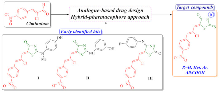
Based on our previous research, we have established that the combination of a thiazolidinone moiety and a structural fragment of the Ciminalum in one hybrid molecule is an effective approach for the design of potential anticancer agents [6,7]. Ciminalum (p-nitro-α-chlorocinnamic aldehyde or (2Z)-2-chloro-3-(4-nitrophenyl)prop-2-enal, CAS 3626-97-9) is an active antimicrobial agent against Gram-positive and Gram-negative microorganisms. Ciminalum was used as a drug in medical practice in the former Soviet Union (Figure 1) [8]. Ciminalum–thiazolidinone hybrid molecules (namely 5-[(Z,2Z)-2-chloro-3-(4-nitrophenyl)-2-propenylidene]-4-thiazolidinones) showed a significant cytotoxic effect on tumor cells. It is important to note that the presence of a Ciminalum moiety in position 5 of the thiazolidinone ring is key to the manifestation of biological activity. Thus, early hits I and II (Figure 1) possessed a selectively high effect on leukemia, melanoma, lung, colon, CNS, ovarian, renal, prostate, and breast cancers cell lines at micro- and submicromolar levels that is probably associated with the immunosuppressive activity [7]. Early anticancer hit III has induced and activated specific signaling apoptotic pathways in tumor cells [6]. Thus, compound III leads to weak caspase-7 activation and a weak cleavage of PARP-1 and DFF45 in the Jurkat T-cells. However, this Ciminalum-thiazolidinone hybrid may be involved in the caspase-independent, AIF-mediated apoptosis. AIF (apoptosis-inducing factor) is known to induce the mitochondria-mediated caspase-independent apoptosis. Derivative III leads to the activation of intrinsic apoptotic pathways, mediated by mitochondria, and caspases seem to play a minor role here.
Figure 1.
Background of the target compounds design.
Our research was aimed at optimization of anticancer activity profile of 5-[(Z,2Z)-2-chloro-3-(4-nitrophenyl)-2-propenylidene]-4-thiazolidinones and SAR analysis within these series in accordance with our systematic study of anticancer activity of thiazolidinone-related derivatives [9,10].
2. Results and Discussion
2.1. Chemistry
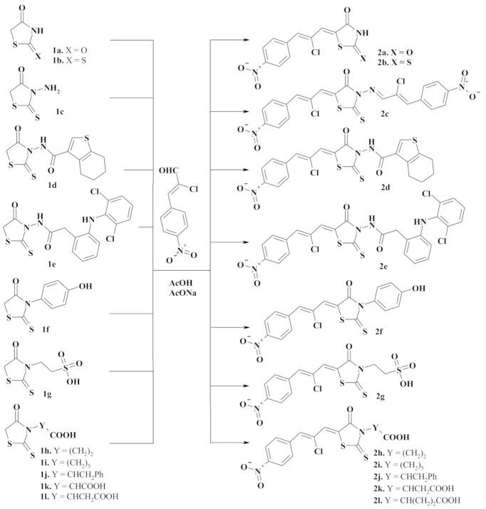
The synthetic approach to target compounds design was based on 4-thiazolidinone derivatives as active methylene heterocycles in Knoevenagel reaction with (2Z)-2-chloro-3-(4-nitrophenyl)prop-2-enal (Ciminalum) as an oxo-compound (Figure 2).
Figure 2.
Synthesis of 5-[(Z,2Z)-2-chloro-3-(4-nitrophenyl)-2-propenylidene]-4-thiazolidinones 2a-m. Reagents and conditions: appropriate 4-thiazolidinone 1a-l (0.01 mol), (2Z)-2-chloro-3-(4-nitrophenyl)prop-2-enal (0.010 mol, in the case of 3-aminorhodanine 1e 0.02 mol), AcONa (0.01 mol), AcOH (20 mL), reflux, 3 h, 68–83%.
The starting 2,4-thiazolidinedione 1a and 2-thioxo-4-thiazolidinone (rhodanine) derivatives 1b-l were obtained according to described procedures. We used three synthetic approaches to this end: (1) [2+3]-cyclocondensation of chloroacetic acid with thiourea (1a [11]) or ammonium thiocyanate (1b [12]); (2) dithiocarbamate method of 3-substituted 2-thioxo-4-thiazolidinone (rhodanine) derivatives synthesis (1c [13], 1g–l [14,15,16,17,18,19]); (3) the reaction of trithiocarbonyl diglycolic acid with amino compounds (1d–f [20,21]).
Target Ciminalum-thiazolidinone hybrid molecules 2a–l were synthesized via the Knoevenagel condensation of (2Z)-2-chloro-3-(4-nitrophenyl)prop-2-enal and appropriate 4-thiazolidinone in the presence of sodium acetate under reflux in acetic acid (Figure 2). In the case of 3-aminorhodanine 1c, in parallel with the Knoevenagel condensation, the reaction with amino group and formation of appropriate azomethine derivative 2c was observed. The data characterizing synthesized novel 4-thiazolidinones are presented in the experimental part. Analytical and spectral data (1H and 13C-NMR, LCMS) confirmed the structure of the synthesized compounds. The 1H-NMR spectra of the synthesized compounds are characterized by the signals of the Cyminalum residue in the form of two singlets at 7.55–8.01 and 7.88–8.07 ppm for the CH=CCl-CH= group as well as two doublets of the p-nitrophenyl substituent at 8.00 and 8.30 ppm. For compound 2g, these signals were slightly shifted into a strong magnetic field and appeared as two singlets at 6.89 and 7.19 ppm and two doublets at 7.23 and 7.51 ppm. In the 13C-NMR spectra of rhodanine derivatives signals of C=O and C=S groups of the core heterocycle were characteristic and appeared at 158.7–174.4 and 193.5–199.3 ppm, respectively.
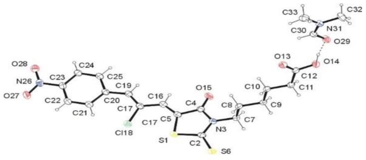
Structural features of the synthesized Ciminalum–thiazolidinone hybrid molecules were confirmed by single-crystal X-ray diffraction study of compound 2i. As follows from the X-ray analysis, the investigated compound has the structure of 6-{5-[(Z,2Z)-2-chloro-3-(4-nitrophenyl)-2-propenylidene]-4-oxo-2-thioxothiazolidin-3-yl}hexanoic acid (2i) and crystallizes as dimethylformamide solvate in a molar ratio of 1:1 (Figure 3). 5-Carboxypentyl group located at N-3 atom adopts anticlinal conformation with respect to C2–N3 bond belonging to 2-thioxo-4-thiazolidinone moiety. This arrangement is confirmed by the torsion angle C2–N3–C7–C12 [−101.19(15)°]. The 3-(4-nitrophenyl)-2-chloroprop-2-en-1-ylidene residue at the C-5 atom assumes Z configuration with respect to the S1–C5 bond. The torsion angle S1–C5–C16–C17 has the value of 2.0(3)°. The system formed by the named residue and 2-thioxo-1,3-thiazolidin-4-one system is approximately planar [r.m.s.d. = 0.0949 Å].
Figure 3.
ORTEP view of 2i·DMF showing the atomic labeling scheme. Non-H atoms are drawn as 30% probability displacement ellipsoids and H atoms are drawn as spheres of an arbitrary radius.
The conformation of the molecule of 2i is stabilized by the intermolecular hydrogen bonding. The hydrogen bonds C22–H···S6iii and C25–H25···O29iv (Table 1) stabilize the almost coplanar arrangement of the 3-(4-nitrophenyl)-2-chloroprop-2-en-1-ylidene and 2-thioxo-4-thiazolidinone moieties. Moreover, the hydrogen bonds O14–H14···O29, C11–H11A···O13i, and C11–H11B···O15ii (Table 1) stabilize the spatial arrangement of the 5-carboxypentyl group. The C5═C16 [1.345(2) Å] and C17═C19 [1.351(2) Å] bond lengths confirmed the occurrence of a double bonds between these carbon atoms (Figure 3).
Table 1.
Hydrogen-bond geometry (Å, °) for 2i·DMF.
| D—H∙∙∙A | D—H | H∙∙∙A | D∙∙∙A | D—H∙∙∙A |
|---|---|---|---|---|
| O14—H14∙∙∙O29 | 0.95 (3) | 1.63 (3) | 2.5758 (16) | 171 (3) |
| C11—H11A∙∙∙O13 i | 0.99 | 2.55 | 3.5287 (19) | 172 |
| C11—H11A∙∙∙O15 ii | 0.99 | 2.47 | 3.135 (2) | 125 |
| C21—H22∙∙∙C118 | 0.95 | 2.51 | 3.2049 (15) | 130 |
| C22—H22∙∙∙S6 iii | 0.95 | 2.75 | 3.6734 (16) | 164 |
| C25—H25∙∙∙O29 iv | 0.95 | 2.50 | 3.4332 (18) | 169 |
Symmetry codes: (i) x, 1 + y, z; (ii) 1/2 − x, 1/2 + y, 1/2 − z; (iii) 1 − x, 2 − y, 1 − z; (iv) 1/2 − x, −1/2 + y, 1/2 − z.
2.2. In Vitro Evaluation of the Anticancer Activity
At the first stage of biological activity study, the antitumor activity screening of the selected compounds 2b, 2c, 2f, 2h, and 2j was performed according to the NCI DTP (USA) standard protocol at the concentrations ranging from 10−4 to 10−8 M toward 60 tumor cell lines [22,23,24,25]. The percentage of growth was evaluated spectrophotometrically versus controls not treated with test agents after 48 h exposure and using SRB protein assay to estimate cell viability or growth. Dose–response parameters were calculated for each cell line: GI50—molar concentration of the compound that inhibits 50% net cell growth; TGI—molar concentration of the compound leading to the total inhibition; and LC50—molar concentration of the compound leading to 50% net cell death. Furthermore, mean graph midpoints (MG_MID) were calculated for each of the parameters, giving an average activity parameter over all cell lines for the tested compound. For the MG_MID calculation, insensitive cell lines were included with the highest concentration tested.
The obtained results of screening evaluation of Ciminalum–thiazolidinone hybrids confirmed their significant anticancer activity (Table 2). Thus, compounds 2f and 2h inhibited the growth of all tested cancer cell lines at submicromolar and micromolar concentrations. The average meanings of three dose–response parameters GI50, TGI, and LC50 were 2.80/32.3/80.8 µM (2f) and 1.57/13.3/65.0 µM (2h), respectively. It is important to note that the most active compound 2h was active in the GI50 concentration range of < 0.01–0.02 μM toward the following cell lines: MOLT-4, SR (Leukemia); SW-620 (Colon cancer); SF-539 (CNS cancer); SK-MEL-5 (Melanoma). Regarding the preliminary SAR analysis, it is worth mentioning that the presence of the (Z,2Z)-2-chloro-3-(4-nitrophenyl)-2-propenylidene moiety turned out to be a necessary requirement for achieving the anticancer effects. Moreover, the substituent nature at position 3 of the 4-thiazolidinone ring is important. Derivatives with carboxylic acids residues (2h, 2j) and p-hydroxyphenyl substituent (2f) proved to be the most effective. The absence of a substituent in position 3 (2b) or an additional fragment of the Cyminalum (2c) leads to the weakening of anticancer cytotoxicity.
Table 2.
Influence of compounds 2b, 2c, 2f, 2h, and 2j on the growth of individual tumor cell lines.
| Cell line/comp. | 2b | 2c | 2f | 2h | 2j |
|---|---|---|---|---|---|
| GI50/TGI/LC50, μM | GI50/TGI/LC50, μM | GI50/TGI/LC50, μM | GI50/TGI/LC50, μM | GI50/TGI/LC50, μM | |
| Leukemia | |||||
| CCRF-CEM | 1.97/21.5/>50 | 4.42/35.3/>100 | 2.92/>100/>100 | ||
| HL-60(TB) | 1.21/14.4/>50 | 5.32/23.3/87.2 | 0.485/4.48/>100 | 0.347/1.87/>100 | |
| K-562 | 1.44/42.5/>50 | 3.98/25.5/>100 | 0.521/>100/>100 | 0.212/2.83/>100 | 3.01/>100/>100 |
| MOLT-4 | 1.74/14.2/>50 | 3.20/8.95/52.5 | 0.389/>100/>100 | 0.016/19.7/>100 | 3.14/>100/>100 |
| RPMI-8226 | 1.54/20.6/>50 | 2.38/6.32/>100 | <0.01/0.264/>100 | 0.138/1.30/>100 | 3.22/>100/>100 |
| SR | 1.18/7.96/>50 | 3.67/12.0/>100 | 0.01/0.138/>100 | <0.01/1.41/>100 | 2.45/>100/>100 |
| Non-Small Cell Lung Cancer | |||||
| A549/ATCC | 8.18/>50/>50 | 18.9/42.6/96.1 | 2.25/6.47/>100 | 1.76/7.62/>100 | 3.46/>100/>100 |
| EKVX | 2.76/>50/>50 | 23.7/47.7/96.1 | 2.37/16.2/>100 | 0.334/4.01/55.6 | 2.59/7.29/>100 |
| HOP-62 | 15.4/33.1/>50 | 17.2/39.7/91.9 | 2.61/5.22/11.6 | 6.54/19.9/52.8 | 2.31/6.75/>100 |
| HOP-92 | 8.48/>50/>50 | 8.53/39.4/>100 | 4.81/18.3/50.7 | 2.36/5.71/>100 | |
| NCI-H226 | 6.28/20.0/> 50 | 12.0/31.8/84.2 | 1.58/3.27/6.76 | ||
| NCI-H23 | 3.27/13.4/45.5 | 16.6/31.6/60.3 | 1.48/3.11/6.52 | 1.44/3.22/7.19 | 2.13/4.86/>100 |
| NCI-322M | 6.51/18.8/>50 | 87.6/>100/>100 | 2.49/9.02/>100 | 2.83/10.9/63.4 | >100/>100/>100 |
| NCI-H460 | 3.95/18.6/>50 | 15.0/32.3/69.5 | 0.754/>100/>100 | 0.804/3.32/>100 | 2.19/>100/>100 |
| NCI-H522 | 3.19/13.1/42.3 | 16.3/32.9/66.2 | 1.72/4.47/29.5 | ||
| Colon Cancer | |||||
| Colo 205 | 4.04/17.4/>50 | 56.2/>100/>100 | 4.34/>100/>100 | 0.350/>100/>100 | 2.45/>100/>100 |
| HCC-2998 | 5.43/11.7/25.0 | 1.96/3.50/6.27 | |||
| HCT-116 | 2.93/11.0/33.7 | 2.73/6.51/23.3 | 1.27/2.96/6.92 | 0.270/1.19/>100 | 2.01/5.47/56.7 |
| HCT-15 | 1.79/8.81/25.4 | 16.6/33.0/65.7 | 0.409/>100/>100 | 0.230/2.96/>100 | 1.92/4.58/>100 |
| HT-29 | 1.48/9.48/>50 | 2.14/>100/>100 | 1.25/2.77/>100 | 2.84/8.47/>100 | |
| KM12 | 5.27/14.6/40.4 | 20.6/59.1/>100 | 2.85/>100/>100 | 0.639/29.5/>100 | 2.06/4.35/9.16 |
| SW-620 | 1.94/7.62/23.5 | 7.24/>100/>100 | 0.037/1.52/>100 | <0.01/3.25/>100 | 2.10/4.39/>100 |
| CNS Cancer | |||||
| SF-268 | 5.24/18.9/>50 | 17.0/41.3/>100 | 2.55/34.5/>100 | 2.12/9.74/>100 | 2.21/>100/>100 |
| SF-295 | 8.46/37.9/>50 | 18.0/37.3/77.1 | 2.26/13.8/>100 | 3.36/21.6/>100 | 2.59/7.85/>100 |
| SF-539 | 9.39/23.1/>50 | 25.9/57.4/>100 | 0.0252/0.242/28.1 | <0.01/0.267/25.8 | 2.74/>100/>100 |
| SNB-19 | 8.10/21.4/>50 | 48.3/>100/>100 | 10.0/32.0/97.5 | 0.658/26.3/83.7 | 1.85/> 100/>100 |
| SNB-75 | 7.59/23.5/>50 | 25.1/>100/>100 | 12.5/46.0/>100 | 1.96/5.37/>100 | |
| U251 | 3.09/9.73/22.1 | 8.03/24.5/65.8 | 0.368/3.06/38.9 | 0.149/2.00/>100 | 1.72/3.52/7.21 |
| Melanoma | |||||
| LOX IMVI | 6.60/15.4/36.0 | 13.0/28.8/63.8 | 1.17/3.70/>100 | 0.161/0.515/>100 | 1.64/3.26/6.46 |
| MALME-3M | 4.93/14.9/44.6 | 25.0/60.4/>100 | 6.53/27.6/94.5 | 9.70/61.9/>100 | 2.13/4.82/>100 |
| M14 | 8.87/32.2/>50 | 16.2/35.7/78.5 | 1.90/3.73/7.32 | 1.66/3.58/7.74 | 2.53/>100/>100 |
| SK-MEL-2 | 8.07/26.9/>50 | 19.0/36.4/69.8 | 0.802/4.93/65.1 | 1.87/4.50/>100 | |
| SK-MEL-28 | 5.87/12.3/25.9 | 4.49/47.7/>100 | 3.59/23.6/89.5 | 2.16/4.68/11.6 | |
| SK-MEL-5 | 3.82/10.6/24.0 | 19.4/38.3/75.6 | <0.01/<0.01/3.41 | 0.0193/0.0849/1.88 | 1.68/3.06/5.60 |
| UACC-62 | 5.75/12.5/27.2 | 13.6/28.1/58.0 | 1.69/3.85/8.78 | 1.23/2.97/7.18 | 2.00/4.85/30.2 |
| Ovarian Cancer | |||||
| IGROV1 | 7.28/21.1/>50 | 13.3/28.0/58.9 | 1.63/6.13/>100 | 0.794/3.68/43.1 | 2.99/>100/>100 |
| OVCAR-3 | 1.65/8.08/26.6 | 0.821/12.7/41.1 | 0.977/41.8/>100 | 0.135/6.05/66.6 | 1.76/3.51/6.98 |
| OVCAR-4 | 1.95/11.7/>50 | 2.63/>100/>100 | 2.66/14.9/56.2 | 3.21/>100/>100 | |
| OVCAR-5 | 11.1/19.5/32.4 | 15.1/36.2/86.6 | 2.82/7.91/>100 | ||
| OVCAR-8 | 5.08/15.6/47.9 | 1.98/4.08/8.38 | 1.14/22.9/>100 | 0.244/11.5/>100 | 3.13/>100/>100 |
| SK-OV-3 | 13.0/>50/>50 | 59.6/>100/>100 | 10.2/41.5/>100 | 6.83/27.4/77.6 | 4.13/>100/>100 |
| Renal Cancer | |||||
| 786-0 | 9.40/18.8/37.6 | 12.1/30.7/77.8 | 2.04/3.83/7.18 | 1.13/2.47/5.42 | 2.10/5.01/>100 |
| A498 | 4.57/12.2/30.9 | 13.0/29.6/67.3 | 1.65/3.24/6.37 | ||
| ACHN | 3.64/11.8/31.7 | 15.6/29.7/56.4 | 1.70/3.51/7.27 | 1.02/2.42/5.76 | 1.80/3.30/6.04 |
| CAKI-1 | 7.14/25.2/>50 | 3.11/11.2/>100 | 4.44/>100/>100 | 0.485/6.14/47.1 | 2.28/5.09/16.7 |
| RXF 393 | 0.105/0.341/1.73 | 1.54/3.02/5.93 | |||
| SN12C | 4.11/18.0/>50 | 29.0/>100/>100 | 1.98/4.41/>100 | 1.05/2.99/8.48 | 2.84/>100/>100 |
| TK-10 | 10.6/23.2/>50 | 28.0/69.6/>100 | 4.87/29.0/>100 | 2.33/16.9/92.0 | 2.43/>100/>100 |
| UO-31 | 4.12/21.8/>50 | 17.5/32.2/59.1 | 2.08/3.98/>100 | 0.812/2.43/6.31 | 1.59/3.10/6.05 |
| Prostate Cancer | |||||
| PC-3 | 4.68/>50/>50 | 8.10/45.8/>100 | 2.29/84.8/>100 | 0.712/13.7/41.9 | 2.53/7.98/>100 |
| DU-145 | 7.09/14.2/28.4 | 21.7/52.8/>100 | 0.666/5.81/50.1 | 0.421/3.48/52.3 | 2.06/4.56/11.7 |
| Breast Cancer | |||||
| MCF-7 | 2.14/38.0/>50 | 4.08/19.8/83.7 | 0.401/28.0/>100 | 0.239/14.4/>100 | 2.38/10.7/>100 |
| NCI/ADR-Res | 5.71/18.3/>50 | 6.55/20.8/61.7 | 2.17/6.13/>100 | 0.407/2.11/6.43 | |
| MDA-MB-231/ATCC | 8.16/28.1/>50 | 10.0/35.4/>100 | 2.18/12.2/92.1 | 1.08/4.86/25.9 | 3.35/>100/>100 |
| HS 578T | 1.42/12.1/>50 | 29.5/>100/>100 | 4.37/>100/>100 | ||
| MDA-MB-435 | 8.57/47.9/>50 | 20.3/41.9/86.3 | 3.50/7.71/>100 | 1.25/4.12/29.3 | |
| BT-549 | 1.47/4.74/27.2 | 27.3/76.0/>100 | 1.55/7.71/>100 | 0.247/0.920/36.3 | 1.79/4.17/>100 |
| T-47D | 3.21/>50/>50 | 24.5/65.3/>100 | 1.55/7.71/>100 | 0.363/>100/>100 | 1.50/3.71/>100 |
| MDA-MB-468 | 1.39/3.28/7.71 | ||||
The selectivity index (SI) obtained by dividing the full panel MG-MID (mM) of the tested compound by their individual subpanel MG-MID (mM) was considered as a measure of selectivity of anticancer activity (Table 3). Ratios between 3 and 6 mean moderate selectivity, ratios greater than 6 indicate high selectivity toward the corresponding cell line, while compounds not meeting either of these criteria are rated nonselective [26]. The most active compounds 2f and 2h in the present study were found to be high selective toward the leukemia subpanel at GI50 levels (selectivity indices 9.89 and 10.73, respectively). Compound 2j possessed high selectivity toward the CNS cancer subpanel at both the TGI and LC50 levels (selectivity index 11.53 and 10.25, respectively). In general, it is worth noting the selectivity of action against leukemia cell lines for the studied class of heterocyclic compounds.
Table 3.
Influence of 2b, 2c, 2f, 2h, and 2j on the growth of tumor panels (GI50, TGI, LC50) and selectivity index (SI) values.
| Compound/Disease | 2b | 2c | 2f | 2h | 2j | ||||||
|---|---|---|---|---|---|---|---|---|---|---|---|
| MG_MID, μM |
SI | MG_MID, μM |
SI | MG_MID, μM |
SI | MG_MID, μM |
SI | MG_MID, μM |
SI | ||
| Leukemia | GI50 | 1.51 | 3.17 | 3.83 | 4.70 | 0.283 | 9.89 | 0.145 | 10.83 | 2.95 | 1.39 |
| TGI | 20.2 | 1.00 | 18.6 | 2.32 | 41.0 | 0.78 | 5.42 | 2.45 | >100 | <0.41 | |
| LC50 | >50 | <0.87 | 90 | 0.91 | >100 | <0.81 | >100 | <0.65 | >100 | <0.74 | |
| Non-Small Cell Lung Cancer | GI50 | 3.79 | 1.26 | 25.9 | 0.69 | 2.93 | 0.96 | 2.65 | 0.59 | 13.1 | 0.31 |
| TGI | 29.7 | 0.68 | 44.8 | 0.96 | 26.6 | 1.21 | 9.61 | 1.38 | 36.9 | 1.10 | |
| LC50 | 48.6 | 0.90 | 83.0 | 0.98 | 74.0 | 1.09 | 61.4 | 1.06 | 92.9 | 0.80 | |
| Colon Cancer | GI50 | 3.27 | 1.46 | 20.7 | 0.87 | 1.84 | 1.52 | 0.458 | 3.43 | 2.19 | 1.87 |
| TGI | 11.5 | 1.77 | 59.7 | 0.72 | 67.4 | 0.48 | 23.3 | 0.57 | 18.7 | 2.17 | |
| LC50 | 35.4 | 1.23 | 77.8 | 1.05 | 84.5 | 0.96 | >100 | <0.65 | 67.4 | 1.10 | |
| CNS Cancer | GI50 | 6.98 | 0.69 | 23.4 | 0.77 | 6.72 | 0.42 | 3.13 | 0.50 | 2.18 | 1.88 |
| TGI | 22.4 | 0.91 | 52.1 | 0.83 | 30.6 | 1.05 | 17.7 | 0.75 | 3.52 | 11.53 | |
| LC50 | 45.4 | 0.96 | 88.6 | 0.92 | 77.4 | 1.04 | 84.9 | 0.77 | 7.21 | 10.25 | |
| Melanoma | GI50 | 5.57 | 0.86 | 17.7 | 1.02 | 2.63 | 1.06 | 2.45 | 0.64 | 2.00 | 2.05 |
| TGI | 16.1 | 1.26 | 38.0 | 1.13 | 14.4 | 2.23 | 13.9 | 0.96 | 17.9 | 2.27 | |
| LC50 | 36.8 | 1.18 | 74.3 | 1.10 | 52.3 | 1.54 | 53.1 | 1.22 | 50.6 | 1.46 | |
| Ovarian Cancer | GI50 | 6.68 | 0.72 | 18.2 | 0.99 | 3.32 | 0.84 | 2.13 | 0.74 | 3.00 | 1.36 |
| TGI | 21.0 | 0.97 | 36.2 | 1.19 | 42.5 | 0.76 | 12.7 | 1.05 | 68.6 | 0.59 | |
| LC50 | 42.8 | 1.02 | 59.0 | 1.38 | >100 | <0.81 | 68.7 | 0.95 | 84.5 | 0.87 | |
| Renal Cancer | GI50 | 6.23 | 0.77 | 16.9 | 1.07 | 2.85 | 0.98 | 0.99 | 1,59 | 2.03 | 2.01 |
| TGI | 18.7 | 1.09 | 43.3 | 1.00 | 24.1 | 1.34 | 4.81 | 2.77 | 27.8 | 1.46 | |
| LC50 | 42.9 | 1.01 | 80.1 | 1.02 | 69.1 | 1.17 | 23.8 | 2.73 | 42.6 | 1.73 | |
| Prostate Cancer | GI50 | 5.89 | 0.81 | 14.9 | 1.21 | 1.48 | 1.89 | 0.567 | 2.77 | 2.30 | 1.78 |
| TGI | 32.1 | 0.63 | 45.3 | 0.95 | 45.3 | 0.71 | 8.59 | 1.55 | 6.27 | 6.48 | |
| LC50 | 39.2 | 1.11 | 75.1 | 1.09 | 75.1 | 1.08 | 47.1 | 1.38 | 55.9 | 1.32 | |
| Breast Cancer | GI50 | 4.38 | 1.09 | 17.5 | 1.03 | 1.89 | 1.48 | 0.598 | 2.63 | 24.6 | 0.17 |
| TGI | 28.4 | 0.71 | 51.3 | 0.84 | 11.6 | 2.78 | 21.1 | 0.63 | 37.0 | 1.10 | |
| LC50 | 46.7 | 0.93 | 90.2 | 0.91 | 98.7 | 0.82 | 49.6 | 1.31 | 84.6 | 0.87 | |
| 60 lines MG_MID | GI50 | 4.79 | 18.0 | 2.80 | 1.57 | 4.09 | |||||
| TGI | 20.3 | 43.1 | 32.2 | 13.3 | 40.6 | ||||||
| LC50 | 43.5 | 81.7 | 80.8 | 65.0 | 73.9 | ||||||
In the second stage of the research, Ciminalum-thiazolidinone hybrids were investigated for antitumor activity on the lines of gastric cancer (AGS), human colon cancer (DLD-1), and breast cancers (MCF-7 and MDA-MB-231). The study was performed in the MTT assay according to the method described previously [27]. The studied cancer line was sensitive to the action of the studied compounds that inhibited its growth in micromolar ranges of GI50. The hit compounds that inhibited the growth of all four cancer lines with the lowest GI50 values were [5-[2-chloro-3-(4-nitrophenyl)prop-2-enylidene]-rhodanines 2c, 2d, 2h, and 2i (Table 4). Moreover, it is important to note the high cytotoxic effect of rhodanine-3-carboxylic acid derivatives 2h and 2i toward breast cancer lines MCF-7 and MDA-MB-231 at the GI50 level of 0.95–1.74 μM, which is consistent with previous data obtained according to DTP NCI protocol (Table 2).
Table 4.
Influence of compounds 2a–e, 2g, 2h, 2k, and 2l on the growth of AGS, DLD-1, MCF-7, and MDA-MB-231 cell lines.
| Compound | Cell line, GI50, μM | |||
|---|---|---|---|---|
| AGS | DLD-1 | MCF-7 | MDA-MB-231 | |
| 2a | 18.71 | 13.98 | 18.03 | 13.89 |
| 2b | 7.86 | 8.39 | 4.79 | 10.56 |
| 2c | 4.43 | 6.34 | 3.60 | 1.59 |
| 2d | 4.08 | 5.47 | 4.45 | 3.11 |
| 2e | 17.05 | 16.00 | 17.92 | 15.83 |
| 2g | 17.99 | 27.49 | 26.40 | 16.84 |
| 2h | 2.69 | 3.67 | 3.62 | 1.63 |
| 2i | 3.20 | 9.22 | 1.73 | 0.95 |
| 2k | 13.05 | 10.00 | 18.08 | 15.30 |
| 2l | 12.57 | 9.19 | 21.23 | 17.50 |
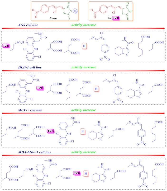
Regarding the SAR analysis (Figure 4), the significant role of the 5-[2-chloro-3-(4-nitrophenyl)prop-2-enylidene (Ciminalum) substituent in the anticancer cytotoxicity appearance was confirmed. Moreover, the presence of a thioxo group in position 2 of the core heterocycle is more important than the oxo group, as evidenced by the lower activity of the thiazolidinedione 2b compared to a structurally close rhodanine derivative 2a. The role of the substituents nature in position 3 of the rhodanine core on the level of anticancer cytotoxicity level is interesting and important for further in-depth research and the design of drug-like molecules. Thus, the most effective is the presence of carboxylic acids residues, among which fragments of propanoic (2h) and hexanoic (2i) acids are considered to be important for cytotoxicity toward AGS, DLD-1, MCF-7, and MDA-MB-231 cell lines. The introduction of an additional carboxylic group reduced the effect of derivatives by about 10 times (compounds 2k and 2l). Replacing the carboxyl group with a sulfo group had reduced the activity more significantly (compound 2g). In addition to the carboxylic acid residues, an additional Ciminalum fragment (2c) or 4,5,6,7-tetrahydrobenzo[b]thiophen-3-ylcarboxamide moiety (2d) at position 3 of the rhodanine cycle were also important for the anticancer activity.
Figure 4.
Impact of different substituents in N3 position of the rhodanine core on the anticancer activity levels.
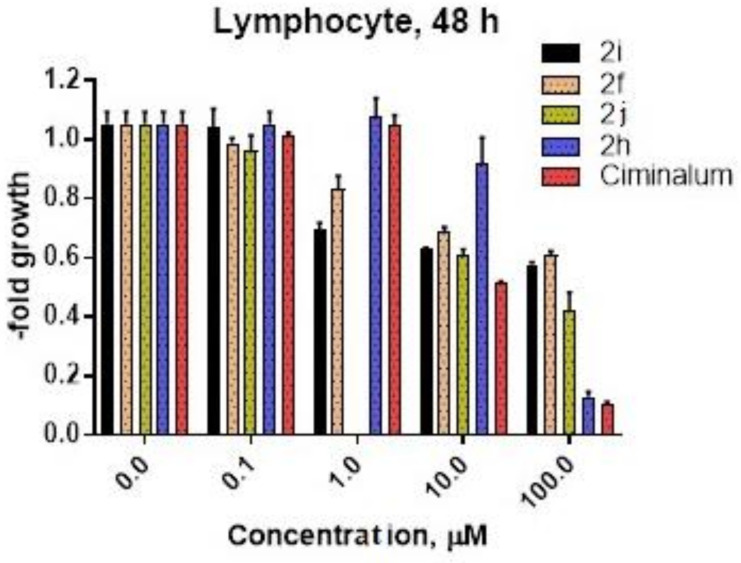
Another part of our study was to determine the influence of compounds 2f, 2i, 2j, and 2h on normal human blood lymphocytes (Figure 5). GI50 values for compounds 2j and 2h were 48.97 μM and 54.54 μM correspondingly. Compounds 2i and 2f do not reach GI50 up to 100 μM after 48 h incubation. The pure Ciminalum has the lowest IC50 value (GI50 = 10.4 μM) for human normal lymphocytes. Thus, normal blood lymphocytes are blood cells, as well as cells of leukemia cell lines, therapeutic index (TI) of compounds 2f, 2h, and 2j was calculated as GI50 (normal blood lymphocyte)/GI50 (leukemia cell line) (Table 5).
Figure 5.
Human lymphocyte viability after 48 h of 2f, 2i, 2j, 2h, and Ciminalum drug exposure was estimated by MTT assay.
Table 5.
Therapeutic index (TI) for compounds 2f, 2h, and 2j regarding diversity leukemia cell lines.
| Compound | Leukemia Cell Line TI (Therapeutic Index) | |||||
|---|---|---|---|---|---|---|
| HL-60(TB) | K-562 | MOLT-4 | RPMI-8226 | SR | Leukemia Panel | |
| 2f | >206.19 | >191.94 | >257.07 | >10,000 | >10,000 | >353.36 |
| 2h | 157.18 | 257.26 | 3408.75 | 395.22 | 5454 | 376.14 |
| 2j | n/a | 16.27 | 15.59 | 15.21 | 19.99 | 16.60 |
3. Materials and Methods
3.1. General Information
All reagents and solvents were purchased from commercial suppliers and were used directly without further purification. NMR spectra were determined with Varian Unity Plus 400 (400 MHz) and Bruker 170 Avance 500 (500 MHz) spectrometers, in DMSO-d6 using tetramethylsilane (TMS) as an internal standard. Melting points were measured on a Kofler hot-stage and are uncorrected. LC-MS was performed using a system with an Agilent 1100 Series HPLC equipped with diode-array detector and Agilent LC\MSD SL mass-selective detector using chemical ionization at atmospheric pressure (APCI). The NMR and LCMS spectra of compounds 2a–l are presented in Figures S1–S32.
3.2. Synthesis of 5-[(Z,2Z)-2-chloro-3-(4-nitrophenyl)-2-propenylidene]-thiazolidinone derivatives (2a-l)
A mixture of (2Z)-2-chloro-3-(4-nitrophenyl)prop-2-enal (0.01 mol) and appropriate 4-thiazolidinone (0.01 mol) in the medium of acetic acid (20 mL) and the presence of sodium acetate (0.01 mol) was refluxed for 3 h. Obtained solid product was collected after cooling by filtration and recrystallized from the mixture DMF-ethanol (1:2).
5-[(Z,2Z)-2-Chloro-3-(4-nitrophenyl)-2-propenylidene]-2,4-thiazolidinedione (2a). Yield: 78%, mp >270 °C. 1H-NMR (400 MHz, DMSO-d6): δ (ppm) 7.70 (s, 1H, CH=), 7.88 (s, 1H, CH=), 8.00 (d, 2H, J = 7.5 Hz, arom.), 8.31 (d, 2H, J = 8.0 Hz, arom.), 12.70 (br.s, 1H, NH). LCMS (ESI): m/z 309.9/312.0 (95.58%, [M + H]+). Anal. Calc. for C12H7ClN2O4S: C 46.39%; H 2.27%; N 9.02%. Found: C 46.50%; H 2.40%; N 8.90%.
5-[(Z,2Z)-2-Chloro-3-(4-nitrophenyl)-2-propenylidene]-2-thioxo-4-thiazolidinone (2b). Yield: 81%, mp 251–253 °C. 1H-NMR (400 MHz, DMSO-d6): δ (ppm) 7.55 (s, 1H, CH=), 7.94 (s, 1H, CH=), 8.01 (d, 2H, J = 8.5 Hz, arom.), 8.31 (d, 2H, J = 8.2 Hz, arom.), 13.91 (br.s, 1H, NH). LCMS (ESI): m/z 324.9/326.9 (100%, [M + H]+). Anal. Calc. for C12H7ClN2O3S2: C 44.11%; H 2.16%; N 8.57%. Found: C 44.00%; H 2.25%; N 8.70%.
5-[(Z,2Z)-2-Chloro-3-(4-nitrophenyl)-2-propenylidene]-3-[(Z,2Z)-2-chloro-3-(4-nitrophenyl)-2-propenylideneamino]-2-thioxo-4-thiazolidinone (2c). Yield: 74%, mp >260 °C. 1H-NMR (400 MHz, DMSO-d6): δ (ppm) 7.79 (s, 1H, =CH), 8.01 (s, 1H, =CH), 8.06 (d, 2H, J = 8.1 Hz, arom.), 8.06 (s, 1H, =CH), 8.07 (s, 1H, =CH), 8.18 (d, 2H, J = 8.8 Hz, arom.), 8.33 (d, 2H, J = 8.8 Hz, arom.), 8.37 (d, 2H, J = 8.1 Hz, arom.), 8.98 (s, 1H, CH=N). LCMS (ESI): m/z 535.0 (95.05%, [M + H]+). Anal. Calc. for C21H12Cl2N4O5S2: C 47.11%; H 2.26%; N 10.46%. Found: C 47.00%; H 2.15%; N 10.65%.
5-[(Z,2Z)-2-Chloro-3-(4-nitrophenyl)-2-propenylidene]-4-oxo-3-(4,5,6,7-tetrahydrobenzo[b]thiophen-3-ylcarboxamido)-2-thioxo-4-thiazolidinone (2d). Yield: 80%, mp >230 °C. 1H-NMR (400 MHz, DMSO-d6): δ (ppm) 1.68–1.74 (m, 4H, 2*CH2), 2.67–2.78 (m, 4H, 2*CH2), 7.87 (s, 1H, s, 1H, CH=), 8.03–8.09 (m, 3H, arom., CH=), 8.10 (s, 1H, s, 1H, thiophene), 8.33 (d, 2H, J = 8.3 Hz, arom.), 11.46 (s, 1H, NH). 13C-NMR (100 MHz, DMSO-d6): δ (ppm) 27.2, 27.8, 29.8, 30.4, 128.2, 129.1, 134.8, 136.2, 137.9, 140.3, 142.5, 144.3, 144.8, 145.0, 152.7, 168.7 (C=O), 177.9 (C=O), 196.4 (C=S). LCMS (ESI): m/z 504.0/506.0 (100%, [M-H]+). Anal. Calc. for C21H16ClN3O4S3: C 49.85%; H 3.19%; N 8.30%. Found: C 50.00%; H 3.15%; N 8.35%.
N1-{5-[(Z,2Z)-2-Chloro-3-(4-nitrophenyl)-2-propenylidene]-4-oxo-2-thioxothiazolidin-3-yl}-2-[2-(2,6-dichloroanilino)phenyl]acetamide (2e). Yield: 74%, mp 257–258 °C. 1H-NMR (400 MHz, DMSO-d6): δ (ppm) 3.85 (d, 1H, J = 14.8 Hz, CH2), 3.90 (d, 1H, J = 14.8 Hz, CH2), 6.30 (d, 1H, J = 7.6 Hz, arom.), 7.08 (t, 1H, J = 7.5 Hz, arom.), 7.19 (t, 1H, J = 8.0 Hz, arom.) 7.29 (s, 1H, NH), 7.34 (d, 1H, J = 7.3 Hz, arom.), 7.53 (d, 2H, J = 8.0 Hz, arom.), 7.83 (s, 1H, =CH), 8.05 (d, 2H, J = 7.7 Hz, arom.), 8.06 (s, 1H, =CH), 8.33 (2H, arom., J = 8.4 Hz, arom.), 11.68 (s, 1H, NH). 13C-NMR (100 MHz, DMSO-d6): δ (ppm) 41.6 (CH2), 121.4, 121.5, 126.1, 126.2, 129.1, 129.2, 130.8, 132.9, 134.4, 134.8, 135.3, 135.6, 136.2, 137.7, 142.3, 144.8, 152.7, 168.3 (C=O), 177.0 (C=O), 195.9 (C=S). LCMS (ESI): m/z 618.8/621.6 (96.2%, [M - H]+). Anal. Calc. for C27H16Cl3N4O4S2: C 50.37%; H 2.76%; N 9.04%. Found: C 50.20%; H 2.85%; N 9.15%.
5-[(Z,2Z)-2-Chloro-3-(4-nitrophenyl)-2-propenylidene]-3-(4-hydroxyphenyl)-2-thioxo-4-thiazolidinone (2f). Yield: 76%, mp >260 °C. 1H-NMR (400 MHz, DMSO-d6): δ (ppm) 6.89 (d, 2H, J = 8.4 Hz, arom.), 7.16 (d, 2H, J = 8.4 Hz, arom.), 7.72 (s, 1H, =CH), 8.02 (s, 1H, =CH), 8.05 (d, 2H, J = 8.5 Hz, arom.), 8.32 (d, 2H, J = 8.5 Hz, arom.), 9.89 (s, 1H, OH). 13C-NMR (100 MHz, DMSO-d6): δ (ppm) 116.3, 119.5, 122.9, 124.4, 127.8, 130.2, 130.4, 131.37, 131.4, 138.2, 149.2, 152.9, 158.7 (C=O), 199.3 (C=S). LCMS (ESI): m/z 419.0/421.0 (97.1%, [M + H]+). Anal. Calc. for C18H11ClN2O4S2: C 51.61%; H 2.65%; N 6.69%. Found: C 51.80%; H 2.85%; N 6.80%.
2-{5-[(Z,2Z)-2-Chloro-3-(4-nitrophenyl)-2-propenylidene]-4-oxo-2-thioxothiazolidin-3-yl}-1-ethanesulfonic acid (2g). Yield: 83%, mp >260 °C. 1H-NMR (400 MHz, DMSO-d6): δ (ppm) 1.95 (t, 2H, J = 7.9 Hz, CH2), 3.47 (t, 2H, J = 7.9 Hz, CH2), 6.89 (s, 1H, CH=), 7.19 (s, 1H, CH=), 7.23 (d, 2H, J = 8.9 Hz, arom.), 7.51 (d, 2H, J = 8.8 Hz, arom.). 13C-NMR (100 MHz, DMSO-d6): δ (ppm) 41.7 (CH2), 47.6 (CH2), 124.3, 127.1, 130.3, 130.9, 131.3, 138.2, 140.3, 147.7, 167.1 (C=O), 194.3 (C=S). LCMS (ESI): m/z 432.8/435.0 (100%, [M + H]+). Anal. Calc. for C14H11ClN2O6S3: C 38.67%; H 2.55%; N 6.44%. Found: C 38.80%; H 2.45%; N 6.60%.
3-{5-[(Z,2Z)-2-Chloro-3-(4-nitrophenyl)-2-propenylidene]-4-oxo-2-thioxothiazolidin-3-yl}propanoic acid (2h). Yield: 75%, mp 254–256 °C. 1H-NMR (400 MHz, DMSO-d6): δ (ppm) 2.63 (t, 2H, J = 6.8 Hz, CH2), 4.22 (t, 2H, J = 6.8 Hz, CH2), 7.73 (s, 1H, CH=), 8.02 (s, 1H, CH=), 8.04 (d, 2H, J = 8.9 Hz, arom.), 8.32 (d, 2H, J = 8.9 Hz, arom.), 12.29 (br.s, 1H, COOH). LCMS (ESI): m/z 399.0/401.0/402.0 (100%, [M + H]+). Anal. Calc. for C15H11ClN2O5S2: C 45.17%; H 2.78%; N 7.02%. Found: C 45.00%; H 2.65%; N 6.90%.
6-{5-[(Z,2Z)-2-Chloro-3-(4-nitrophenyl)-2-propenylidene]-4-oxo-2-thioxothiazolidin-3-yl}hexanoic acid (2i). Yield: 75%, mp >220 °C. 1H-NMR (400 MHz, DMSO-d6): δ (ppm) 1.30 (quint, 2H, J = 6.7 Hz, CH2), 1.52 (quint, 2H, J = 7.1 Hz, CH2), 1.62 (quint, 2H, J = 6.7 Hz, CH2), 2.20 (quint, 2H, J = 7.0 Hz, CH2), 3.99 (quint, 2H, J = 6.9 Hz, CH2), 7.69 (s, 1H, CH=), 7.98 (s, 1H, CH=), 8.02 (d, 2H, J = 8.5 Hz, arom.), 8.30 (d, 2H, J = 8.5 Hz, arom.), 12.00 (s, 1H, COOH). 13C-NMR (100 MHz, DMSO-d6): δ (ppm) 24.5 (CH2), 26.1 (CH2), 26.5 (CH2), 33.8 (CH2), 44.5 (CH2), 124.3, 126.8, 130.2, 131.2, 131.3, 138.4, 140.2, 147.8, 167.4, 174.7 (C=O), 194.6 (C=S). LCMS (ESI): m/z 441.0/443.1 (100%, [M + H]+). Anal. Calc. for C18H17ClN2O5S2: C 49.03%; H 3.89%; N 6.35%. Found: C 49.10%; H 3.85%; N 6.40%.
2-{5-[(Z,2Z)-2-Chloro-3-(4-nitrophenyl)-2-propenylidene]-4-oxo-2-thioxothiazolidin-3-yl}-3-phenylpropanoic acid (2j). Yield: 70%, mp >220 °C. 1H-NMR (400 MHz, DMSO-d6): δ (ppm) 3.49 (t, 2H, CH2), 5.87 (br.s, 1H, CH), 7.10–7.25 (m, 5H, arom.), 7.71 (s, 1H, s, 1H, CH=), 8.00 (s, 1H, s, 1H, CH=), 8.02 (d, 2H, J = 8.0 Hz, arom.), 8.31 (d, 2H, J = 8.0 Hz, arom.), 13.59 (br.s, 1H, COOH). 13C-NMR (100 MHz, DMSO-d6): δ (ppm) 38.2 (CH2), 63.3 (CH), 129.1, 132.0, 133.5, 134.2, 134.7, 136.2, 137.1, 137.2, 141.6, 144.1, 152.6, 171.6 (C=O), 173.7 (C=O), 198.7 (C=S). LCMS (ESI): m/z 324.9/326.9 (100%, [M + H]+). Anal. Calc. for C21H15ClN2O5S2: C 53.11%; H 3.18%; N 5.90%. Found: C 53.00%; H 3.15%; N 5.80%.
2-{5-[(Z,2Z)-2-Chloro-3-(4-nitrophenyl)-2-propenylidene]-4-oxo-2-thioxothiazolidin-3-yl}succinic acid (2k). Yield: 68%, mp 220–222 °C. 1H-NMR (400 MHz, DMSO-d6): δ (ppm) 2.89 (d, 1H, J = 15.5 Hz, CH2), 3.23 (dd, J = 7.6, 15.6 Hz, 1H, CH2), 5.94 (br.s, 1H, CH), 7.75 (s, 1H, CH=), 8.02 (s, 1H, CH=), 8.04 (d, 2H, J = 8.9 Hz, arom.), 8.32 (d, 2H, J = 8.8 Hz, arom.), 12.68 (br.s, 2H, 2*COOH). 13C-NMR (100 MHz, DMSO-d6): δ (ppm) 33.0 (CH2), 53.2 (CH), 115.1 (C-Cl), 123.9, 129.6 (=CH), 131.0, 131.7 (=CH), 138.6, 139.7, 147.4, 166.5 (C=O), 168.6 (COOH), 171.2 (COOH), 193.5 (C=S). LCMS (ESI): m/z 442.8/444.7 (100%, [M + H]+). Anal. Calc. for C16H11ClN2O7S2: C 43.40%; H 2.50%; N 6.33%. Found: C 43.54%; H 2.48%; N 6.45%.
2-{5-[(Z,2Z)-2-Chloro-3-(4-nitrophenyl)-2-propenylidene]-4-oxo-2-thioxothiazolidin-3-yl}pentanedioic acid (2l). Yield: 72%, mp 205–207 °C. 1H-NMR (400 MHz, DMSO-d6): δ (ppm) 2.25–2.45 (m, 4H, CH2CH2), 5.59 (br.s, 1H, CH), 7.72 (s, 1H, CH=), 8.01 (s, 1H, CH=), 8.04 (d, 2H, J = 8.9 Hz, arom.), 8.32 (d, 2H, J = 8.9 Hz, arom.), 12.59 (br.s, 2H, 2*COOH). 13C-NMR (100 MHz, DMSO-d6): δ (ppm) 23.3 (CH2), 30.7 (CH2), 57.2 (CH), 119.3 (C-Cl), 124.3, 125.7 (=CH), 131.4, 131.8 (=CH), 138.8, 140.2, 147.8, 162.5 (C=O), 169.3 (COOH), 174.1 (COOH), 194.8 (C=S). LCMS (ESI): m/z 455.0/456.9 (100%, [M + H]+). Anal. Calc. for C17H13ClN2O7S2: C 44.69%; H 2.87%; N 6.13%. Found: C 44.56%; H 2.78%; N 6.05%.
3.3. Crystal Structure Determination of 6-{5-[(Z,2Z)-2-chloro-3-(4-nitrophenyl)-2-propenylidene]- 4-oxo-2-thioxothiazolidin-3-yl}hexanoic Acid Dimethylaminoformamide Solvate (2i·DMF)
Compound 2i was recrystallized from DMF by slow evaporation at room temperature.
Crystal data. C18H17ClN2O5S2, C3H7NO2, Mr = 514.00, monoclinic, space group P21/n, a = 13.20068(11), b = 5.12876(4), c = 35.3537(3) Å, β = 94.7348(6)°, V = 2385.39(3) Å3, Z = 4 (Z’ = 1), Dcalc = 1.431 g/cm3, μ = 3.425 mm−1, T = 130.0(1) K.
Data collection. An orange lath crystal (DMF) of 0.40 × 0.10 × 0.07 mm was used to record 18,412 (Cu Kα-radiation, θmax = 76.22°) intensities on a Rigaku SuperNova Dual Atlas diffractometer [28] using mirror monochromatized Cu Kα-radiation from a high-flux microfocus source (λ = 1.54178 Å). Accurate unit cell parameters were determined by least-squares techniques from the θ values of 12,519 reflections, θ range 3.47–76.02°. The data were corrected for Lorentz, polarization and for absorption effects [28]. The 4955 total unique reflections (Rint = 0.0175) were used for structure determination.
Structure solution and refinement. The structure was solved by a dual space algorithm (SHELXT) [29] and refined against F2 for all data (SHELXL) [30]. The position of the H atom bonded to the O atom was obtained from the difference Fourier map and was refined freely. The remaining H atoms were positioned geometrically and were refined within the riding model approximation: C–H = 0.98 Å (CH3), 0.99 Å (CH2), 0.95 Å (Csp2H), and Uiso(H) = 1.2Ueq(C) or 1.5Ueq(C) for methyl H atoms. The methyl groups were refined as a rigid group, which were allowed to rotate. Final refinement converged with R = 0.0319 (for 4729 data with F2 > 4σ(F2), wR = 0.0864 (on F2 for all data), and S = 1.052 (on F2 for all data). The largest difference peak and hole was 0.281 and -0.275 eÅ3.
The molecular illustration was drawn using ORTEP-3 for Windows [31]. Software used to prepare material for publication was WINGX [31], OLEX2 [32], and PLATON [33].
The supplementary crystallographic data are deposited at the Cambridge Crystallographic Data Centre (CCDC), 12 Union ROAD, Cambridge CB2 1EZ (UK) [phone, (+44) 1223/336-408; fax, (+44) 1223/336-033; e-mail, deposit@ccdc.cam.ac.uk; World Wide Web, http://www.ccdc.cam.ac.uk, accessed on 18 April 2021 (deposition no. CCDC 2082064)].
3.4. In Vitro Evaluation of the Anticancer Activity According DTP NCI Protocol
Primary anticancer assay was performed on a panel of approximately sixty human tumor cell lines derived from nine neoplastic diseases, in accordance with the protocol of the Drug Evaluation Branch, National Cancer Institute, Bethesda [22,23,24,25]. Tested compounds were added to the culture at a single concentration (10−5 M) and the cultures were incubated for 48 h. End point determinations were made with a protein binding dye, sulforhodamine B (SRB). Results for each tested compound were reported as the percent of growth of the treated cells when compared to the untreated control cells. The percentage growth was evaluated spectrophotometrically versus controls not treated with test agents. The cytotoxic and/or growth inhibitory effects of the most active selected compounds were tested in vitro against the full panel of human tumor cell lines at concentrations ranging from 10−4 to 10−8 M. A 48 h continuous drug exposure protocol was followed, and an SRB protein assay was used to estimate cell viability or growth.
Using absorbance measurements (time zero (Tz), control growth in the absence of drug (C), and test growth in the presence of drug (Ti)), the percentage growth was calculated for each drug concentration. Percentage growth inhibition was calculated as:
| [(Ti − Tz)/(C − Tz)] × 100 for concentrations for which Ti ≥ Tz | (1) |
| [(Ti − Tz)/Tz] × 100 for concentrations for which Ti < Tz. | (2) |
Dose–response parameters (GI50, TGI, LC50) were calculated for each compound. Growth inhibition of 50% (GI50) was calculated from [(Ti − Tz)/(C − Tz)] × 100 = 50 (1), which is the drug concentration resulting in a 50% lower net protein increase in the treated cells (measured by SRB staining) as compared to the net protein increase seen in the control cells. The drug concentration resulting in total growth inhibition (TGI) was calculated from Ti = Tz. The LC50 (concentration of drug resulting in a 50% reduction in the measured protein at the end of the drug treatment as compared to that at the beginning) indicating a net loss of cells following treatment was calculated from [(Ti − Tz)/Tz] × 100 = −50 (2). Values were calculated for each of these parameters if the level of activity was reached; however, if the effect was not reached or was excessive, the value for that parameter was expressed as more or less than the maximum or minimum concentration tested. The lowest values were obtained with the most sensitive cell lines. Compounds having GI50 values ≤ 100 μM were declared to be active.
3.5. Cell Viability Assay (AGS, DLD-1, MCF-7 and MDA-MB-231 Cell Lines; Human Blood Lymphocytes)
The assay was performed by using 3-(4,5-dimethylthiazole-2-yl)- 2,5-diphenyltetrazolium bromide (MTT). Confluent cells, cultured for 24 h with 0.1, 1, 5, 10, 20, 30, and 100 µM concentrations of studied compounds in 24-well plates were washed with PBS. MTT was dissolved in PBS, and 25 µL were added to each well. Plates were incubated for 4 h at 37 °C in 5% CO2 in an incubator. The medium with MTT was removed, and 1 mL of DMSO was added to the attached cells. Furthermore, cells were incubated for 5–10 min in RT and then 10 µl of Sorensen buffer was added to each well. The absorbance of converted dye in living cells was measured at a wavelength of 570 nm. The cell viability of breast cancer cells, gastric cancer cells, and human colon cancer cells cultured in the presence of ligands was calculated as percent of control cells.
3.6. Isolation of Human Blood Lymphocytes and Their Activation
First, 20 mL of venous blood was taken from volunteers (Ethical protocol number 2, 27 January 2019) and collected in the presence of 200 μL of undiluted fresh heparin (1/100). Sterile blood was diluted 2 times with 0.9% NaCl under the sterile conditions. Isolation of lymphocytes was performed in a density gradient of ficol-verografin using the protocol of the manufacturer (Lympoprep, NYCOMED PHARMA AS, Oslo Norway). The resulting lymphocytes were resuspended in the RPMI-1640 medium and cultured for several days (up to 10 days). To separate the lymphocytes from the monocytes, cell suspension was left for 24 h. After 24 h of culture, monocytes were attached, while lymphocytes were transferred to a fresh Falcon tube (15 mL). To stimulate the proliferation of lymphocytes, they were cultured on CD3+ antibody-coated plastic plate in the RPMI-1640 medium supplemented with 20% FBS.
4. Conclusions
In the presented paper, new 5-[(Z,2Z)-2-chloro-3-(4-nitrophenyl)-2-propenylidene]- 4-thiazolidinones (Ciminalum-thiazolidinone hybrid molecules) are described. NCI 60-Cell-line antitumor activity assay allowed identifying a highly active compound 2h with the mean GI50 1.57 μM and TGI 13.3 μM with a certain sensitivity profile in the GI50 concentration range of < 0.01–0.02 μM toward leukemia (MOLT-4, SR), colon cancer (SW-620), CNS cancer (SF-539) and melanoma (SK-MEL-5) cell lines. High cytotoxicity of 5-[(Z,2Z)-2-chloro-3-(4-nitrophenyl)-2-propenylidene]-2-thioxo-4-thiazolidinone-3-carboxylic acids against cell lines of gastric cancer (AGS), human colon cancer (DLD-1), and breast cancers (MCF-7 and MDA-MB-231) was established. The hit compounds 2f, 2i, 2g, and 2h have been found to have low toxicity toward normal human blood lymphocytes and a fairly wide therapeutic range—TI for leukemia panel > 353.36 (2f), 376.14 (2h) and 16.60 (2j). The SAR analysis allowed confirming the crucial role of 2-chloro-3-(4-nitrophenyl)prop-2-enylidene (Ciminalum) substituent in position 5 for 4-thiazolidinones and establish the dependence of the anticancer activity of the synthesized compounds on the nature of the substituents in N3 position of the core heterocycle. Further investigations on the Ciminalum–thiazolidinone hybrid molecules could lead to more potent compounds as promising candidates for the development of new anticancer chemotherapy. The levels of their anticancer activity cause the need for the in-depth study of their mechanisms of action.
Acknowledgments
We are grateful to G. Morris from Drug Synthesis and Chemistry Branch, National Cancer Institute, Bethesda, MD, USA, for in vitro evaluation of anticancer activity.
Supplementary Materials
The following are available online, Figures S1–S32: Copies of NMR and LCMS spectra of compounds 2a–l.
Author Contributions
Conceptualization, A.B., K.B. (Krzysztof Bielawski) and R.L.; methodology, A.B. and R.L.; validation, K.B. (Krzysztof Bielawski), A.K.-D. and O.R.; formal analysis, K.B. (Krzysztof Bielawski) and A.G.; investigation, K.B. (Kamila Buzun), A.K.-D., J.S. and A.G.; resources, R.L.; data curation, K.B. (Krzysztof Bielawski) and J.S.; writing—original draft preparation, R.L.; writing—review and editing, A.B. and R.L.; visualization, J.S.; supervision, A.B. and R.L.; project administration, A.B. and R.L. All authors have read and agreed to the published version of the manuscript.
Funding
This work was financially supported by Grant of Ministry of Healthcare of Ukraine 0121U100690 and the National Research Foundation of Ukraine, under the project number: 2020.02/0035.
Institutional Review Board Statement
The study was conducted according to the guidelines of the Declaration of Helsinki, and approved by the Ethics Committee of Institute of Cell Biology of National Academy of Sciences of Ukraine (protocol number 2, 27 January 2019).
Informed Consent Statement
Informed consent was obtained from all subjects involved in the study.
Data Availability Statement
Data available in a publicly accessible repository.
Conflicts of Interest
The authors declare no conflict of interest.
Sample Availability
Samples of the compounds 2a–l are available from the authors.
Footnotes
Publisher’s Note: MDPI stays neutral with regard to jurisdictional claims in published maps and institutional affiliations.
References
- 1.Fortin S., Berube G. Advances in the development of hybrid anticancer drugs. Expert Opin. Drug Discov. 2013;8:1547–1577. doi: 10.1517/17460441.2013.798296. [DOI] [PubMed] [Google Scholar]
- 2.Gediya L.K., Njar V.C. Promise and challenges in drug discovery and development of hybrid anticancer drugs. Expert Opin. Drug Discov. 2009;4:1099–1111. doi: 10.1517/17460440903341705. [DOI] [PubMed] [Google Scholar]
- 3.Nepali K., Sharma S., Sharma M., Bedi P.M.S., Dhar K.L. Rational approaches, design strategies, structure activity relationship and mechanistic insights for anticancer hybrids. Eur. J. Med. Chem. 2014;77:422–487. doi: 10.1016/j.ejmech.2014.03.018. [DOI] [PubMed] [Google Scholar]
- 4.Kaminskyy D., Kryshchyshyn A., Lesyk R. 5-Ene-4-thiazolidinones—An efficient tool in medicinal chemistry. Eur. J. Med. Chem. 2017;140:542–594. doi: 10.1016/j.ejmech.2017.09.031. [DOI] [PMC free article] [PubMed] [Google Scholar]
- 5.Kaminskyy D., Kryshchyshyn A., Lesyk R. Recent developments with rhodanine as a scaffold for drug discovery. Expert Opin. Drug Discov. 2017;12:1233–1252. doi: 10.1080/17460441.2017.1388370. [DOI] [PubMed] [Google Scholar]
- 6.Panchuk R.R., Chumak V.V., Fil’ M.R., Havrylyuk D.Y., Zimenkovsky B.S., Lesyk R.B., Stoika R.S. Study of molecular mechanisms ofproapoptotic action of novel heterocyclic 4-thiazolidone derivatives. Biopolym. Cell. 2012;28:121–128. doi: 10.7124/bc.00003D. [DOI] [Google Scholar]
- 7.Subtelna I., Atamanyuk D., Szymańska E., Kieć-Kononowicz K., Zimenkovsky B., Vasylenko O., Gzella A., Lesyk R. Synthesis of 5-arylidene-2-amino-4-azolones and evaluation of their anticancer activity. Bioorg. Med. Chem. 2010;18:5089–5101. doi: 10.1016/j.bmc.2010.05.073. [DOI] [PubMed] [Google Scholar]
- 8.Antypenko L., Gladysheva S. Development and validation of UV-spectrophotometric determination of ciminalum in drug. Recipe. 2017;20:153–160. [Google Scholar]
- 9.Lesyk R. Drug design: 4-thiazolidinones applications. Part 1. Synthetic routes to the drug-like molecules. JMS. 2020;89:e406. doi: 10.20883/medical.406. [DOI] [Google Scholar]
- 10.Lesyk R. Drug design: 4-thiazolidinones applications. Part 2. Pharmacological profiles. JMS. 2020;89:e407. doi: 10.20883/medical.407. [DOI] [Google Scholar]
- 11.Turkevych N.M., Vvedenskij V.M., Petlichnaya L.P. Method of obtaining pseudothiohydantoin and thiazolidinedione-2,4. Ukr. Khim. Zh. (Russ. Ed.) 1961;27:680–681. [Google Scholar]
- 12.Nencki M. Ueber die Einwirkung der Monochloressigsäure auf Sulfocyansäure und ihre Salze. J. Prakt. Chem. 1877;16:1–17. doi: 10.1002/prac.18770160101. [DOI] [Google Scholar]
- 13.Petlichnaya L.I., Turkevich N.M. Synthesis of new arylidene derivatives of 3-aminorhodanine. Chem. Heterocycl. Compd. 1970;4:57–59. doi: 10.1007/BF00478079. [DOI] [Google Scholar]
- 14.Langhals E., Balli H. Novel dimethine merocyanine dyes undergoing J-aggregation in highly dilute solution. Helv. Chim. Acta. 1985;68:1782–1797. doi: 10.1002/hlca.19850680633. [DOI] [Google Scholar]
- 15.Stawoska I., Tejchman W., Mazuryk O., Lyčka A., Nowak-Sliwinska P., Żesławska E., Nitek W., Kania A. Spectral Characteristic and Preliminary Anticancer Activity in vitro of Selected Rhodanine-3-carboxylic Acids Derivatives. J. Heterocycl. Chem. 2017;54:2889–2897. doi: 10.1002/jhet.2897. [DOI] [Google Scholar]
- 16.Horishny V., Kartsev V., Geronikaki A., Matiychuk V., Petrou A., Glamoclija J., Ciric A., Sokovic M. 5-(1H-Indol-3-ylmethylene)-4-oxo-2-thioxothiazolidin-3-yl)alkancarboxylic acids as antimicrobial agents: Synthesis, biological evaluation and molecular docking studies. Molecules. 2020;25:1964. doi: 10.3390/molecules25081964. [DOI] [PMC free article] [PubMed] [Google Scholar]
- 17.Nitsche C., Schreier V.N., Behnam M.A.M., Kumar A., Bartenschlager R., Klein C.D. Thiazolidinone-Peptide Hybrids as Dengue Virus Protease Inhibitors with Antiviral Activity in Cell Culture. J. Med. Chem. 2013;56:8389–8403. doi: 10.1021/jm400828u. [DOI] [PubMed] [Google Scholar]
- 18.Ali Muhammad S., Ravi S., Thangamani A. Synthesis and evaluation of some novel N-substituted rhodanines for their anticancer activity. Med. Chem. Res. 2016;25:994–1004. doi: 10.1007/s00044-016-1545-7. [DOI] [Google Scholar]
- 19.Brahmbhatt H., Molnar M., Pavić V., Vesna Rastija V. Synthesis, Characterization, Antibacterial and Antioxidant Potency of N-Substituted-2-Sulfanylidene-1,3-Thiazolidin-4-one Derivatives and QSAR Study. Med. Chem. 2019;15:840–849. doi: 10.2174/1573406415666181205163052. [DOI] [PubMed] [Google Scholar]
- 20.Liu K., Lu H., Hou L., Qi Z., Teixeira C., Barbault F., Fan B.-T., Liu S., Jiang S., Xie L. Design, Synthesis, and Biological Evaluation of N-Carboxyphenylpyrrole Derivatives as Potent HIV Fusion Inhibitors Targeting gp41. J. Med. Chem. 2008;51:7843–7854. doi: 10.1021/jm800869t. [DOI] [PMC free article] [PubMed] [Google Scholar]
- 21.Shepeta Y.L., Lelyukh M.I., Zimenkovsky B.S., Nektegayev I.O., Lesyk R.B. Synthesis and anti-inflammatory activity evaluation of rhodanine derivatives with 2-(2,6-dichlorophenylamino)-phenylacetamide fragment in molecules. Pharm. Rev. 2018;1:6–15. doi: 10.11603/2312-0967.2018.1.8601. (In Ukrainian) [DOI] [Google Scholar]
- 22.Monks A., Scudiero D., Skehan P., Shoemaker R., Paull K., Vistica D., Hose C., Langley J., Cronise P., Vaigro-Wolff A., et al. Feasibility of a high-flux anticancer drug screen using a diverse panel of cultured human tumor cell lines. J. Nat. Cancer Inst. 1991;83:757–766. doi: 10.1093/jnci/83.11.757. [DOI] [PubMed] [Google Scholar]
- 23.Boyd M.R., Paull K.D. Some practical considerations and applications of the national cancer institute in vitro anticancer drug discovery screen. Drug Dev. Res. 1995;34:91–109. doi: 10.1002/ddr.430340203. [DOI] [Google Scholar]
- 24.Boyd M.R. In: Cancer Drug Discovery and Development. Teicher B.A., editor. Volume 2. Humana Press; Totowa, NJ, USA: 1997. pp. 23–43. [Google Scholar]
- 25.Shoemaker R.H. The NCI60 human tumour cell line anticancer drug screen. Nat. Rev. Cancer. 2006;6:813–823. doi: 10.1038/nrc1951. [DOI] [PubMed] [Google Scholar]
- 26.Rostom S.A.F. Synthesis and in vitro antitumor evaluation of some indeno [1,2-c]pyrazol(in)es substituted with sulfonamide, sulfonylurea(-thiourea) pharmacophores, and some derived thiazole ring systems. Bioorg. Med. Chem. 2006;14:6475–6485. doi: 10.1016/j.bmc.2006.06.020. [DOI] [PubMed] [Google Scholar]
- 27.Carmichael J., DeGraff W.G., Gazdar A.F., Minna J.D., Mitchell J.B. Evaluation of a Tetrazolium-based Semiautomated Colorimetric Assay: Assessment of Chemosensitivity Testing. Cancer Res. 1987;47:936–942. [PubMed] [Google Scholar]
- 28.Rigaku Oxford Diffraction . CrysAlis, PRO. Rigaku Oxford Diffraction; Yarnton, UK: 2020. Version 1.171.40.84a. [Google Scholar]
- 29.Sheldrick G.M. SHELXT-Integrated space-group and crystal-structure determination. Acta Cryst. A. 2015;71:3–8. doi: 10.1107/S2053273314026370. [DOI] [PMC free article] [PubMed] [Google Scholar]
- 30.Sheldrick G.M. Crystal structure refinement with SHELXL. Acta Cryst. C. 2015;71:3–8. doi: 10.1107/S2053229614024218. [DOI] [PMC free article] [PubMed] [Google Scholar]
- 31.Farrugia L.J. WinGX and ORTEP for windows: An update. J. Appl. Cryst. 2012;45:849–854. doi: 10.1107/S0021889812029111. [DOI] [Google Scholar]
- 32.Dolomanov O.V., Bourhis L.J., Gildea R.J., Howard J.A.K., Puschmann H. OLEX2: A complete structure solution, refinement and analysis program. J. Appl. Cryst. 2009;42:339–341. doi: 10.1107/S0021889808042726. [DOI] [Google Scholar]
- 33.Spek A.L. Structure validation in chemical crystallography. Acta Cryst. 2009;65:148–155. doi: 10.1107/S090744490804362X. [DOI] [PMC free article] [PubMed] [Google Scholar]
Associated Data
This section collects any data citations, data availability statements, or supplementary materials included in this article.
Supplementary Materials
Data Availability Statement
Data available in a publicly accessible repository.


