Abstract
Background
Proteus mirabilis is the second most common pathogen that causes urinary tract infections after Escherichia coli. In rare cases, it is associated with vertebral osteomyelitis. The underlying mechanism of this relationship may be related to the retrograde dissemination of bacteria through the paravertebral venous plexus.
Case presentation
We report a case of an 80-year-old Taiwanese woman who had recurrent episodes of fever and chronic back pain for 1 year. All blood cultures were positive for P. mirabilis. Inflammation scans and magnetic resonance imaging revealed a previously undetected vertebral lesion between the seventh and eighth thoracic vertebra. She responded well to treatment with antibiotics, reporting considerable relief of back pain and no fever recurrence at the 4-month follow-up.
Conclusions
Chronic back pain is a common but often dismissed symptom among the older population; osteomyelitis should be considered in patients with recurrent fever or neurological symptoms. Old age, chronic renal failure, and diabetes mellitus are possible predisposing factors for osteomyelitis. Our findings suggest that long-term treatment with antibiotics is effective for osteomyelitis caused by P. mirabilis,, although surgery is required for abscess formation or serious vertebral destruction.
Keywords: Proteus mirabilis, Osteomyelitis, Thoracic vertebrae, Bacteremia, Case report
Background
Spinal osteomyelitis is usually a monomicrobial infection most commonly caused by Staphylococcus aureus [1]. In rare cases, spinal osteomyelitis can be caused by Proteus mirabilis; vertebral osteomyelitis caused by this organism has been reported in fewer than 30 individuals over the last 75 years [2]. The underlying mechanism of infection remains unclear. P. mirabilis is the second most common pathogen in urinary tract infections (UTIs). Therefore, some researchers have proposed a transmission pathway involving retrograde dissemination of the pathogen from the urinary tract to the vertebrae through the Batson plexus [3]. Research indicates that the Batson plexus, which connects the deep pelvic veins to the internal vertebral venous plexuses, may be responsible for bone metastases of pelvic organ cancers, such as prostate cancer [4].
Vertebral osteomyelitis often presents with varied symptoms and is therefore a diagnostic challenge for clinicians. In the present case report, we explain our clinical process for identifying the infection source and causative pathogen in the case of an older adult woman who had recurrent fevers. In addition to describing conservative treatment plans for P. mirabilis vertebral osteomyelitis, we comprehensively reviewed previous reports of similar cases and their respective diagnostic approaches.
Case presentation
An 80-year-old Taiwanese woman was admitted to the emergency room of our hospital with intermittent fever and colic abdominal pain lasting for 3 days. She also expressed concerns about severe back pain lasting for over 1 year, and she had no prior trauma or history of surgery. She had type 2 diabetes mellitus and hypertension, which had been under medical control for over 10 years. She was [1] taking a calcium channel blocker once daily as an antihypertensive drug and [2] having an insulin pen injection twice daily for diabetes control. She also had hepatitis B infection, for which she was undergoing treatment with tenofovir, and mild dementia. The patient had no history of alcohol consumption or smoking. Her husband had died several years previously, and she had two sons and one daughter. Before retiring, she worked as a deputy section manager in a local farm alliance. In the emergency department, she had a fever (body temperature, 38.2 ℃), sinus tachycardia (heart rate, 107 beats/minute), and high blood pressure (146/84 mmHg). After admission to ward, her body temperature returned to normal (36.7 ℃), but her sinus tachycardia (108 beats/minute) and high blood pressure (136/84 mmHg) persisted. We noted bilateral proximal weakness of the lower limbs without urinary sphincter involvement. According to the patient’s caregiver, this weakness symptom had progressed over the last few years. The patient could walk short distances but was dependent on a wheelchair most of the time. Physical examination and sonography ruled out pyelonephritis and hydronephrosis. The patient’s other neurological and physical examinations reveal no abnormalities. This was the patient’s third admission to our hospital; she had been admitted twice in the previous 6 months for intermittent fever. Mid-portion urine was collected during each admission, and routine urine analysis revealed pyuria, positive urine nitrates, and bacteriuria. Urine aerobic culture showed mixed growth of P. mirabilis and Escherichia coli during the first hospital stay and E. coli alone during the second admission. Three sets of aerobic blood cultures were taken from the patient at both admissions, and the results indicated gram negative bacilli later identified as P. mirabilis bacteremia. Anaerobic blood culture was also conducted during her second hospital stay, and the results were negative. Laboratory examination results during her first and second admissions (written on the left and right, respectively) were as follows: white blood cell count, 8.3 × 10³/µL and 11.0 × 10³/µL; C-reactive protein (CRP) level, 5.1 mg/dL and 6.8 mg/dL; neutrophil percentage, 80.6% and 68.3%; blood urea nitrogen, 13 mg/dL and 20 mg/dL; and serum creatinine value, 0.8 mg/dL and 0.98 mg/dL, respectively. Flomoxef was prescribed, and fever subsided within 1 day in her previous admissions. During her third admission, laboratory tests also revealed leukocytosis (white blood cell count, 8.2 × 10³/µL), high CRP level (5.2 mg/dL), normal neutrophil percentage (77.4%), and pyuria combined with positive urine nitrate and bacteriuria. Urine culture revealed E. coli growth > 104 CFU/mL, and no specific indications were obtained from abdominal sonography, chest X-ray, or computed tomography findings.
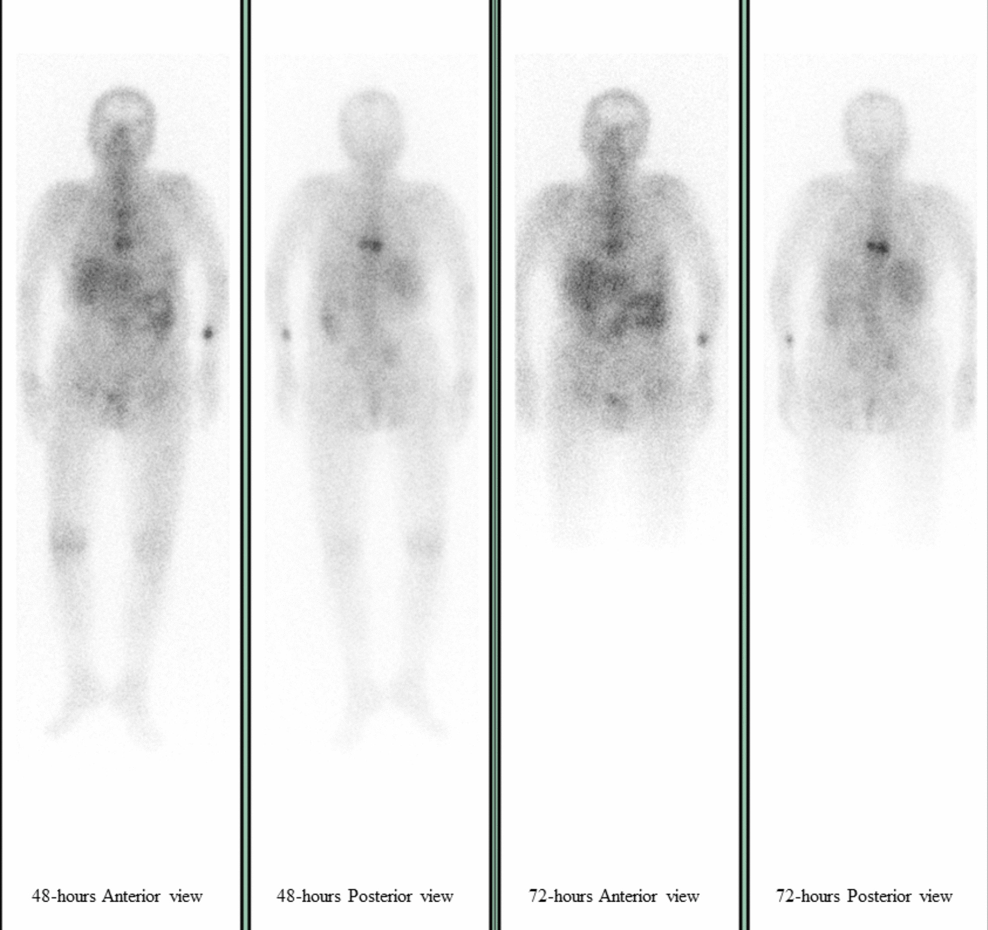
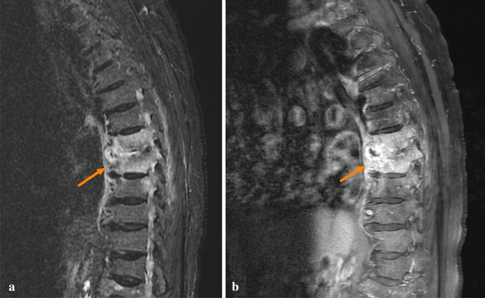
After starting flomoxef empirical treatment on day 1 of admission, the patient’s fever subsided promptly and her abdominal pain was also alleviated. Thus, we downgraded her regimen of antibiotics from flomoxef to cefuroxime on the following day. The patient’s fever did not recur during the remainder of her hospital stay. Although the urine culture indicated a possible UTI, a gallium scan was also conducted to rule out other potential causes of recurrent fever and to determine the infection source of P. mirabilis bacteremia. The scan revealed increased Ga67 citrate uptake in the ninth thoracic vertebra, which aligned with the location of the patient’s back pain (Fig. 1). We also conducted magnetic resonance imaging (MRI) several days later. Both the short T1 inversion recovery (STIR) sequence image (Fig. 2a) and the contrast-enhanced T1 sequence (Fig. 2b) depicted enhanced signals between the seventh and eighth thoracic vertebral segments, which indicated infectious spondylodiscitis. In addition, mild spinal canal stenosis was noted in the same area, which may have caused the patient’s neurological symptoms. We consulted orthopedic specialists, and they suggested conservative treatment. The patient’s family was also opposed to surgery. We did not assess whether the patient had tuberculosis (TB) infection; however, the possibility of TB infection was low because the patient reported no TB-associated symptoms in the past decade, and no TB-like lesions were identified in her chest X-rays.
Fig. 1.

Gallium scan of the patient showing increased Ga67 citrate uptake in the ninth thoracic vertebra area
Fig. 2.

Magnetic resonance imaging of short T1 inversion recovery sequence (a) and contrast-enhanced T1 sequence (b). Enhanced signals between the seventh and eighth thoracic vertebra segments (arrows )
Intravenous cefuroxime injections during the entire course of hospitalization stabilized the patient’s clinical condition, and white blood cell count was normal during hospitalization. Because of the considerable improvement in her condition, she was discharged on the 19th day of hospitalization with oral cefuroxime as her take-home medication. She reported to the outpatient department for follow-up once a month for 4 months. Her erythrocyte sedimentation rate (ESR) had decreased from 91 mm/hour to approximately 30 mm/hour by her fourth outpatient department visit. After the initial 4-month follow-up period, the patient came to the outpatient department once every 3 months. At her last follow-up, which was approximately 10 months after discharge, the patient had no complaints of back pain or fever. According to the patient and her caregivers, she became more energetic and was no longer experiencing proximal weakness of the lower limbs.
Discussion and conclusions
In this case report, an elderly woman with recurrent fever was admitted to the hospital and eventually diagnosed with P. mirabilis vertebral osteomyelitis. She had a clinical history of two previous P. mirabilis infections and bacteremia episodes. In her previous hospital admissions, her fever resolved within 1 day after starting antibiotics but resurged after termination of antibiotic treatment, a perplexing clinical presentation that has not been reported in previous P. mirabilis vertebral osteomyelitis cases. However, the patient also exhibited continuous UTI, progressive back pain, and deteriorating neurological functions, with the latter two being well-described clinical signs of P. mirabilis vertebral osteomyelitis in the literature.
Vertebral osteomyelitis is an infectious disease that affects the spine. It is frequently misdiagnosed because of its insidious onset and unclear clinical course. Older adults are particularly vulnerable to this disease. Its incidence is approximately 2.2–5.8 per 100,000 person years [1]. The most common symptoms of vertebral osteomyelitis are back pain, fever, and local tenderness. Severe cases involve neurologic impairments, such as sensory loss, weakness, and radiculopathy, due to bone destruction and subsequent spinal cord compression [5]. Other means of diagnosis include laboratory findings of increased ESR, CRP, and leukocytosis. MRI is the most sensitive mode of examination for detecting vertebral osteomyelitis, and its diagnostic accuracy can be maximized if it is performed using contrast agents, fat-suppressed sequences, and subtraction images to prevent bone marrow and fat tissue from emerging in T1-weighted sequences [6].
Vertebral osteomyelitis caused by P. mirabilis is rare. A retrospective cohort study reported that, among 253 episodes of vertebral osteomyelitis, only 5 cases were secondary to P. mirabilis infections [7]. The first case of vertebral osteomyelitis caused by P. mirabilis was published in 1934, in which the patient was infected after undergoing cystoscopy [8]. In the subsequent years, several case reports of vertebral osteomyelitis caused by P. mirabilis infection have been published [2, 3, 9–13]. Diagnoses have mostly been based on blood or urine culture results combined with radiological evidence, and some studies have confirmed P. mirabilis infection through direct biopsy and pus culture. The majority of patients have recovered completely after long-term treatment with antibiotics. The regimens and treatment durations have varied across studies; patients have been prescribed different antibiotics, including quinolone, cephalosporin, and aminoglycoside, with treatment periods lasting from 5 weeks to over 3 months. Surgery is occasionally necessary, particularly in cases involving abscesses formation or severe bone destruction. Immediate drainage of abscess, disc excision, and internal vertebral stabilization have been performed in these patients [3, 12].
Our patient initially presented with recurrent fever and P. mirabilis bacteremia, which was confirmed by three sets of positive P. mirabilis blood culture results in both of her previous admissions. We initially suspected a UTI to be the source of infection based on the patient’s quick response to antibiotic treatment. However, her fever recurred soon after discharge. This was an atypical clinical presentation of UTI. In addition, her persistent back pain and bilateral lower limbs weakness prompted us to consider the possibility of an undetected infectious source of bacteremia, such as an infective vertebral disease. Consequently, we investigated the matter more thoroughly. Vertebral osteomyelitis was detected through inflammation scans and MRI images. Although TB-induced vertebral osteomyelitis could not be ruled out, we judged that TB infection was not likely considering the patient’s past history, chest X-rays, and significant improvements with cefuroxime treatment. We did not perform surgical aspiration of the lesion because of opposition from the patient’s family; however, considering that P. mirabilis was the only pathogen detected in the blood cultures, vertebral osteomyelitis caused by P. mirabilis is the most likely final diagnosis.
Conclusions
Because chronic back pain in the older adult population is often dismissed as a common ailment, the diagnosis of spondylodiscitis requires a high index of suspicion. Physicians should consider the possibility of undiscovered osteomyelitis when the patient presents with concurrent neurologic symptoms and bacteremia. Once TB osteomyelitis is ruled out, pyogenic osteomyelitis of other pathogens should be considered. Vertebral osteomyelitis caused by P. mirabilis is extremely rare, and old age, chronic renal failure, and diabetes mellitus are possible predisposing factors. Previous case reports have also indicated recurrent UTI episodes despite sufficient antibiotic treatment; this symptom, along with the presence of permanent indwelling urinary catheter, should alert clinicians to the possibility of causative pathogenic organisms other than S. aureus [2]. Vertebral osteomyelitis caused by P. mirabilis can be sufficiently treated with long-term antibiotics administrations; the duration of treatment was around 4 months in the present case, with intravenous injection for the first 3 weeks and oral admission for the remainder of treatment. Overall, the disease has a favorable prognosis.
Acknowledgements
This manuscript was edited by Wallace Academic Editing
Abbreviations
- CRP
C-reactive protein
- ESR
Erythrocyte sedimentation rate
- MRI
Magnetic resonance imaging
- UTI
Urinary tract infections
- TB
Tuberculosis
Authors’ contributions
M-HC, the first author, took the responsibility of conceptualization, data gathering, and manuscript drafting. M-HL, took the responsibility of literature search, analysis of the data, and manuscript drafting. Y-CL, contributed significantly to manuscript revision. C-HL, the corresponding author, took the responsibility of conceptualization, revision of the current manuscript, and manuscript submission. All authors read and approved the final manuscript.
Funding
The authors declare that this study received no financial support.
Availability of data and materials
All data generated or analyzed during this study are included in this published article.
Declarations
Ethics approval and consent to participate
For this case, no ethical approval was sought. Informed consent was obtained from the patient.
Consent for publication
Written informed consent was obtained from the patient for publication of this case report and any accompanying images. A copy of the written consent is available for review by the Editor-in-Chief of this journal.
Competing interests
All authors confirmed that this work had no potential financial or nonfinancial conflicts of interest.
Footnotes
Publisher’s Note
Springer Nature remains neutral with regard to jurisdictional claims in published maps and institutional affiliations.
Contributor Information
Ming-Hsiu Chiang, Email: b101103050@tmu.edu.tw.
Mei-Hui Lee, Email: 16286@s.tmu.edu.tw.
Yung-Ching Liu, Email: yungching@tmu.edu.tw.
Chi-Hung Lee, Email: 15526@s.tmu.edu.tw.
Reference
- 1.Kehrer M, Pedersen C, Jensen TG, Lassen AT. Increasing incidence of pyogenic spondylodiscitis: a 14-year population-based study. J Infect. 2014;68(4):313–320. doi: 10.1016/j.jinf.2013.11.011. [DOI] [PubMed] [Google Scholar]
- 2.Yong TY, Li JY. Proteus vertebral osteomyelitis. Int J Rheum Dis. 2009;12(2):155–157. doi: 10.1111/j.1756-185X.2009.01397.x. [DOI] [PubMed] [Google Scholar]
- 3.Redfern RM, Cottam SN, Phillipson AP. Proteus infection of the spine. Spine (Phila Pa 1976). 1988;13(4):439–41. doi: 10.1097/00007632-198804000-00014. [DOI] [PubMed] [Google Scholar]
- 4.Batson OV. The function of the vertebral veins and their role in the spread of metastases. Ann Surg. 1940;112(1):138–149. doi: 10.1097/00000658-194007000-00016. [DOI] [PMC free article] [PubMed] [Google Scholar]
- 5.Zimmerli W. Clinical practice vertebral osteomyelitis. N Engl J Med. 2010;362(11):1022–1029. doi: 10.1056/NEJMcp0910753. [DOI] [PubMed] [Google Scholar]
- 6.Hogan A, Heppert VG, Suda AJ. Osteomyelitis. Arch Orthop Trauma Surg. 2013;133(9):1183–1196. doi: 10.1007/s00402-013-1785-7. [DOI] [PubMed] [Google Scholar]
- 7.McHenry MC, Easley KA, Locker GA. Vertebral osteomyelitis: long-term outcome for 253 patients from 7 Cleveland-area hospitals. Clin Infect Dis. 2002;34(10):1342–1350. doi: 10.1086/340102. [DOI] [PubMed] [Google Scholar]
- 8.Selig S. Bacillus proteus osteomyelitis infection of the spine. J Bone Joint Surg. 1934;16B:189–192. [Google Scholar]
- 9.Fafalak M, Bryll-Perzan E, Bobde R, Bhattarai B. Proteus mirabilis: a rare but dangerous cause of osteomyelitis. p. A6560-A. 10.1164/ajrccm-conference.2019.199.1_MeetingAbstracts.A65602019.
- 10.Digby JM, Kersley JB. Pyogenic non-tuberculous spinal infection: an analysis of thirty cases. J Bone Joint Surg British Volume. 1979;61(1):47–55. doi: 10.1302/0301-620x.61b1.370121. [DOI] [PubMed] [Google Scholar]
- 11.de Weerd W, Kimpen JL, Miedema CJ. Spinal osteomyelitis caused by Proteus mirabilis in a child. Eur J Pediatr. 1997;156(1):33–34. doi: 10.1007/s004310050547. [DOI] [PubMed] [Google Scholar]
- 12.Henriques CQ. Osteomyelitis as a complication in urology; with special reference to the paravertebral venous plexus. Br J Surg. 1958;46(195):19–28. doi: 10.1002/bjs.18004619505. [DOI] [PubMed] [Google Scholar]
- 13.Smits JP, Peltenburg HG, Mooi-Kokenberg EA, Koster T. Proteus mirabilis spondylodiscitis complicating a urinary tract infection. Scand J Infect Dis. 2006;38(6–7):575–576. doi: 10.1080/00365540500504141. [DOI] [PubMed] [Google Scholar]
Associated Data
This section collects any data citations, data availability statements, or supplementary materials included in this article.
Data Availability Statement
All data generated or analyzed during this study are included in this published article.


