Abstract
Background
Ventricular septal defects (VSDs) are the most common congenital cardiac defect in horses.
Objectives
To identify prevalence, age, breed, and sex distribution of VSD and to describe associated clinical and ultrasonographic findings.
Animals
Hospital‐based population of 21 136 horses presented to the equine internal medicine department.
Methods
Medical records over a 12‐year period were reviewed for VSD confirmed by ultrasonography. Age, breed, sex, sport discipline, murmur, clinical signs, outcome, VSD type, VSD size, shunt velocity, cardiac dimensions, concomitant cardiac anomalies, and valvular regurgitations were recorded.
Results
From 1894 horses that underwent echocardiography, 54 had a VSD: 42 as an isolated lesion and 12 as part of complex congenital heart disease (CHD). Median age was 5 years (range, 0‐26) and 1 year (range, 0‐8), respectively. Warmbloods and males were overrepresented. In the isolated VSD group, only 15% had associated clinical signs and most horses had a perimembranous VSD (pmVSD; 36/42). Horses with a pmVSD and clinical signs showed a significantly lower maximal shunt velocity (3.77 vs 5.20 m/s; P < .001), higher VSD/Aortic root (Ao) diameter (0.52 vs 0.38; P = .05), higher left atrium/Ao diameter (1.94 vs 1.22; P < .001), and higher pulmonary artery/Ao diameter (1.15 vs 0.88; P = .005) compared to horses without clinical signs. All horses with complex CHD had clinical signs and abnormal cardiac dimensions.
Conclusion and Clinical Importance
Most isolated VSD were diagnosed only at a later age and were not associated with clinical signs. Horses with complex CHD were more likely to have or develop clinical signs at younger age.
Keywords: cardiology, congenital heart disease, echocardiography, horses, interventricular septum
Abbreviations
- Ao
aorta
- AoV
aortic valve
- ASD
atrial septal defect
- CHD
congenital heart disease
- infVSD
infundibular ventricular septal defect(s)
- LA
left atrium
- LADd
left atrial diameter at end‐diastole
- LADs
left atrial diameter at systole
- LVID
left ventricular internal diameter at end‐diastole
- LVOT
left ventricular outflow tract
- muscVSD
muscular ventricular septal defect(s)
- PA
pulmonary artery
- PDA
patent ductus arteriosus
- pmVSD
perimembranous ventricular septal defect(s)
- PV
pulmonary valve
- RVOT
right ventricular outflow tract
- TV
tricuspid valve
- VSD
ventricular septal defect
1. INTRODUCTION
Ventricular septal defects (VSDs) are the most common congenital cardiac defect in horses, 1 , 2 , 3 , 4 , 5 with an increased prevalence in certain breeds such as Standardbreds, Welsh Mountain ponies, and Arabian horses. 1 , 5 , 6 , 7 They may occur in conjunction with other congenital cardiac malformations as part of complex congenital heart disease (CHD) 5 although they also may occur as an isolated defect. Congenital cardiac malformations occur secondary to errors during cardiac development, 3 and have different consequences depending on the extent of anomaly, leading to variable clinical signs. In contrast to humans, 8 the heritability of VSD in horses is unknown, but affected horses should be considered unsuitable for breeding. 7 To make a diagnosis, ultrasonographic sequential segmental analysis 9 is recommended to identify unusual arrangement of the cardiac chambers and major arteries, and altered topographic cardiovascular anatomy. It is not uncommon for isolated VSD to be diagnosed at a later age. Literature is scarce regarding case series of congenital cardiac defects, especially isolated VSD. 1 , 2 , 5 The purpose of this retrospective study was to identify the prevalence, age, breed, and sex distribution of VSD as an isolated defect and as part of a complex congenital anomaly, to describe the associated cardiac murmur and to correlate the ultrasonographic characteristics of the VSD with cardiac dimensions, clinical signs, and sport discipline, in an equine internal medicine hospital population over a 12‐year period (2008‐2019).
2. MATERIAL AND METHODS
Medical records from horses, admitted to the Large Animal Internal Medicine Department of Ghent University between 2008 and 2019, were reviewed. The breed and sex distribution of the entire population was recorded. For each horse with an ultrasonographic diagnosis of VSD, age, breed, sex, sport discipline (pleasure riding, dressage, or jumping), and outcome (discharged or euthanasia after diagnosis) were obtained. Clinical signs and heart murmurs at the time of VSD diagnosis and electrocardiographic and echocardiographic findings, including measurements and presence of concomitant structural abnormalities, were recorded. All owners were contacted 1 to 12 years after initial examination and follow‐up data were obtained.
2.1. Echocardiography and color flow Doppler examination
Sequential segmental analysis 9 was used to identify the presence of a VSD with or without other congenital cardiac malformations. Standard 2‐dimensional (2D), color flow Doppler and continuous wave Doppler examinations together with continuous electrocardiographic recording 10 were obtained by trained observers using a dedicated ultrasonographic unit (GE Vivid 7 Dimension, Vivid IQ or Vivid E95, GE Healthcare, Diegem, Belgium). Oblique views from multiple angles were taken to optimize visualization of congenital defects. Data were digitally stored and off‐line analysis was performed using dedicated software (Echopac version BT12 or 203, GE Medical System, Diegem, Belgium). The images were reassessed by a single person (G. van Loon, Dip. ECEIM, assoc. member ECVDI, PhD) for this study.
Horses were divided into 2 groups, depending on the presence of an isolated VSD or a VSD as part of complex CHD. Horses with CHD without simultaneous presence of a VSD were not included in the study.
The VSD type was recorded as perimembranous (pmVSD), infundibular (infVSD), or muscular (muscVSD). The VSD long‐axis and short‐axis size was measured from 2D images (Figures 1 and 2) and the average of both values was used in ratio calculations. Maximal shunt velocity (Vmax) was obtained by continuous wave Doppler and the pressure gradient was calculated using the modified Bernoulli equation. 11 Color flow, pulsed wave and continuous wave Doppler were used to diagnose shunting of blood. The end‐systolic aortic and left atrial (LA) diameter were measured using the right parasternal short‐axis view at aortic valve (AoV) level to obtain the VSD/aortic root diameter ratio (VSD/Ao) and left atrial/aortic root diameter ratio (LA/Ao). The end‐systolic pulmonary artery (PA) diameter was measured using the right parasternal left ventricular outflow tract (LVOT) view and the pulmonary artery/Ao root diameter ratio (PA/Ao) was calculated. The left atrial diameter, parallel with the mitral valve, was measured at end‐systole (LADs) and end‐diastole (LADd) using a right parasternal 4‐chamber view. End‐diastolic left ventricular internal diameter at chordal level (LVIDd) was measured using an M‐mode from a right parasternal short‐axis view or from a long‐axis 4‐chamber view. All valves were evaluated for structural abnormalities and regurgitation, using different scanning planes and color flow Doppler. Aortic valve prolapse was diagnosed when a distinct inflection point from the natural curve of the sinus of Valsalva was found and part of the base of the cusp was dragged into the defect. 12 , 13 Prolapse of the AoV was assessed using a right parasternal LVOT view for a pmVSD and using a right parasternal right ventricular outflow tract (RVOT) view and left parasternal cranial view of pulmonary valve (PV) and PA for an infVSD. Dextroposition with override of the aortic root and right ventricular (RV) hypertrophy were subjectively identified on the right parasternal LVOT view and 4‐chamber view, respectively. Stenosis of the PV was morphologically identified, including abnormal valve structure, abnormal valve opening, and abnormal annulus diameter using the right parasternal RVOT view and confirmed using a left parasternal view of the RVOT and PA. Continuous wave Doppler maximal flow velocity (Vmax; m/s), maximal pressure gradient (Pmax; mm Hg), and pulmonic velocity time integral (cm) were used to identify flow across the stenotic valve. A patent ductus arteriosus (PDA) was identified using the right parasternal RVOT view or left parasternal long‐axis view of the PA with its bifurcation. 14 To confirm the diagnosis, systolic or continuous flow between aorta and PA was documented by color flow Doppler. An atrial septal defect (ASD) was identified by angling the ultrasound beam in multiple directions from a right and left parasternal approach. Shunting of blood through the defect was confirmed by color flow Doppler.
FIGURE 1.
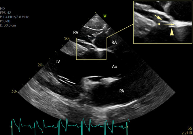
An isolated perimembranous ventricular septal defect (pmVSD), viewed from a right parasternal left ventricular outflow image, shows discrete bulging of the sinus of Valsalva (arrow) and the opened aortic valve cusp (arrowhead) into the defect. The double arrow shows the pmVSD longitudinal diameter. Ao, aorta; LV, left ventricle; PA, pulmonary artery; RA, right atrium; RV, right ventricle
FIGURE 2.
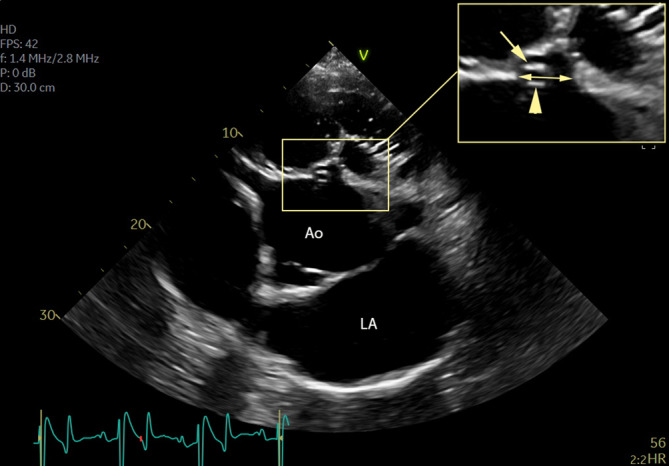
Short‐axis view of the perimembranous ventricular septal defect, adjacent to the commissure of the right and noncoronary cusp, of the same horse as in Figure 1. The sinus of Valsalva (arrow) and opened aortic valve cusp (arrowhead) are shown. The double arrow shows the pmVSD short‐axis diameter. Ao, aorta; LA, left atrium; pmVSD, perimembranous ventricular septal defect
When horses were admitted for reevaluation, examinations were repeated as mentioned above.
2.2. Data analysis
Normality of all variables was graphically assessed using Q‐Q plots. Within the group of pmVSD, a multivariate analysis of variance (SPSS Statistics 26) was used to compare VSD average diameter, Vmax, VSD/Ao, LA/Ao, PA/Ao, LADs, LADd, and LVIDd between horses with and without clinical signs. P values ≤.05 were considered significant.
3. RESULTS
3.1. General characteristics of the study sample
During the 12‐year study period, 21 136 horses were admitted to the Department of Large Animal Internal Medicine, and consisted of 60% males (25% stallions) and 60% Warmbloods. Only 7% were racehorses (5% Trotters and 2% Thoroughbreds). Six percent of the entire study sample were younger than 1 month and 89% were older than 6 months. Almost all underwent cardiac auscultation. Nine percent (1894/21136) received an echocardiographic examination. Fifty‐four (54/1894; 2.9%) horses had a VSD confirmed by echocardiography and their median age was 5 years (range, 0‐26). None of the horses had ˃1 VSD. Forty‐two of the horses with VSD (78%) had no other congenital cardiac anomaly, whereas 12 horses (22%) had a VSD combined with another cardiac malformation (ASD, dextroposition with override of the aortic root, RV hypertrophy, pulmonary stenosis, PDA, tricuspid valve [TV] atresia). In the entire equine internal medicine patient population, this resulted in a prevalence of 0.2% (42/21136) for isolated VSD and 0.05% (12/21136) for VSD in combination with other cardiac anomaly.
3.2. Characteristics of the isolated VSD
The median age at presentation for these 42 horses was 5 years (range, 0‐26), 67% were males (38% stallions) and 60% were Warmbloods. Only 1 Thoroughbred and 1 Trotter were present in the VSD population. Twenty‐nine horses had a right‐sided (grade 3/6 to 6/6) and 8 a left‐sided (grade 4/6 to 6/6) pansystolic murmur with a holosystolic murmur on the contralateral side. Three horses had a holosystolic murmur on the right and left side and in 2 the murmur type had not been documented. In 3 horses (3/42), atrial fibrillation was present. On echocardiography, 86% (36/42) of the horses with an isolated VSD had a pmVSD (Figure 3, Video S1) of which 64% (23/36) had AoV prolapse into the defect (Figure 4, Video S2), which was associated with AoV regurgitation in 79% of those cases (19/23) and with a diastolic murmur on auscultation in 26% (6/23). Ten percent (4/42) had an infVSD and 4% (2/42) a muscVSD. Detailed information can be found in Table 1. Only 15% (6/42) of the isolated VSD horses showed clinical signs such as dullness, poor body condition score, poor growth, chronic diarrhea, tachypnea, fever, ventral edema, or jugular vein pulsation. Seventy‐one percent (30/42) exercised on a regular basis, most of them for jumping, dressage, or pleasure riding. Of the 3 horses with atrial fibrillation, 2 had clinical signs with left and right heart dilatation. In the third horse, only the left heart was markedly dilated.
FIGURE 3.
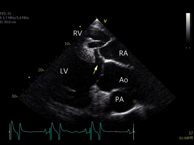
An isolated perimembranous ventricular septal defect (arrow), viewed from a right parasternal left ventricular outflow image. There is no prolapse of the aortic valve. Ao, aorta; LV, left ventricle; PA, pulmonary artery; RA, right atrium; RV, right ventricle
FIGURE 4.
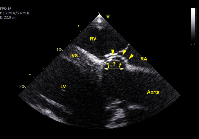
A large isolated perimembranous ventricular septal defect (pmVSD), viewed from a right parasternal left ventricular outflow image in a 15‐year‐old warmblood horse. The marked bulging of the sinus of Valsalva (arrows) into the defect reduces the VSD shunt area but also causes aortic valve regurgitation. The double arrow shows the pmVSD longitudinal diameter. Arrowheads indicate the septal leaflet of the tricuspid valve. IVS, interventricular septum; LV, left ventricle; PA, pulmonary artery; RA, right atrium; RV, right ventricle
TABLE 1.
Breed, median age, sex, and ventricular septal defect type of the affected horses
| Isolated VSD with clinical signs (n = 6) | Isolated VSD without clinical signs (n = 36) | VSD as part of complex congenital cardiac anomaly (n = 12) | |
|---|---|---|---|
| Breed |
5 Warmbloods 1 Pony |
20 Warmbloods 5 Ponies 5 Draft horses 2 Welsh ponies 1 Arabian horse 1 Lusitano 1 Thoroughbred 1 Trotter |
6 Warmbloods 1 Draft horse 2 Welsh ponies 2 Arabian horses 1 Arabian x Friesian |
| Median (range) age | 4 (0‐14) years | 6 (0‐26) years | 1 (0‐8) years |
| Sex |
2 Stallions 1 Gelding 3 Mares |
14 Stallions 11 Geldings 11 Mares |
4 Stallions 3 Geldings 5 Mares |
| VSD type |
5 Perimembranous 1 Infundibular |
31 Perimembranous 3 Infundibular 2 Muscular |
11 Perimembranous 1 Muscular |
Abbreviations: n, number of horses; VSD, ventricular septum defect(s).
Within the group of pmVSD, horses with and horses without clinical signs showed a significantly different Vmax (3.77 vs 5.20; P < .001), VSD/Ao (0.52 vs 0.38; P = .05), LA/Ao (1.94 vs 1.22; P < .001), and PA/Ao (1.15 vs 0.88; P = .005), respectively (Table 2). Within the same group, no significant difference was found between horses with and without clinical signs in pmVSD diameter (2.80 vs 2.18; P = .37), LADs (11.0 vs 10.8; P = .88), LADd (10.2 vs 9.82; P = .8), and LVIDd (12.4 vs 10.7; P = .25). Concerning the pmVSD group with clinical signs, 1 horse had a small VSD diameter, but also had a chordal rupture, a severely dilated left heart and PA, and lung edema. In the pmVSD group without clinical signs, 6 horses had a pmVSD diameter ˃3 cm, of which 5 had AoV prolapse that decreased the size of the shunt (Figure 4).
TABLE 2.
Mean, SD, and P value of horses with and without clinical signs within the isolated perimembranous ventricular septal defect (pmVSD) group
| Isolated pmVSD with clinical signs (n = 5) | Isolated pmVSD without clinical signs (n = 30 a ) | P value | |
|---|---|---|---|
| VSD diameter (cm) | 2.80 ± 0.75 | 2.18 ± 0.90 | .37 |
| Vmax (m/s) | 3.77 ± 0.70 | 5.20 ± 0.61 | <.001 |
| VSD/Ao | 0.52 ± 0.1 | 0.38 ± 0.13 | .05 |
| LA/Ao | 1.94 ± 0.54 | 1.22 ± 0.14 | <.001 |
| PA/Ao | 1.15 ± 0.25 | 0.88 ± 0.16 | .005 |
| LADs (cm) | 11.00 ± 1.52 | 10.80 ± 2.48 | .88 |
| LADd (cm) | 10.15 ± 1.05 | 9.82 ± 2.57 | .8 |
| LVIDd (cm) | 12.38 ± 1.60 | 10.71 ± 2.73 | .25 |
Note: P values ≤.05 were considered statistically significant.
Abbreviations: Ao, aorta diameter; LA, left atrium; LADd, left atrial diameter at end‐diastole; LADs, left atrial diameter at end‐systole; LVIDd, end‐diastolic left ventricle internal diameter; n, number; PA, pulmonary artery diameter; Vmax, maximal VSD shunt velocity.
No measurements were available from one Draft horse due to poor image quality.
For the horses with infVSD (4/42; Figure 5), the average infVSD diameter in 2 horses was 1.75 cm and 3.60 cm and the VSD/Ao was 0.36 cm and 0.57 cm, respectively. In the third horse, only a short‐axis diameter was available, which was 2.6 cm. The average maximal shunt velocity of all 3 was 4.02 ± 0.59 m/s (range, 3.36‐4.48 m/s). In 1 Draft horse, no measurements could be obtained because of insufficient image quality. One horse with an infVSD had clinical signs and was simultaneously diagnosed with AoV prolapse in the defect (Video S3) and subsequent valve tear.
FIGURE 5.
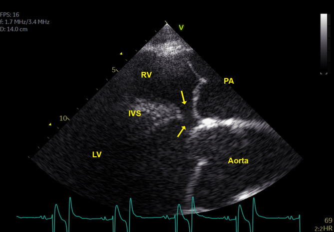
Left parasternal cranial view of the pulmonary valve in a 3‐year‐old Warmblood horse shows an infundibular septal defect (arrows) ventral to the pulmonary valve, connecting left (LV) and right (RV) ventricle. IVS, interventricular septal defect; PA, pulmonary artery
Concerning the horses diagnosed with a muscVSD (2/42; Figure 6), average VSD diameter was 0.9 and 1.1 cm, maximal shunt velocity 4.18 and 6.15 m/s and VSD/Ao ratio 0.18 and 0.20, respectively. None of them had clinical signs.
FIGURE 6.
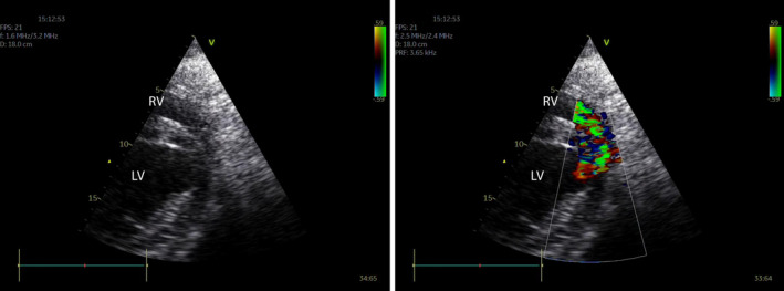
A right parasternal short‐axis view near the ventricular apex (cranial to the right) without (left) and with (right) color flow Doppler. The small muscular ventricular septal defect is difficult to visualize on 2D because it is located ventrally and near the junction of septum and free wall. With color flow Doppler the systolic turbulent flow (green) into the right ventricle can be identified. LV, left ventricle; RV, right ventricle
Eighty‐one percent (34/42) of the horses with an isolated VSD were discharged. Four horses with a pmVSD and 1 with an infVSD could be followed over several years. Echocardiography was repeated 6 months (1 horse) or approximately 1 year (4 horses) after diagnosis. In addition, 3 were re‐evaluated 3 to 4 times over the 3 to 5 years after initial diagnosis. In all 5 horses, VSD diameter and Vmax remained similar at re‐examination. In 3 of 4 horses with pmVSD and AoV prolapse, mild to severe AoV regurgitation was diagnosed at initial examination and remained stable during follow‐up examinations. The fourth horse did not have AoV prolapse at the initial examination but it developed 1 year after initial diagnosis. Three of the 4 horses diagnosed with pmVSD continued their regular exercise (from recreational riding to 135 cm jumping).
Telephone contact 1 to 12 years after initial diagnosis allowed follow‐up information to be obtained in 62% of the cases. Twenty‐six percent of the horses followed‐up exercised at recreational (low) level exercise to more intense jumping competition on a regular basis without noticeable exercise intolerance. One 8‐year‐old horse with additional severe mitral and tricuspid regurgitation and cardiomegaly, for which it had been advised to no longer ride the horse, collapsed and died during a forest walk. Twenty‐nine percent of the horses were euthanized within 3 years after initial diagnosis because of the VSD or other health problems.
3.3. Characteristics of the complex congenital malformations
In 22% (12/54) of the horses with a VSD, the VSD was part of complex CHD with the presence of ≥1 of the following abnormalities: ostium primum ASD, dextroposition with override of the aortic root, RV hypertrophy, pulmonary stenosis, PDA, and TV atresia. This represents 0.6% (12/1894) of the population that underwent an echocardiographic examination. Detailed information can be found in Table 1. Eleven of the 12 horses had a pmVSD; 1 horse had a muscVSD. The median age at presentation was 1 year (range, 0‐8), 58% were males and 50% were Warmbloods. Two Arabians, 1 Arabian cross‐breed, 1 Draft horse, and 2 Welsh ponies were affected. No racehorses were affected. Almost all horses had a left‐ and right‐sided systolic murmur ranging from holosystolic (grade 4‐5/6) to pansystolic (grade 4‐5/6). Two also had a holodiastolic murmur. Murmur intensity was highest on the right side of the thorax or similar on both sides. In 2 horses, the murmur was incompletely or not recorded. Two horses had a continuous murmur associated with a PDA. One horse had AF and tachycardia (>52 bpm) at rest was recorded in 7 horses. At the time of examination, all horses had clinical signs, some more pronounced than others. Nasal discharge, cough, tachypnea, dyspnea, fever, collapse, mild colic, exercise intolerance, ventral edema, and jugular vein pulsation were reported. In 4 cases, arterial oxygen tension was measured and was decreased (range, 20‐92 mm Hg). Three horses (3/12) exercised on a regular basis, 1 of them frequently participated in low level endurance competitions (40 km), but all of them had exercise intolerance. The mean ± SD VSD diameter was 2.93 ± 1.44 cm, mean maximal shunt velocity 2.90 ± 0.88 m/s, and mean VSD/Ao was 0.59 ± 0.13. Because of insufficient image quality in a Draft horse, the VSD/Ao could not be calculated. In 2/12 horses, no measurements of the left heart were obtained, whereas the left heart was enlarged in 8/10. Eight of 12 horses had a VSD in combination with dextroposition with override of the aortic root (Figure 7, Video S4), PV stenosis (Figure 8, Video S5), and RV hypertrophy, fulfilling the criteria of tetralogy of Fallot (TOF), of which 4 had a dilated PA. One of the TOF horses also was diagnosed with an ostium primum ASD and was referred to as pentalogy of Fallot. In almost all horses with complex CHD (10/12), PV stenosis was present. In all, a predominant left‐to‐right shunting of blood through the VSD was found. Two horses had an ostium primum ASD and 1 also had TV atresia, a cleft mitral valve, a VSD and a difference in left ventricle (LV) and RV size, which was compatible with the diagnosis of atrioventricular septal defect with tricuspid atresia and ventricular imbalance. 15
FIGURE 7.
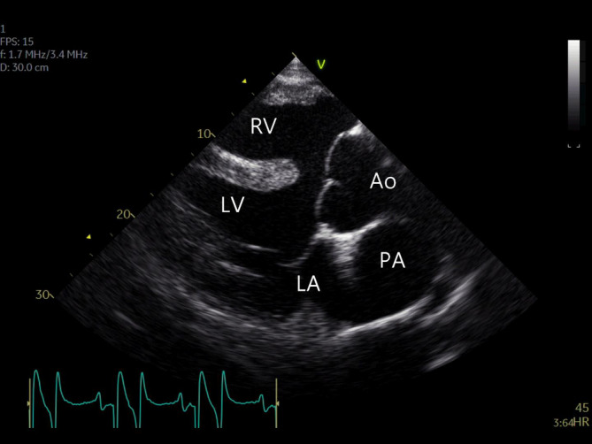
A right parasternal left ventricular outflow view shows right ventricular hypertrophy, a large perimembranous ventricular septal defect and dextroposition with override of the aortic root in this horse with Tetralogy of Fallot. Ao, aorta; LA, left atrium; LV, left ventricle; PA, pulmonary artery; RV, right ventricle
FIGURE 8.
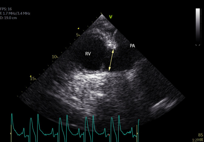
A left parasternal cranial view of the pulmonary valve shows marked pulmonary valve stenosis (double arrow). PA, pulmonary artery; RV, right ventricle
Ten horses (10/12) were euthanized and 2 were discharged. Despite the advice not to ride both discharged horses, 1 died during recreational exercise several weeks after diagnosis.
4. DISCUSSION
In this retrospective study, we describe 54 horses presented with VSD as an isolated defect or as part of a CHD. Most cases were isolated pmVSD, often with AoV prolapse into the defect. In this predominant non‐racehorse population, most were Warmbloods and males with a median age of 5 years old (range, 0‐26). All horses had loud, widely radiating cardiac murmurs. Concerning the isolated pmVSD, most horses (85%) did not have any clinical signs and exercised on a regular basis in contrast to horses affected with complex CHD. In the latter population, some had a tetralogy of Fallot and almost all were euthanized. In horses with an isolated pmVSD, a significant difference was found in several ultrasonographic variables between horses with and without clinical signs. Follow‐up indicated that 26% of the horses continued to exercise on a regular basis.
Although it has been reported that VSD often accompany more complicated malformations, 5 in our population only 22% of the VSD were associated with another cardiac malformation. This represents a prevalence of 0.05% of the entire referred equine internal medicine hospital population (21 136 horses) over a 12‐year period. In the literature, a prevalence of 0.1% to 0.5% has been reported for congenital cardiac anomalies in general. 5 Previous studies 1 reported a high prevalence of VSD in Standardbreds and Arabians. In our study, isolated VSDs were predominantly found in Warmbloods, which reflected the breed distribution within our study sample. The mean age in our study was higher than previously reported. 1 Similar to an older study, 1 more horses with an isolated VSD were males, which also reflected the sex distribution within our study sample. The group with complex CHD included fewer males and fewer Warmbloods compared to the study sample. All owners were advised not to breed their horses.
The phenotypic presentation in our study was the same as reported in literature, with a pmVSD being most commonly reported. 1 , 3 , 4 Because color flow Doppler might slightly overestimate the size of a defect, we chose to asses VSD size from 2D images (Figures 1 and 2). Anatomically, the pmVSD (Figure 3, Video S1) is located below the right and noncoronary cusp of the AoV and encompassing or contiguous with the fibrous part of the interventricular septum. 16 , 17 The infVSD, also called a “subpulmonic” (Figure 5), “subarterial,” “supracristal,” “doubly committed,” or “juxta‐arterial” defect, is in fibrous continuity between the AoV and PV. 3 Both the pmVSD and infVSD can be identified on ultrasonography using standardized scanning planes. 1 , 6 , 11 The muscVSD, also called “trabecular defect,” is surrounded by muscular tissue. 3 This VSD type may be located anywhere along the interventricular septum and may be difficult to identify. Based on our experience, it is useful to examine the entire RV with color flow Doppler from long‐ and short‐axis planes to identify small defects, especially those located apically or near the junction between septum and free wall (Figure 6).
As described in previous reports, 1 , 4 , 6 , 11 , 18 in our study all isolated VSD were associated with a harsh holo‐ or pan‐systolic murmur on the right or left or both thoracic sides. Concerning the pmVSD, the murmur usually is loudest just below the TV region. In contrast, the murmur of an infVSD is loudest cranially on the left side, below the PV area, and a muscVSD may create a left‐ or right‐sided systolic murmur with a different radiation pattern. 7 , 11 , 18 In our study, 64% of the pmVSD cases had AoV prolapse (Figure 4, Video S2), which is higher than the 25% previously reported. 1 , 4 , 6 , 11 Factors such as breed, anatomical position of the aorta in relation to other cardiac structures, and better image quality today might have played a role in the high prevalence of AoV prolapse identified in our study. In contrast to human patients, AoV prolapse seemed to occur less frequently in infVSD, but the numbers in our study were too small to draw conclusions. 12 , 13 Aortic valve prolapse usually involved part of the aortic root. Indeed, also in human medicine, AoV prolapse is defined as a distinct inflection point from the natural curve of the sinus of Valsalva, with part of the base of the cusp dragged into the defect. 12 , 13 , 19 , 20 The AoV cusp is thin and mobile and especially vulnerable to be drawn into the high‐velocity low‐pressure jet as blood shunts left to right through the VSD. Over time, this can result in elongation of the nadir of the adjacent AoV cusp and dilatation of the sinus of Valsalva with outward and downward displacement of the aortic annulus. This often will lead to a lack of coaptation between the AoV cusps, resulting in AoV regurgitation. 20 One should be aware that concurrent presence of aortic regurgitation may result in more rapid deterioration of cardiac function.
A pmVSD may lead to a discrete systolic murmur over the PV area caused by relative pulmonary stenosis with occurrence of turbulence. 18
Small isolated VSD generally are well tolerated. 7 , 18 Larger isolated VSD lead to increased pulmonary flow and increased venous return to the LA and LV. This eventually causes LA enlargement and LV dilatation and eccentric hypertrophy. 11 Literature mentions that horses without clinical signs have pmVSD size <2.5 cm, maximal shunt velocity >4 m/s, and VSD/Ao <0.4, 1 which was the case for most of our horses without clinical signs. However, the pmVSD diameter was not significantly different between horses with and without clinical signs. This could be explained by the fact that a small VSD could be associated with clinical signs because of the presence of other important cardiac abnormalities, such as severe acquired valvular regurgitation. And on the other hand, several of the horses without clinical signs had a relatively large defect that was restrictive because of AoV prolapse into the defect, narrowing the actual shunt size. Nevertheless, some horses without clinical signs showed lower shunt velocity compared to the literature, a higher VSD/Ao ratio and increased LA, LV and PA size without noticeable exercise intolerance. These horses were used in less physically demanding sport disciplines, such as jumping, dressage, or pleasure riding, in contrast to a previous study of racehorses. 1 Our data therefore suggest that horses not engaged in racing may better tolerate larger VSDs. Also, increased cardiac chamber diameters in some of the horses might have been a consequence of valvular regurgitation rather than the septal defect. A few horses developed atrial fibrillation, which was presumed to be secondary to atrial dilatation. No ultrasonographic reference data, were found for infVSD or muscVSD.
In people, spontaneous closure of a pmVSD by a thin layer of fibrous tissue has been reported on rare occasions. 21 Also, muscVSD may close by apposition of the muscular borders of the VSD by muscular in‐growth and fibrous proliferation or by hypertrophy of the myocardium. In 1 horse with a 1.09 cm muscVSD, the VSD could no longer be identified on color flow Doppler when the horse was 25 months old. 21 Spontaneous closure was not encountered in our study, but not enough follow‐up data were available to draw any conclusions. Our study only included CHD cases when they were associated with a VSD. In many case reports, CHD defects are fatal early in life 6 and the diagnosis is made at that time. 5 , 14 , 15 , 16 , 17 , 18 , 19 , 20 , 21 , 22 In our study, horses with complex CHD were younger than those with isolated VSD, but still, half of them were ˃1 year old. An association between complex CHD and the Arabian breed has been confirmed. 5 In our study, most horses were Warmbloods but the Arabian breed was underrepresented in our overall internal medicine hospital population of 21 136 horses. As reported before and as seen in our study, foals affected with complex CHD often have a loud systolic murmur with the point of maximal intensity over the PV area. 6
Some horses with complex CHD exercised on regular basis, 1 frequently participated in low level endurance competitions and only had mild exercise intolerance, an occasional cough and increased sweating. Pulmonary valve stenosis may have an important effect on RV pressure and volume overload, which is compensated for by RV hypertrophy, but also decreases Vmax of the shunt flow and limits PA and LA volume overload. 6 As such, a mild degree of stenosis actually may be protective, and fewer clinical signs might be seen, than with a large pmVSD. 6
Eight horses were diagnosed with a Tetralogy of Fallot which consists of RV outflow obstruction associated with a VSD, dextroposition with override of the aortic root, and RV hypertrophy. In human medicine, 22 the term dextroposition of the aortic root is used when a pathological rightward displacement of the aortic root is present. 22 This is in contrast to overriding of the aorta without dextroposition, which can occur in an otherwise normal heart because of a sigmoid interventricular septum or RV hypertrophy. 22 One foal did have a muscVSD together with a reversed PDA, which has been described in a previous case report. 23 Both recorded ASDs were ostium primum ASDs, where the defect is situated in the ventral portion of the atrial septum and above the inlet ventricular septum, 15 and an atrioventricular septal defect was diagnosed in 1 horse.
Our study had several limitations. Because the study consisted of an internal medicine hospital population, it does not allow conclusions to be drawn regarding the general horse population. Horses admitted to other departments for reproduction and obstetrics, medical imaging and orthopedics, and surgery and anesthesiology were not included. The mean VSD size, as well as LA, and LV diameters were calculated for all patients and included different breeds and ages, resulting in a wide range. Especially for the complex CHD group, the mean VSD diameter might have been underestimated because it included more young animals compared to the isolated VSD group. Bubble studies using injection of agitated saline, and measurement of hematocrit and arterial oxygen saturation were only performed occasionally. Our study focused on cardiac findings and does not allow conclusions to be drawn regarding other noncardiac congenital abnormalities that might have been associated with the CHD.
5. CONCLUSION
Horses with a VSD as part of complex CHD are more likely to develop clinical signs at a younger age. Isolated pmVSD are found most commonly. Size of the VSD and shunt flow velocity assist in judging their clinical importance, but concomitant aortic regurgitation caused by valvular prolapse in the defect complicates this judgment. Compared to a racehorse population, this predominant Warmblood population included horses without clinical signs with a larger isolated VSD size and lower shunt flow velocity than previously reported. For infVSD and muscVSD, no specific criteria currently exist to judge the effect on performance.
CONFLICT OF INTEREST DECLARATION
Authors declare no conflict of interest.
OFF‐LABEL ANTIMICROBIAL DECLARATION
Authors declare no off‐label use of antimicrobials.
INSTITUTIONAL ANIMAL CARE AND USE COMMITTEE (IACUC) OR OTHER APPROVAL DECLARATION
Authors declare no IACUC or other approval was needed. The animals were in‐clinic admissions, the examinations performed were routine examinations performed while working up those cases.
HUMAN ETHICS APPROVAL DECLARATION
Authors declare human ethics approval was not needed for this study.
Supporting information
Supplementary Table 1 Age, gender, breed, VSD type and other cardiac findings, history of exercise, follow‐up and clinical signs associated with isolated ventricular septal defect cases. AR: aortic regurgitation; Arab: Arabian horse; D: Died; E: Euthanized; G: gelding; inf: infundibular; HD: holodiastolic; HS: holosystolic; m: months M: Mare; musc: muscular; NA: not available; PS: pansystolic; pm: perimembranous; S: stallion; Sx: clinical signs; Thb: Thoroughbred; VSD: ventricular septal defect; Vmax: maximal VSD shunt velocity (m/s); Wbl: warmblood; y: years; +: present; −: absent; a: simultaneous presence of a chordae tendineae rupture; b: simultaneous presence of an aortic valve tear.
Supplementary Table 2 Age, gender, breed, VSD type and other cardiac findings associated with complex congenital heart disease cases. Ao: aorta; ASD: ostium primum atrial septal defect; Arab: Arabian horse; C: Continuous; d: days; G: gelding; M: mare; m: months musc: muscular; NA: not available; HD: holodiastolic; HS: holosystolic; PA/Ao: pulmonary artery/aortic root diameter ratio; PDA: patent ductus arteriosus; pm: perimembranous; PS: pansystolic; PVS: pulmonary valve stenosis; RVH: right ventricular hypertrophy; S: stallion; TOF: tetralogy of Fallot; TV: tricuspid valve; Vmax: maximal velocity; VSD: ventricular septal defect; VTI: velocity time integral; Wbl: warmblood; y: years; *: concomitant presence of tricuspid valve atresia; **: reversed PDA; ***: TOF + ASD: referred as pentalogy of Fallot; +: present; −: absent.
Video 1 An isolated perimembranous ventricular septal defect (arrow) is seen on a right parasternal left ventricular outflow view. Aortic valve prolapse is absent. Ao, aorta; LV, left ventricle; PA, pulmonary artery; RA, right atrium; RV, right ventricle.
Video 2 A isolated perimembranous ventricular septal defect (pmVSD), viewed from a right parasternal left ventricular outflow image. The marked bulging of the sinus of Valsalva (yellow arrow) and aortic valve (red arrow) into the defect reduces the VSD shunt area but also causes aortic valve regurgitation. Ao, aorta; LV, left ventricle; RA, right atrium; RV, right ventricle.
Video 3 A right parasternal view of the right ventricular outflow show turbulent flow (green) into the right ventricular outflow tract and pulmonary artery (PA) due to an infundibular ventricular septal defect, adjacent to the right coronary cusp of the aortic valve. The diastolic flow is caused by aortic valve regurgitation. Ao, aorta; RA, right atrium; RV, right ventricle
Video 4 A right parasternal left ventricular outflow view, shows right ventricular hypertrophy, a large perimembranous ventricular septal defect and dextroposition with override of the aortic root in this horse with Tetralogy of Fallot. Ao, aorta; LV, left ventricle; PA, pulmonary artery; RV, right ventricle
Video 5 A left parasternal cranial view of the pulmonary valve (circle) and pulmonary artery (PA). Abnormal pulmonary valve structure causes stenosis with only a small opening for the blood to pass (arrow) from right ventricle (RV) into PA. Ao, aorta; RA, right atrium
ACKNOWLEDGMENT
L. Vera and G. Van Steenkiste are PhD fellows funded by the Research Foundation Flanders (FWO‐Vlaanderen; Grant number 1134919N and 1S56217N, respectively). I. Vernemmen is a PhD fellow funded by the Special Research Fund of Ghent University (Grant number 01D16619). Ultrasound equipment was supported by the Special Research Fund of Ghent University (Grant number 01B05818).
De Lange L, Vera L, Decloedt A, Van Steenkiste G, Vernemmen I, van Loon G. Prevalence and characteristics of ventricular septal defects in a non‐racehorse equine population (2008‐2019). J Vet Intern Med. 2021;35:1573–1581. 10.1111/jvim.16106
Funding information Special Research Fund of Ghent University, Grant/Award Numbers: 01B05818, 01D16619; Research Foundation Flanders, Grant/Award Numbers: 1S56217N, 1134919N
REFERENCES
- 1. Reef VB. Evaluation of ventricular septal defects in horses using two‐dimensional and Doppler echocardiography. Equine Vet J Suppl. 1995;19:86‐96. [DOI] [PubMed] [Google Scholar]
- 2. Leroux AA, Detilleux J, Sandersen CF, et al. Prevalence and risk factors for cardiac diseases in a hospital‐based population of 3,434 horses (1994‐2011). J Vet Intern Med. 2013;27:1563‐1570.24112454 [Google Scholar]
- 3. Scansen BA. Equine congenital heart disease. Vet Clin North Am Equine Pract. 2019;35:103‐117. [DOI] [PubMed] [Google Scholar]
- 4. Reef VB. Cardiovascular disease in the equine neonate. Vet Clin North Am Equine Pract. 1985;1:117‐130. [DOI] [PubMed] [Google Scholar]
- 5. Hall TL, Magdesian KG, Kittleson MD. Congenital cardiac defects in neonatal foals: 18 cases (1992‐2007). J Vet Intern Med. 2010;24:206‐212. [DOI] [PubMed] [Google Scholar]
- 6. Marr CM. Cardiac murmurs: congenital heart disease. Cardiology of the Horse; Second edition. W. B. Saunders; 2010;193‐205. [Google Scholar]
- 7. Reef VB, Bonagura J, Buhl R, et al. Recommendations for management of equine athletes with cardiovascular abnormalities. J Vet Intern Med. 2014;28:749‐761. [DOI] [PMC free article] [PubMed] [Google Scholar]
- 8. Chung IM, Rajakumar G. Genetics of congenital heart defects: the NKX2‐5 gene, a key player. Genes. 2016;7(2):6. [DOI] [PMC free article] [PubMed] [Google Scholar]
- 9. Schwarzwald CC. Sequential segmental analysis ‐ a systematic approach to the diagnosis of congenital cardiac defects. Equine Vet Educ. 2008;20(6):305‐309. [Google Scholar]
- 10. Bomassi E, Misbach C, Tissier R, et al. Signalment, clinical features, echocardiographic findings, and outcome of dogs and cats with ventricular septal defects: 109 cases (1992–2013). J Am Vet Med Assoc. 2015;247:166‐175. [DOI] [PubMed] [Google Scholar]
- 11. Schwarzwald CC. Disorders of the cardiovascular system. Equine Internal Medicine. 4th ed.; St Louis, Missouri: W. B. Saunders; 2018;387‐541. [Google Scholar]
- 12. Tohyama K, Satomi G, Momma K. Aortic valve prolapse and aortic regurgitation associated with subpulmonic ventricular septal defect. Am J Cardiol. 1997;79:1285‐1289. [DOI] [PubMed] [Google Scholar]
- 13. Eroglu AG, Öztunç F, Saltik L, Dedeoglu S, Bakari S, Ahunbay G. Aortic Valve Prolapse and Aortic Regurgitation in Patients with Ventricular Septal Defect. Pediatric Cardiology. 2003;24(1):36‐39. 10.1007/s00246-002-1423-6. [DOI] [PubMed] [Google Scholar]
- 14. Vandecasteele T, Cornillie P, van Steenkiste G, et al. Echocardiographic identification of atrial‐related structures and vessels in horses validated by computed tomography of casted hearts. Equine Vet J. Vol 51; 2019(1):90‐96. [DOI] [PubMed] [Google Scholar]
- 15. Drábková Z, Amory H, Kabeš R, Melková P, van Loon G. Partial atrioventricular septal defect in an adult sport horse. J Vet Cardiol. Vol 31; 2020:8‐14. [DOI] [PubMed] [Google Scholar]
- 16. Franklin RCG, Béland MJ, Colan SD, et al. Nomenclature for congenital and paediatric cardiac disease: the International Paediatric and Congenital Cardiac Code (IPCCC) and the Eleventh Iteration of the International Classification of Diseases (ICD‐11). Cardiol Young. 2017;27:1872‐1938. [DOI] [PubMed] [Google Scholar]
- 17. Mostefa‐Kara M, Houyel L, Bonnet D. Anatomy of the ventricular septal defect in congenital heart defects: a random association? Orphanet J Rare Dis. 2018;13:118. [DOI] [PMC free article] [PubMed] [Google Scholar]
- 18. Young LE, van Loon G. Diseases of the heart and vessels. Equine Sports Medicine and Surgery. 2nd ed. W. B. Saunders; 2013;695–743. [Google Scholar]
- 19. Tweddell JS, Pelech AN, Frommelt PC. Ventricular Septal Defect and Aortic Valve Regurgitation: Pathophysiology and Indications for Surgery. Seminars in Thoracic and Cardiovascular Surgery: Pediatric Cardiac Surgery Annual. 2006;9(1):147‐152. 10.1053/j.pcsu.2006.02.020. [DOI] [PubMed] [Google Scholar]
- 20. Yacoub MH, Khan H, Stavri G, Shinebourne E, Radley‐Smith R. Anatomic correction of the syndrome of prolapsing right coronary aortic cusp, dilatation of the sinus of valsalva, and ventricular septal defect. J Thorac Cardiovasc Surg. 1997;113:253‐261. [DOI] [PubMed] [Google Scholar]
- 21. Short DM, Seco OM, Jesty SA, Reef VB. Spontaneous closure of a ventricular septal defect in a horse. J Vet Intern Med. 2010;24:1515‐1518. [DOI] [PubMed] [Google Scholar]
- 22. Isaaz K, Cloez JL, Marcon F, et al. Is the aorta truly dextroposed in tetralogy of Fallot? A two‐dimensional echocardiographic answer. Circulation. 1986;73:892‐899. [DOI] [PubMed] [Google Scholar]
- 23. Dufourni A, Decloedt A, De Clercq D, et al. Reversed patent ductus arteriosus and multiple congenital malformations in an 8‐day‐old Arabo‐Friesian foal. Equine Vet Educ. 2018;30:315‐321. [Google Scholar]
Associated Data
This section collects any data citations, data availability statements, or supplementary materials included in this article.
Supplementary Materials
Supplementary Table 1 Age, gender, breed, VSD type and other cardiac findings, history of exercise, follow‐up and clinical signs associated with isolated ventricular septal defect cases. AR: aortic regurgitation; Arab: Arabian horse; D: Died; E: Euthanized; G: gelding; inf: infundibular; HD: holodiastolic; HS: holosystolic; m: months M: Mare; musc: muscular; NA: not available; PS: pansystolic; pm: perimembranous; S: stallion; Sx: clinical signs; Thb: Thoroughbred; VSD: ventricular septal defect; Vmax: maximal VSD shunt velocity (m/s); Wbl: warmblood; y: years; +: present; −: absent; a: simultaneous presence of a chordae tendineae rupture; b: simultaneous presence of an aortic valve tear.
Supplementary Table 2 Age, gender, breed, VSD type and other cardiac findings associated with complex congenital heart disease cases. Ao: aorta; ASD: ostium primum atrial septal defect; Arab: Arabian horse; C: Continuous; d: days; G: gelding; M: mare; m: months musc: muscular; NA: not available; HD: holodiastolic; HS: holosystolic; PA/Ao: pulmonary artery/aortic root diameter ratio; PDA: patent ductus arteriosus; pm: perimembranous; PS: pansystolic; PVS: pulmonary valve stenosis; RVH: right ventricular hypertrophy; S: stallion; TOF: tetralogy of Fallot; TV: tricuspid valve; Vmax: maximal velocity; VSD: ventricular septal defect; VTI: velocity time integral; Wbl: warmblood; y: years; *: concomitant presence of tricuspid valve atresia; **: reversed PDA; ***: TOF + ASD: referred as pentalogy of Fallot; +: present; −: absent.
Video 1 An isolated perimembranous ventricular septal defect (arrow) is seen on a right parasternal left ventricular outflow view. Aortic valve prolapse is absent. Ao, aorta; LV, left ventricle; PA, pulmonary artery; RA, right atrium; RV, right ventricle.
Video 2 A isolated perimembranous ventricular septal defect (pmVSD), viewed from a right parasternal left ventricular outflow image. The marked bulging of the sinus of Valsalva (yellow arrow) and aortic valve (red arrow) into the defect reduces the VSD shunt area but also causes aortic valve regurgitation. Ao, aorta; LV, left ventricle; RA, right atrium; RV, right ventricle.
Video 3 A right parasternal view of the right ventricular outflow show turbulent flow (green) into the right ventricular outflow tract and pulmonary artery (PA) due to an infundibular ventricular septal defect, adjacent to the right coronary cusp of the aortic valve. The diastolic flow is caused by aortic valve regurgitation. Ao, aorta; RA, right atrium; RV, right ventricle
Video 4 A right parasternal left ventricular outflow view, shows right ventricular hypertrophy, a large perimembranous ventricular septal defect and dextroposition with override of the aortic root in this horse with Tetralogy of Fallot. Ao, aorta; LV, left ventricle; PA, pulmonary artery; RV, right ventricle
Video 5 A left parasternal cranial view of the pulmonary valve (circle) and pulmonary artery (PA). Abnormal pulmonary valve structure causes stenosis with only a small opening for the blood to pass (arrow) from right ventricle (RV) into PA. Ao, aorta; RA, right atrium


