Abstract
Aim
Alterations in circadian rhythms caused by tumor growth are thought to be clinically relevant as they affect the prognosis and treatment response. We aimed to evaluate the chronotherapeutic approach in rats with ovarian cancer receiving cisplatin intravenously (IV) or with hyperthermic intraperitoneal chemoperfusion (HIPEC) and to assess daily variations in tumor and intestinal epithelium proliferation.
Methods
In the pilot study, we used 12 intact rats and 12 rats with transplantable ovarian cancer, which were euthanized at ZT0 (08:00, lights on), ZT6, ZT12 and ZT18. In the main study, we used 45 rats with transplantable ovarian cancer. Animals were randomized into five groups: control, HIPEC with cisplatin at ZT0 (08:00), HIPEC with cisplatin at ZT12 (20:00), IV cisplatin at ZT0 and IV cisplatin at ZT12. We assessed the proliferation rate of tumor and small intestinal epithelium, apoptosis in small intestinal epithelium, and levels of γ-H2AX (DNA damage/repair marker) in kidneys and liver. Survival was calculated in each group.
Results
Ascitic ovarian cancer disrupted daily variations in intestinal epithelium proliferation and DNA damage/repair in rats. Ovarian carcinoma exhibited no daily variation in mitotic activity. In animals receiving IV cisplatin, massive cell damage in the renal medulla and cystic changes within renal tubules were observed, unlike in rats receiving HIPEC. Tumor mitotic activity was lower in morning-treated groups. The median survival of rats in the control group was 8.5 days (95% CI 6.0–22.0), in HIPEC at ZT0 40.5 days (95% CI 28.0–47.0, p<0.001) and in HIPEC at ZT12 32.0 days (95% CI 28.0–37.0, p<0.001).
Conclusion
In a rat model, ovarian tumor growth disrupted daily variations in intestinal epithelium proliferation and caused genotoxic stress in tumor-free tissues. HIPEC with cisplatin at ZT0 had a better efficacy/toxicity profile than HIPEC with cisplatin at ZT12 and IV administration at both time points.
Keywords: ovarian cancer, circadian rhythm, chronochemotherapy, cisplatin, hyperthermic intraperitoneal chemoperfusion, HIPEC, proliferation, DNA damage
Introduction
Circadian rhythms have emerged as an adaptation to the day/night cycle of environmental changes. In mammals, sleep, activity, body temperature, hormone secretion, cell proliferation, metabolism and gene expression exhibit approximately 24-hour oscillations.
A chronotherapeutic approach is often considered as an emerging field in anticancer therapy, although it was described several decades ago. Variability in the efficacy and toxicity of anticancer drugs throughout the day is linked to circadian oscillations in drug absorption, distribution, intracellular metabolism and elimination.1,2 The main principle of chronomedicine – the right drug at the right time – goes well with the knowledge on how circadian cycles work and with experimental data on chronopharmacology. However, precision timed therapy does not necessarily result in improved outcomes for cancer patients.
Several clinical and experimental studies have revealed that cancer itself may affect the host’s circadian rhythms. Disruptions in the normal regulation of sleep, body temperature and cortisol secretion rhythm, as well as reduced melatonin secretion, have been described in patients with advanced cancer.3–6 Moreover, patients who maintain endogenous circadian rhythmicity tend to have better outcomes.4,7,8 In animal cancer models, tumors may shift or disrupt circadian rhythms in distant cancer-free tissues and the body as a whole.9,10 For instance, the liver circadian metabolome and circadian transcriptome of tumor-bearing mice were shown to be affected by cancer progression.11,12 However, Granda et al did not find changes in circadian patterns in bone marrow cells when comparing mice with breast cancer and intact mice.13 Circadian rhythmicity of proliferation, expression of clock genes and thymidylate synthase activity (5-fluorouracil target) has been shown in transplantable sarcoma.14 Higher efficacy and better tolerability of timed platinum-based systemic chemotherapy have been demonstrated in several experimental models.15–19
Hyperthermic intraperitoneal chemoperfusion (HIPEC) is a type of locoregional chemotherapy which is used in combination with cytoreductive surgery for certain types of cancer affecting the peritoneum.20 Local hyperthermia along with high doses of the drugs enhances the penetration of the drugs into residual tumor nodes left after surgery.21,22 HIPEC is being studied as a therapeutic option for patients with locally advanced and locally recurrent ovarian cancer with few remaining therapeutic options, and it has shown efficacy in combination with cytoreductive surgery.23
In this study, we aimed to evaluate a chronotherapeutic approach in rats with ascitic ovarian cancer treated with cisplatin intravenously (IV) or via HIPEC, and to assess daily variations in intestinal epithelium and tumor proliferation.
Materials and Methods
Animals
A total of 69 4-month-old female Wistar rats were used in the experiments. The animals were quarantined for 1 month prior to the experiments (12:12 light/dark). During the experiment, animals were maintained on a 12:12 light/dark cycle (lights on at 08:00 a.m., lights off at 08:00 p.m.) at 21±2°C with 50±20% average humidity, and with ad libitum access to tap water and PK120 laboratory diet (Laboratorkorm, Moscow, Russia). Animal experiments were performed in compliance with the ethical principles established by the European Convention for the Protection of Vertebrate Animals used for Experimental and Other Scientific Purposes (accepted in Strasbourg 18.03.1986 and confirmed in Strasbourg 15.06.2006), and approved by the local ethical committee of N.N. Petrov National Medical Research Center of Oncology (Protocol #11, dated 21.09.2018).
Ovarian Cancer Model
We used ascitic ovarian cancer to simulate locally advanced ovarian cancer with peritoneal carcinomatosis in rats. Rat transplantable ovarian cancer cells were purchased from the tumor cell bank of N.N. Blokhin National Medical Research Center of Oncology.
We have described the model previously.24 In brief, each animal received rat ascitic fluid containing 1×10⁷ tumor cells intraperitoneally.
Chemotherapy Regimens
Cisplatin was administered on day 5 after ovarian cancer transplantation either intravenously or as HIPEC at 4 or 20 mg/kg correspondingly, which were the maximum tolerated doses.25 The HIPEC protocol has been described elsewhere.24
Study Design
Pilot Study
In the pilot study, 24 rats were randomly assigned either to the intact control group or to the group with transplanted ovarian cancer.
The day of ovarian cancer transplantation was the start of the experiment (day 0). Euthanasia was performed on day 10 at the following time points: 08:00 (ZT0, zeitgeber time 0, lights on, N=6), 14:00 (ZT6, 6 hours after lights on, N=6), 20:00 (ZT12, N=6) and 02:00 (ZT18, N=6). At each time point, three intact and three tumor-bearing animals were euthanized. The small intestine and liver of tumor-bearing rats were sampled and fixed in 10% neutral buffered formalin. To prepare a cell block, 1 mL of ascitic fluid was placed in an Eppendorf tube and centrifuged at 3000 rpm for 10 minutes. The remaining cell pellet after supernatant removal was placed in 10% formalin. Tissue samples and cell pellets were subjected to routine histological processing and embedded in paraffin blocks.
Main Study
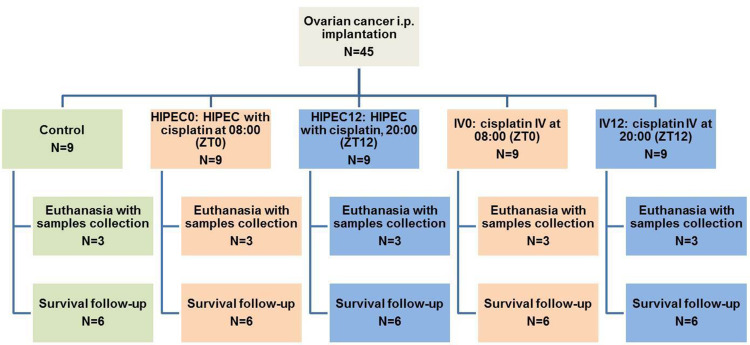
In the main study, 45 rats were randomized to five groups after ovarian cancer implantation:
Control: ovarian tumor, no treatment (N=9)
HIPEC-ZT0: HIPEC with cisplatin at 08:00 (ZT0) (N=9)
HIPEC-ZT12: HIPEC with cisplatin at 20:00 (ZT12) (N=9)
IV-ZT0: intravenous cisplatin at 08:00 (ZT0) (N=9)
IV-ZT12: intravenous cisplatin at 20:00 (ZT12) (N=9).
Six animals from each group were followed up to estimate survival (Figure 1). Three animals from each group were euthanized on day 10 (control) or 12 (treatment groups) to obtain samples for histology and immunohistochemistry. Kidneys, liver and small intestine were dissected; ascitic fluid was collected and centrifuged as described in the “Pilot Study” section, and then fixed in 10% neutral buffered formalin. Tissue samples and cell pellets were subjected to routine histological processing and embedded in paraffin blocks.
Figure 1.
Experimental study design.
Abbreviations: IV, intravenous; HIPEC, hyperthermic intraperitoneal chemoperfusion; ZT, Zeitgeber time.
Tumor Proliferation
Cell block slides were stained with hematoxylin and eosin (H&E) and used for proliferation analysis. The index of mitotic activity of ovarian cancer cells was calculated as the total number of mitotic figures per 10 high-power fields (×400 magnification).
Intestinal Epithelium Proliferation and Apoptosis
Intestinal epithelium proliferation was evaluated in H&E-stained transverse sections of jejunum. The number of mitotic figures was counted in 200 crypts cut longitudinally using a light microscope (magnification ×400). The number of apoptotic cells per crypt was counted similarly.
γ-H2AX Immunohistochemical Evaluation
Levels of γ-H2AX (a marker of DNA double-strand breaks) were measured in kidney and liver samples. Thus, 3–4 μm sections of liver and kidneys were fixed on positively charged slides. After deparaffinization and rehydration of the slides, antigen retrieval was performed in Tris–EDTA buffer (pH 9.0) at 95°C for 30 minutes. Endogenous peroxidase blocking was performed with 3% hydrogen peroxide for 15 minutes. Then, slides were washed in Tris–saline buffer (TSB) and incubated with primary antibody (anti-γ-H2AX (phospho-Ser139), ab81299; Abcam, USA) at room temperature for 1 hour. After three washes in TSB, the slides were incubated with secondary polymerized peroxidase polymer-conjugated antibodies (ab214882; Abcam, USA) for 1 hour. After three washes in TSB, the slides were exposed to chromogen 3ʹ-diaminobenzidine and incubated for 5 minutes. Slides were then counterstained with hematoxylin, dehydrated and mounted.
To assess anti-γ-H2AX staining in hepatocytes, the percentage of stained area of nuclei was calculated (stained nuclei area/total nuclei area). Total nuclei area was measured manually; stained nuclei area was measured using the Colour Deconvolution function in ImageJ software (NIH, USA). The localization and intensity of anti-γ-H2AX staining in kidneys were assessed qualitatively by a trained morphologist in a blinded manner (only in the main study).
Survival Analysis
Six animals from each group were followed up after the intervention. Cachexia, signs of hemorrhage, difficulty with breathing, and not being able to reach food and water were all considered as criteria for euthanasia. Survival was estimated as the number of days from the day of ovarian cancer transplant until death/euthanasia.
Statistical Analysis
GraphPad Prism 8.0.1 software was used for statistical analysis. Data are presented as mean values ± standard error of the mean. The Shapiro–Wilk test was used to test the normality of the data. Differences between groups and/or time points were tested by one-way ANOVA with Tukey’s post-hoc test. The Kaplan–Meier estimator was used in survival analysis, with survival values being compared using log-rank and Gehan–Breslow–Wilcoxon tests. The significance level was taken as 0.05.
Results
Pilot Study
Tumor Proliferation
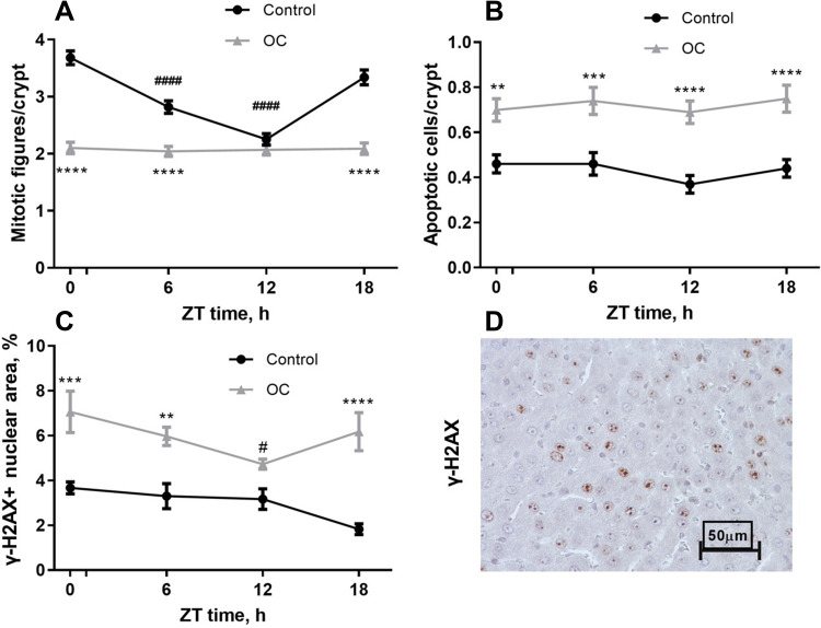
The mean mitotic activity index of ovarian carcinoma reached 107.33±3.84, at ZT0, 108.33±4.70 at ZT6, 110.67±4.84 at ZT12 and 108.67±4.84 at ZT18. No significant differences between the time points were found.
Intestinal Epithelium Proliferation and Apoptosis
In the control group, intestinal epithelium mitotic activity reached a maximum of 3.69±0.12 mitoses per crypt at ZT0, decreased to 2.82±0.11 at ZT6 (p<0.0001 vs ZT0, p<0.001 vs ZT18) and further to 2.25±0.10 at ZT12 (p<0.0001 vs ZT0 and ZT18, p<0.001 vs ZT6), and went up to 3.34±0.13 at ZT18. In animals with ovarian carcinoma, the number of mitoses per crypt was significantly decreased at time points ZT0, ZT6 and ZT18 (p<0.0001 vs control) and there were no differences between time points within the group (Figure 2A).
Figure 2.
Daily variation in proliferation in small intestine crypts, (B) apoptosis in small intestine crypts, and (C) DNA damage/repair in liver in a pilot study. (D) Representative photomicrograph of anti-γ-H2AX stained liver of tumor-bearing rat (×400), ZT6. Control: intact rats; OC: rats with ovarian cancer. **–p<0.01, ***–p<0.001, ****p<0.0001 compared to control at the same time point, #p<0.05 compared to ZT0, ####p<0.0001 compared to ZT0.
The number of apoptotic cells per crypt did not vary throughout the day; it was significantly increased, approximately 1.5-fold, in animals with carcinomatosis at all time points (p<0.01) (Figure 2B).
DNA Damage/Repair in Liver
DNA damage/repair in hepatocytes was increased 1.5–3.0-fold in tumor-bearing rats compared to control animals at time points ZT0, ZT6 and ZT18 (p<0.01) (Figure 2C and D). There was no daily variation in γ-H2AX level in control animals; however, in tumor-bearing rats the γ-H2AX level was significantly lower at ZT12 than at ZT0 (p<0.05).
Main Study
Tumor Proliferation
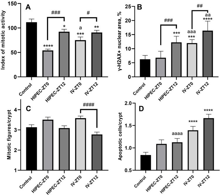
The index of mitotic activity of ovarian carcinoma in the control group (Figure 3A) was 111.66±3.32. The index was significantly reduced in all treatment groups compared to controls: 54.25±2.39 (р<0.0001) in HIPEC-ZT0, 92.3±2.51 (р<0.05) in HIPEC-ZT12, 75.0±3.28 (р<0.001) in IV-ZT0 and 90.66±2.25 (р<0.05) in IV-ZT12.
Figure 3.
Effects of HIPEC and IV cisplatin administration at ZT0 and ZT12 on (A) index of mitotic activity in tumor, (B) DNA damage/repair in liver (γ-H2AX-immunostaining, (C) mitotic activity, and (D)apoptosis in intestinal epithelium (mean ± SEM). *p<0.05, **p<0.01, ***p<0.001, ****p<0.0001 compared to control, ap<0.05, aap<0.01, aaa p<0.001, aaaap<0.0001 compared to different treatment at the same time, #p<0.05, ##p<0.01, ###p<0.001, ####p<0.0001 compared to the same treatment at the different time (Tukey’s test).
Therefore, in groups with both HIPEC (p<0.001) and IV administration (p<0.05), tumor mitotic activity was significantly reduced in animals with morning versus evening treatment.
Furthermore, at ZT0, HIPEC with cisplatin exerted a much more pronounced anti-proliferative effect compared to IV cisplatin administration (p<0.05).
Intestinal Epithelium Proliferation and Apoptosis
The mean number of mitotic figures per intestinal crypt (Figure 3C) in the control group was 3.13±0.13 and this value was at the same level in all groups with cisplatin administration. There was one intergroup difference, with a significantly higher number of mitotic figures in IV-ZT0 (3.58±0.10) compared to IV-ZT12 (2.78±0.11) (р<0.0001).
The number of apoptotic cells per crypt was at the lowest level in the control group (Figure 3D) (0.84±0.06), while in HIPEC-ZT0 and HIPEC-ZT12 groups it was slightly increased. In IV-ZT0 and IV-ZT12 groups, the number of apoptotic cells per crypt was significantly higher compared to the control group (1.39±0.09 and 1.66±0.09, respectively; p<0.0001). In animals treated at ZT12, a more profound toxic effect of cisplatin on the intestinal epithelium was found in the IV-ZT12 group compared to HIPEC-ZT12 (p<0.0001).
DNA Damage/Repair in Liver
The percentage of γ-H2AX-positive nuclear area (nuclei or foci of chromatin) (Figure 3B) in hepatocytes of control animals was 6.25±0.55%; in the HIPEC-ZT0 group there was a trend toward higher numbers (6.80±0.76%), but in the other experimental groups this percentage was significantly higher: 12.27±0.72% in HIPEC-ZT12 (p<0.001), 11.99±0.41% in IV-ZT0 (p<0.001) and 16.41±1.12% in IV-ZT12 (p<0.0001) groups. For both modes of cisplatin treatment, γ-H2AX staining was more intense at ZT12 compared to ZT0 (HIPEC: p<0.01; IV: p<0.01). Within each route of cisplatin administration, the percentage of γ-H2AX-stained nuclear area was significantly higher in evening-treated compared to morning-treated groups (HIPEC: p<0.001; IV: p<0.01).
DNA Damage in Kidney
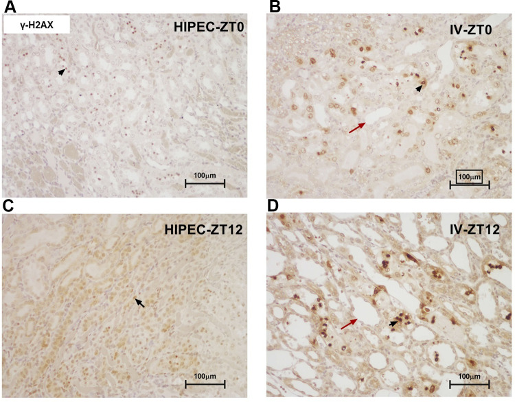
In kidney samples, the most intense anti-γ-H2AX staining was detected in the renal medulla across all groups. In animals receiving IV cisplatin, there was massive cellular damage in the renal medulla, with intense γ-H2AX staining and cystic changes within the renal tubules (Figure 4). In animals receiving HIPEC with cisplatin, post-treatment changes in renal tubules were less pronounced and staining with anti-γ-H2AX was less intense compared to the IV groups. Overall, the least intense staining of renal medulla with γ-H2AX was observed in the HIPEC-ZT0 group.
Figure 4.
Representative microphotographs of anti-γ-H2AX-stained kidney sections, illustrating samples from: (A) group receiving HIPEC with cisplatin at 08:00 (ZT0), (B) group receiving IV cisplatin at 08:00 (ZT0), (C) group receiving HIPEC with cisplatin at 20:00 (ZT12), (D) group receiving IV cisplatin at 20:00 (ZT12). Black arrows: positively stained nuclei; red arrows: cystic tubular changes (×200).
Survival Analysis
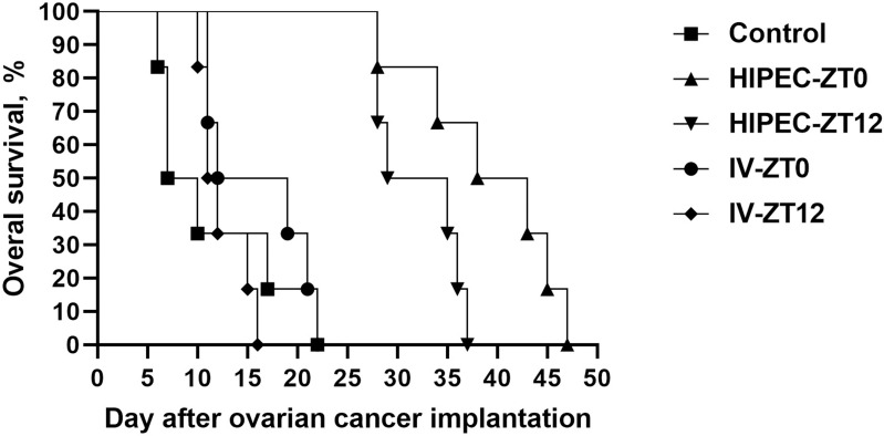
The median length of survival of rats (Figure 5) in the control group was 8.5 days (95% CI 6.0–22.0), while it was significantly higher in the HIPEC-ZT0 (40.5 days, 95% CI 28.0–47.0, p<0.001, log-rank test, p<0.05, Gehan–Breslow–Wilcoxon test) and HIPEC-ZT12 (32.0 days, 95% CI 28.0–37.0, p<0.001, log-rank test, p<0.01, Gehan–Breslow–Wilcoxon test) groups. Furthermore, median survival in the HIPEC-ZT0 group was significantly higher than in HIPEC-ZT12 (p<0.05, log-rank test). Median survival time was also significantly reduced in IV-ZT0 compared to HIPEC-ZT0 (15.5 days, 95% CI 11.0–22.0, p<0.001, log-rank test, p<0.05, Gehan–Breslow–Wilcoxon test) and in IV-ZT12 compared to HIPEC-ZT12 (11.5 days, 95% CI 10.0–16.0, p<0.001, log-rank test, p<0.01, Gehan–Breslow–Wilcoxon test).
Figure 5.
Survival of rats with ovarian cancer after intravenous administration and HIPEC with cisplatin at different time points.
Discussion
In this study, we show that ascitic ovarian cancer disrupts the daily variations in intestinal epithelium proliferation and DNA damage and repair. We observed higher efficacy and better tolerability of cisplatin administered either IV or as HIPEC at the beginning of the light phase (ZT0).
In the pilot study, we found a loss of the daily pattern of intestinal epithelium proliferation and its overall decrease, increased apoptosis in enterocytes and increased γ-H2AX staining in the liver of tumor-bearing rats. All of these changes could be caused by tumor development, which may lead to chronic inflammation and DNA damage in distant tissues.26 Dampening27 or phase shift28 of the intestinal epithelium proliferation rhythm in mammary carcinoma-bearing mice was also described previously. The loss of the peak of proliferation could explain why cisplatin treatment in the morning was no more toxic for enterocytes than evening treatment.
In tumor-bearing rats, the level of γ-H2AX declined at ZT12. γ-H2AX is not only a DNA damage marker but also a starting point for DNA repair molecular machinery assembly,29 so its decreased level at the end of the light phase may be an indicator of less efficient DNA repair. It has been shown that accumulation of the RAD51 gene at DNA lesions is a marker of homologous recombination proficiency.30 So, a lack of RAD51 expression following DNA damage could be considered as a functional biomarker of dysfunctional homologous recombination. γ-H2AX-RAD51 has shown promising results in breast cancer biopsies, predicting PARP inhibitor sensitivity and detecting the emergence of secondary resistance.31
Circadian regulation of DNA repair initially emerged in unicellular organisms to protect the genome from aggressive UV irradiation in the light phase.32 The rhythm of DNA damage response is conserved in animals; in mammalian cells and tissues that are never exposed to light throughout life, DNA repair is more efficient during daytime.33–35
In transplantable ovarian carcinoma, we failed to find daily changes in proliferation rate. According to published data, most poorly differentiated transplantable tumors have disorganized circadian rhythms,10,36 while chemically induced slowly growing tumors may retain rhythmic features such as clock and cell cycle-controlling gene expression.9,37,38 Thus, ovary carcinoma cells seem to divide in a circadian-independent manner; moreover, tumor growth disrupts the daily pattern of healthy tissue proliferation and DNA damage and repair.
In the main study, we revealed that HIPEC and morning cisplatin administration was more effective and tolerable compared to IV and evening administration, respectively. The advantages of HIPEC over systemic drug administration have been confirmed in previous experimental studies.25,39
This is mainly explained by the fact that cisplatin administered intraperitoneally affects tumor cells directly, while the absorbed portion of the drug binds with proteins, which leads to lower toxicity. Higher drug doses may be used compared to systemic administration.25
In this study, morning cisplatin treatment appeared to be more effective and less toxic than evening treatment. These results correspond well to those described by Seto et al, where peripheral neuropathy was less aggravated in morning-treated rats.19 In contrast, it has been shown that in mice or rats, platinum-based drugs are less toxic and more effective when administered at the beginning or in the middle of the dark phase.40–43 Moreover, elimination of cisplatin–DNA adducts reaches its peak in the late afternoon.34,35 It is important to note that in several of these studies,41–43 healthy tumor-free animals were used. In our main experiment, all of the rats had significant tumor burden that could shift or invert the rhythm of drug absorption or elimination, and as well as affect its circadian toxicity pattern. We assume that changes in the daily dynamics of proliferation and DNA damage/repair in tumor-free tissues may explain the reverse cisplatin efficacy/safety pattern.
We observed significantly lower damage to intestinal crypts in animals receiving cisplatin, both as HIPEC and through the IV route in the morning, with minimal damage registered in the group with HIPEC at ZT0. In addition, at this time point, we found only slightly increased expression of DNA damage/repair in the liver compared to the control group. Based on the obtained results, the toxicity of the studied cisplatin treatment modes increases in the following order: HIPEC-ZT0 < HIPEC-ZT12< IV-ZT0 < IV-ZT12.
Conclusion
In the present study, we found that ovarian tumor growth disrupts daily variations in intestinal epithelium proliferation and causes genotoxic stress in tumor-free tissues. HIPEC with cisplatin at ZT0 had a better efficacy/toxicity profile than HIPEC with cisplatin at ZT12 and IV administration at both time points, based on animal survival and tumor mitotic activity.
Funding Statement
This work was supported by the Russian Science Foundation (RSF) (grant number 18-75-10017).
Author Contributions
All authors made a significant contribution to the work reported, whether that is in the conception, study design, execution, acquisition of data, analysis and interpretation, or in all these areas; took part in drafting, revising or critically reviewing the article; gave final approval of the version to be published; have agreed on the journal to which the article has been submitted; and agree to be accountable for all aspects of the work.
Disclosure
The authors declare no conflict of interest.
References
- 1.Ozturk N, Ozturk D, Kavakli IH, Okyar A. Molecular aspects of circadian pharmacology and relevance for cancer chronotherapy. Int J Mol Sci. 2017;18:10. [DOI] [PMC free article] [PubMed] [Google Scholar]
- 2.Lévi F, Altinok A, Clairambault J, Goldbeter A. Implications of circadian clocks for the rhythmic delivery of cancer therapeutics. Philos Trans a Math Phys Eng Sci. 2008;366(1880):3575–3598. [DOI] [PubMed] [Google Scholar]
- 3.Lévi F, Komarzynski S, Huang Q, et al. Tele-monitoring of cancer patients’ rhythms during daily life identifies actionable determinants of circadian and sleep disruption. Cancers (Basel). 2020;12(7):1–21. [DOI] [PMC free article] [PubMed] [Google Scholar]
- 4.Sephton SE, Sapolsky RM, Kraemer HC, Spiegel D. Diurnal cortisol rhythm as a predictor of breast cancer survival. J Natl Cancer Inst. 2000;92(12):994–1000. [DOI] [PubMed] [Google Scholar]
- 5.Innominato PF, Giacchetti S, Bjarnason GA, et al. Prediction of overall survival through circadian rest-activity monitoring during chemotherapy for metastatic colorectal cancer. Int J Cancer. 2012;131(11):2684–2692. [DOI] [PubMed] [Google Scholar]
- 6.Innominato PF, Komarzynski S, Palesh OG, et al. Circadian rest-activity rhythm as an objective biomarker of patient-reported outcomes in patients with advanced cancer. Cancer Med. 2018;7(9):4396–4405. [DOI] [PMC free article] [PubMed] [Google Scholar]
- 7.Lévi F, Dugué PA, Innominato P, et al. Wrist actimetry circadian rhythm as a robust predictor of colorectal cancer patients survival. Chronobiol Int. 2014;31(8):891–900. [DOI] [PubMed] [Google Scholar]
- 8.Ortiz-Tudela E, Innominato PF, Rol MA, Lévi F, Madrid JA. Relevance of internal time and circadian robustness for cancer patients. BMC Cancer. 2016;16:1. [DOI] [PMC free article] [PubMed] [Google Scholar]
- 9.Soták M, Polidarová L, Ergang P, Sumová A, Pácha J. An association between clock genes and clock-controlled cell cycle genes in murine colorectal tumors. Int J Cancer. 2013;132(5):1032–1041. [DOI] [PubMed] [Google Scholar]
- 10.Huisman SA, Oklejewicz M, Ahmadi AR, et al. Colorectal liver metastases with a disrupted circadian rhythm phase shift the peripheral clock in liver and kidney. Int J Cancer. 2015;136(5):1024–1032. [DOI] [PubMed] [Google Scholar]
- 11.Masri S, Papagiannakopoulos T, Kinouchi K, et al. Lung adenocarcinoma distally rewires hepatic circadian homeostasis. Cell. 2016;165(4):896–909. [DOI] [PMC free article] [PubMed] [Google Scholar]
- 12.Hojo H, Enya S, Arai M, et al. Remote reprogramming of hepatic circadian transcriptome by breast cancer. Oncotarget. 2017;8(21):34128–34140. [DOI] [PMC free article] [PubMed] [Google Scholar]
- 13.Granda TG, Liu X-H, Smaaland R, et al. Circadian regulation of cell cycle and apoptosis proteins in mouse bone marrow and tumor. FASEB J. 2005;19(2):304–306. [DOI] [PubMed] [Google Scholar]
- 14.Wood PA, Du-Quiton J, You S, Hrushesky WJM Circadian clock coordinates cancer cell cycle progression, thymidylate synthase, and 5-fluorouracil therapeutic index. [DOI] [PubMed]
- 15.Li XM, Tanaka K, Sun J, et al. Preclinical relevance of dosing time for the therapeutic index of gemcitabine–cisplatin. Br J Cancer. 2005;92(9):1684–1689. [DOI] [PMC free article] [PubMed] [Google Scholar]
- 16.Granda TG, D’Attino RM, Filipski E, et al. Circadian optimisation of irinotecan and oxaliplatin efficacy in mice with Glasgow osteosarcoma. Br J Cancer. 2002;86(6):999–1005. [DOI] [PMC free article] [PubMed] [Google Scholar]
- 17.Sothern RB, Lévi F, Haus E, Halberg F, Hrushesk WJM. Control of a murine plasmacytoma with doxorubicin-cisplatin: dependence on circadian stage of treatment. J Natl Cancer Inst. 1989;81(2):135–145. [DOI] [PubMed] [Google Scholar]
- 18.Chakarov S, Petkova R, Russev G, Zhelev N. DNA damage and the circadian clock. Biodiscovery. 2014;13(13):1. [Google Scholar]
- 19.Seto Y, Okazaki F, Horikawa K, Zhang J, Sasaki H, To H. Influence of dosing times on cisplatin-induced peripheral neuropathy in rats. BMC Cancer. 2016;16:1. [DOI] [PMC free article] [PubMed] [Google Scholar]
- 20.Auer RC, Sivajohanathan D, Biagi J, Conner J, Kennedy E, May T. Indications for hyperthermic intraperitoneal chemotherapy with cytoreductive surgery: a systematic review. Eur J Cancer. 2020;127. [DOI] [PubMed] [Google Scholar]
- 21.Chan DL, Morris DL, Rao A, Chua TC. Intraperitoneal chemotherapy in ovarian cancer: a review of tolerance and efficacy. Cancer Manag Res. 2012;4. [DOI] [PMC free article] [PubMed] [Google Scholar]
- 22.Cristea M, Han E, Salmon L, Morgan RJ. Review: practical considerations in ovarian cancer chemotherapy. Ther Adv Med Oncol. 2010;2. [DOI] [PMC free article] [PubMed] [Google Scholar]
- 23.Boussios S, Pavlidis N. Ovarian cancer: state of the art and perspectives of clinical research. Ann Transl Med. 2020;8:24. [DOI] [PMC free article] [PubMed] [Google Scholar]
- 24.Bespalov VG, Kireeva GS, Belyaeva OA, et al. Both heat and new chemotherapeutic drug dioxadet in hyperthermic intraperitoneal chemoperfusion improved survival in rat ovarian cancer model. J Surg Oncol. 2016;113(4):438–442. [DOI] [PubMed] [Google Scholar]
- 25.Kireeva G, Kruglov S, Maydin M, et al. Modeling of Chemoperfusion vs Intravenous Administration of Cisplatin in Wistar Rats. Adsorpt Tissue Distribution Mol. 2020;25(20):4733. [DOI] [PMC free article] [PubMed] [Google Scholar]
- 26.Redon CE, Dickey JS, Nakamura AJ, et al. Tumors induce complex DNA damage in distant proliferative tissues in vivo. Proc Natl Acad Sci U S A. 2010;107(42):17992–17997. [DOI] [PMC free article] [PubMed] [Google Scholar]
- 27.Gubareva EA, Maydin MA, Tyndyk ML, Vìnogradova IA, Panchenko AV. Circadian rhythm of proliferation in intestinal epithelium and mammary tumors in HER-2/neu transgenic and FVB/N wild type mice; their correction with melatonin. Vopr Onkol. 2019;65(1):154–158. [Google Scholar]
- 28.Reyna JC, Barbeito CG, Badrán AF, Moreno FR. [Mitotic activity of duodenal-crypt enterocytes in mice with hepatocarcinoma]. Medicina (B Aires). 1997;57(6):708–712. [PubMed] [Google Scholar]
- 29.Turinetto V, Giachino C, Multiple facets of histone variant H2AX: a DNA double-strand-break marker with several biological functions. Nucleic Acids Research. 2015;5(43): 2489–2498. [DOI] [PMC free article] [PubMed] [Google Scholar]
- 30.Cruz C, Castroviejo-Bermejo M, Gutiérrez-Enríquez S, et al. RAD51 foci as a functional biomarker of homologous recombination repair and PARP inhibitor resistance in germline BRCA-mutated breast cancer. Ann Oncol. 2018;29:5. [DOI] [PMC free article] [PubMed] [Google Scholar]
- 31.Boussios S, Abson C, Moschetta M, et al. Poly (ADP-Ribose) polymerase inhibitors: talazoparib in ovarian cancer and beyond. Drugs R D. 2020;20. [DOI] [PMC free article] [PubMed] [Google Scholar]
- 32.Shostak A. Circadian clock, cell division, and cancer: from molecules to organism. Int J Mol Sci. 2017;18:4. [DOI] [PMC free article] [PubMed] [Google Scholar]
- 33.Palombo P, Moreno-Villanueva M, Mangerich A. Day and night variations in the repair of ionizing-radiation-induced DNA damage in mouse splenocytes. DNA Repair (Amst). 2015;28:37–47. [DOI] [PubMed] [Google Scholar]
- 34.Yang Y, Adebali O, Wu G, et al. Cisplatin-DNA adduct repair of transcribed genes is controlled by two circadian programs in mouse tissues. Proc Natl Acad Sci U S A. 2018;115(21):E4777–85. [DOI] [PMC free article] [PubMed] [Google Scholar]
- 35.Kang T-H, Lindsey-Boltz LA, Reardon JT, Sancar A. Circadian control of XPA and excision repair of cisplatin-DNA damage by cryptochrome and HERC2 ubiquitin ligase. Proc Natl Acad Sci U S A. 2010;107(11):4890–4895. [DOI] [PMC free article] [PubMed] [Google Scholar]
- 36.de Assis L, Moraes M, Magalhães-Marques K, Kinker G, Da Silveira Cruz-machado S, de Lauro Castrucci A. Non-metastatic cutaneous melanoma induces chronodisruption in central and peripheral circadian clocks. Int J Mol Sci. 2018;19(4):1065. [DOI] [PMC free article] [PubMed] [Google Scholar]
- 37.Yang K, Ye H, Tan X-M, Fu X-J, Li H-X. Daily rhythm variations of the clock gene PER1 and cancer-related genes during various stages of carcinogenesis in a golden hamster model of buccal mucosa carcinoma. Onco Targets Ther. 2015;1419. [DOI] [PMC free article] [PubMed] [Google Scholar]
- 38.Tan X-M, Ye H, Yang K, et al. Circadian variations of clock gene Per2 and cell cycle genes in different stages of carcinogenesis in golden hamster buccal mucosa. Sci Rep. 2015;5(1):9997. [DOI] [PMC free article] [PubMed] [Google Scholar]
- 39.Pelz JOW, Doerfer J, Dimmler A, Hohenberger W, Meyer T. Histological response of peritoneal carcinomatosis after hyperthermic intraperitoneal chemoperfusion (HIPEC) in experimental investigations. BMC Cancer. 2006;6:162. [DOI] [PMC free article] [PubMed] [Google Scholar]
- 40.Dakup PP, Porter KI, Little AA, et al. The circadian clock regulates cisplatin-induced toxicity and tumor regression in melanoma mouse and human models. Oncotarget. 2018;9(18):14524–14538. [DOI] [PMC free article] [PubMed] [Google Scholar]
- 41.Levi FA, Hrushesky WJM, Blomquist CH, et al. Reduction of cis-diamminedichloroplatinum nephrotoxicity in rats by optimal circadian drug timing. Cancer Res. 1982;42:3. [PubMed] [Google Scholar]
- 42.Boughattas NA, Lévi F, Fournier C, et al. Circadian rhythm in toxicities and tissue uptake of 1,2-diamminocyclohexane(trans-1)oxalatoplatinum(II) in mice. Cancer Res. 1989;49(12):3362–3368. [PubMed] [Google Scholar]
- 43.Boughattas NA, Lévi F, Hecquet B, et al. Circadian time dependence of murine tolerance for carboplatin. Toxicol Appl Pharmacol. 1988;96(2):233–247. [DOI] [PubMed] [Google Scholar]


