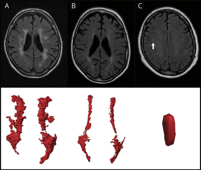
Figure 1. White Matter Hyperintensities (WMH) on Fluid-Attenuated Inversion Recovery (FLAIR) Images With Corresponding Visualizations in the Automated Algorithm.
Examples of confluent (A), periventricular (B), and deep (C) WMH on FLAIR images with the corresponding visualizations in our algorithm shown below. The deep WMH lesion (arrow) is reconstructed in the coronal view, while the periventricular and confluent WMH are viewed from a transverse perspective. Note that the coronal reconstruction of the deep WMH lesion (C) may be influenced by the slice thickness and the lesion may be more punctiform. The confluent WMH lesion in (A) showed a volume of 11.57 mL with an accompanying deep WMH volume of 0.25 mL. The periventricular WMH lesion in (B) showed a volume of 4.98 mL without any accompanying deep WMH lesions. The deep WMH lesion in (C) showed a volume of 0.02 mL with an accompanying periventricular and deep WMH volume of 2.12 and 0.49 mL, respectively.

