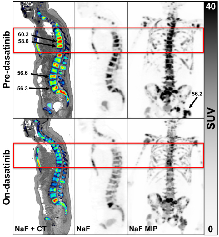
Figure 1.
Example of an 82-year-old patient scan 1 (top row) and scan 2 (bottom row). Panels left to right, NaF overlaid on CT, NaF alone and with NaF PET maximal image projection (MIP) of the entire WB volume. The red box is the single FOV for the dynamic scan. Three of the 5 hottest tumors were not located in the single dynamic FOV, the results of which were reported previously [8]. An example WB patient with none of the hottest tumors in the dynamic FOV appears in Supplementary Materials Figure S3.

