Southeast Asian ovalocytosis (SAO) is an autosomal dominant inherited red blood cell (RBC) membrane disorder caused by the heterozygous deletion of codons 400–408 in SLC4A1/band 3/anion exchanger 1 (AE1).1 This deletion leads to misfolding of the protein, creating an inactive anion-transporter and altering the mechanical stability of the RBC. Heterozygous SAO is characterized by the presence of stomatocytes, theta cells (RBC with two stomas), macro-ovalocytes and ≥25% ovalocytes in the peripheral blood smears.2 Although heterozygous SAO carriers are generally asymptomatic, homozygous SAO was considered to be lethal.3 However, one successful birth of a homozygous SAO individual, born to asymptomatic heterozygous SAO Comorian parents, was reported in 2014 showing an association with severe dyserythropoietic anemia.4 Here, we report the birth of five unrelated homozygous SAO babies in Malaysia showing that homozygous SAO is not as rare as previously thought. These babies belong to an SAO cohort collected from January 2007 to March 2020 at University Sains Malaysia (USM), which includes a total of 68 patients. The study was instigated to further understand the physiological implications of heterozygous SAO within the Malay population. The detection of numerous homozygous SAO births has highlighted the need for the development of good practice to support the survival of these babies.
SAO has a distinctive geographical distribution, occurring mostly in areas of Southeast Asia including neighboring regions of the Malay Peninsula, coastal regions of Papua New Guinea, Thailand, Indonesia, Taiwan and even Madagascar in the western Indian Ocean.5,6 Investigations into the high incidence of SAO in malarious areas revealed that the SAO mutation confers protection from severe malaria caused by Plasmodium vivax and Plasmodium falciparum.7,8
AE1 is the most abundant RBC membrane protein and mediates the efficient transport of carbon dioxide in the blood.1 Heterozygous SAO individuals have only 50% normal RBC anion transport activity and thus less efficient gas transport. The heterozygous expression of the SAO AE1 in the red cell membrane not only affects the folding and structure of AE1 but also affects the structure of other red cell membrane proteins depressing a relatively high number of red cell antigens.9 Neonatal anemia has also been reported in SAO families.2
A truncated form of AE1 is expressed in the α-intercalated cells of the kidneys and is essential for acid secretion in the urine.1 Loss of AE1 anion-transport activity in the kidney causes distal renal tubular acidosis (dRTA) and occurs when AE1 expression is severely reduced as in hereditary spherocytosis (HS)10,11 or when kidney AE1 activity is severely reduced1 or trafficking of kidney AE1 is impeded in the α-intercalated cell.1 Many dRTA mutations are recessive. When SAO AE1 is inherited in trans to a recessive dRTA AE1 mutation then the compound heterozygote will completely lack AE1 anion transport activity. This phenomenon explains the prevalence of dRTA in South East Asia where there are a number of recessive dRTA AE1 mutations maintained in the population. 12
Homozygous SAO individuals, with no functioning AE1, and homozygous HS individuals, with no AE1, suffer from hemolytic anemia, dRTA and failure to thrive.3,10,11 An in-depth study of homozygous SAO cells,13 showed expression of SAO AE1 during erythropoiesis (in vitro) affected trafficking and cytokinesis resulting in numerous multinucleated erythroblasts and reduced proliferation and enucleation. Unexpectedly, even the in vitro cultured heterozygous SAO erythroblasts displayed multinuclearity and reduced enucleation, with an intermediate phenotype part way between the homozygous and control cells,13 suggesting that heterozygous SAO individuals may have a mild dyserythropoiesis. Mature homozygous SAO RBC show signs of altered membrane composition, oxidative damage, are very large, oval and unstable.4,13 AE1 null HS individuals sometimes have a mild dyserythropoiesis10 and their RBC are unstable.11 Extensive clinical management of AE1 null individuals, including splenectomy, has improved the survival of these children, some of whom have now reached adolescence. Similarly, the Comorian homozygous SAO child is now 10 years old and quite well, receiving regular transfusions, iron chelation and acidosis treatment.4,13 From Malaysia, a live-born homozygous SAO neonate with dRTA was reported in 2015 but the child failed to thrive after 2 years.14
In Malaysia, where the prevalence of SAO is around 4% within the Malay ethnic group,15 SAO diagnostic tests are performed mostly upon still birth or recurring miscarriages of the mother. Several heterozygous SAO mothers in this cohort have a history of past miscarriages and complications during pregnancy such as hydrops fetalis as well as intrauterine death (Figure 1). Furthermore, SAO was identified in 18 of 22 dRTA patients (81.8%) when the association between SAO and dRTA was studied in the Malaysian dRTA cohort.15 Here, we provide an overview of a Malaysian heterozygous SAO cohort and describe the clinical characteristics of five live-born homozygous SAO cases in Malaysia.
This SAO cohort includes a total of 107 patients whose blood samples were tested for the 27 base pair deletion of AE1 using polymerase chain reaction (PCR) and blood smears (Online Supplementary Appendix; Online Supplementary Table S1). Among the 107 tested patients, 68 cases were positive where 93% (63 of 68 cases) of SAO positive cases belonged to Malay ethnicity. Within this Malaysian SAO cohort, 81 cases studied belong to a total of 28 families. Among these 28, 23 families had at least one heterozygous SAO family member. The remaining 26 samples were from individual cases, of which 15 were heterozygous SAO. The majority of the SAO positive cases (43%, n=29) are from Penang, followed by Kuala Lumpur (22%, n=15), Johor (10%, n=7), Sabah (9%, n=6), Kedah (9%, n=6), and Melaka (7%, n=5) (Online Supplementary Figure S3). A variety of RBC morphologies were observed from peripheral blood film examinations of the SAO positive cases, which includes macro-ovalocytes, stomatocytes, theta cells, spherocytes and target cells (Online Supplementary Table S1; Online Supplementary Figure S1).
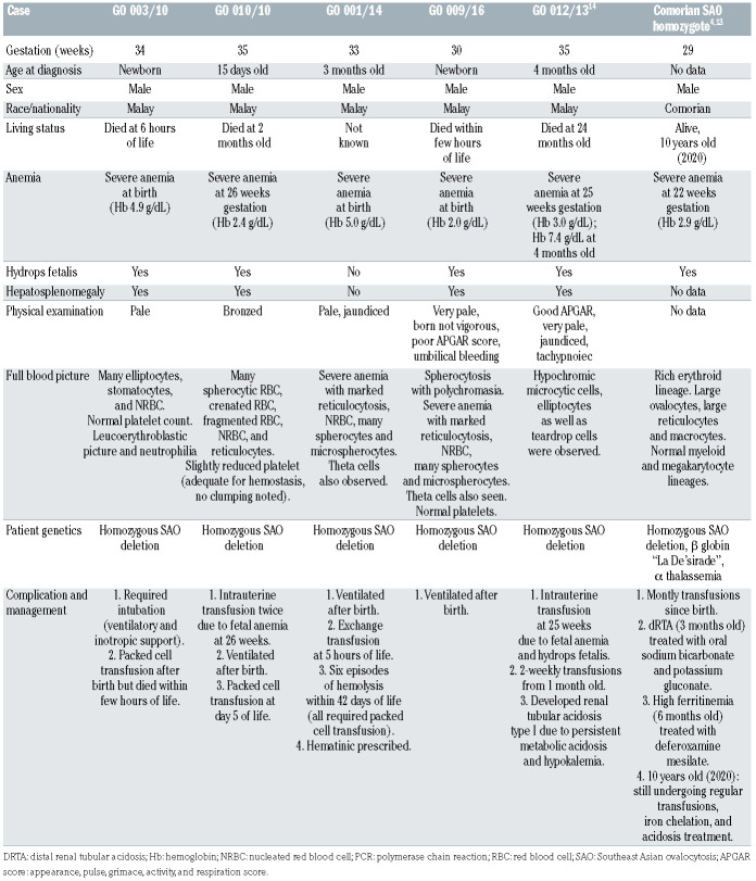
Surprisingly, a total of six homozygous SAO were found in our cohort, five of whom were live-born (Figure 1, Table 1, Online Supplementary Appendix, and Homozygous SAO case histories). All parents of liveborn homozygous SAO cases, with the exception of one (GO 009/16), were confirmed heterozygous for the SAO AE1 deletion; case GO 012/19 was a homozygous fetus that suffered intra-uterine death at 29 weeks. All the five live-born cases were delivered prematurely, at 30 to 35 weeks of gestation. Similar to previous homozygous SAO live-borns, our five homozygous SAO live-borns suffered a severe phenotype requiring intra-uterine transfusions, post-delivery transfusions and ventilation support. Despite efforts to manage their condition, two of the five patients reported here died, one during the neonatal period and one at 2 months. Even though the other two cases survived during the neonatal period, one of them (GO 012/13) succumbed during infancy due to complications of anemia and the other is not traceable after 2 months. Further details of the obstetric history of these families are provided in Figure 1 and the Online Supplementary Table S2.
Among the five live-born homozygous SAO cases, three of them had severe anemia at birth and two of them developed it at 26 weeks (GO/01/10) and 25 weeks (GO 012/13). All except GO 001/14 had hydrops fetalis and hepatosplenomegaly (Table 1). Physical examination revealed pale appearance, distended abdomen due to hepatosplenomegaly, and jaundice in all five cases. One of the five cases was reported to have dRTA diagnosed in the third month14 but the other four were not tested. Despite medical interventions, all five homozygous SAO children failed to thrive. This could be due to hepatosplenomegaly, severe anemia or lack of appropriate clinical management. It is difficult to gain more insights into the implications of the SAO homozygous cases as incomplete clinical data of the SAO cohort was collected, lacking data on hemoglobin abnormalities and blood group compatibility. It is also not known whether prenatal diagnosis and termination of pregnancy were offered. These services are not always available, and/or not acceptable to parents. It is now evident that careful clinical management can improve the life of homozygous SAO individuals.4 The long-standing assumptions that homozygous SAO is lethal, and that heterozygous SAO is asymptomatic should be re-assessed.
Table 1.
Comparison of five Malaysian live-born homozygous Southeast Asian ovalocytosis (SAO) cases with a previously reported Comorian homozygous SAO case.
Figure 1.
Family pedigree charts of five homozygous Southeast Asian ovalocytosis cases. Families of each homozygous Southeast Asian ovalocytosis (SAO) cases are represented: (A) GO 003/10, (B) GO 001/14, (C) GO 010/10, (D) GO 012/19, and (E) GO 012/13. Squares and circles denote male and females, respectively, with filled ones (grey or black) showing members affected with SAO. The numbered triangle (C) shows number of spontaneous abortions that occurred in the couple’s past pregnancies. Parents of two SAO homozygous children (GO 012/19 and GO 010/10) had multiple unsuccessful pregnancies. The mother of GO 012/19 suffered two intra-uterine deaths (including GO 012/19), while the mother of GO 010/10 suffered three spontaneous abortions. None of these other unsuccessful pregnancies were investigated further. On the other hand, parents of GO 012/13 have two living children among whom one is SAO heterozygous, while parents of GO 012/19 have three living children with unknown SAO status (see the Online Supplementary Table S2). d: died; IUD: intra-uterine death.
Several point mutations in AE1 that result in a wide range of complications have been reported, many of them leading to anemia and dRTA.2,12 Hemolytic anemia is recorded in children with homozygous G701D or compound heterozygous G701D/SAO, V850/SAO, A858D/SAO and V850/A858D mutations.1 Therefore, homozygous SAO and other AE1 null individuals, who show disparity in survival, may be indicating the cumulative result of hemolysis, acidosis and other hematological abnormalities influenced by the co-occurrence of additional mutations.
To our knowledge, this preliminary Malaysian SAO cohort study, for the first-time reports five homozygous SAO live-born cases. Since Malaysia and Southeast Asia have a high prevalence of SAO it would be beneficial to screen for SAO at antenatal clinics, at least for parents with a history of hydropic or stillborn fetuses. It is also essential to investigate the impact of heterozygous mutant SAO AE1 on other pathological conditions and ageing. Development of international and national guidelines for assessing a number of hematological, renal, and hepatic conditions in combination with genetic mutation markers to improve survival of homozygous AE1 null probands is needed.
Supplementary Material
Acknowledgments
the authors would like to thank the patients and their families for their full cooperation without which this report would not have been possible.
References
- 1.Wrong O, Bruce LJ, Unwin RJ, Toye AM, Tanner MJA. AE1 mutations, distal renal tubular acidosis, and Southeast Asian ovalocytosis. Kidney Int. 2002;62(1):10-19. [DOI] [PubMed] [Google Scholar]
- 2.Laosombat V, Dissaneevate S, Wongchanchailert M, Satayasevanaa B. Neonatal anemia associated with Southeast Asian ovalocytosis. Int J Hematol. 2005;82(3):201-205. [DOI] [PubMed] [Google Scholar]
- 3.Liu SC, Jarolim P, Rubin HL, et al. The homozygous state for the AE1 protein mutation in Southeast Asian ovalocytosis may be lethal. Blood. 1994;84(10):3590-3591. [PubMed] [Google Scholar]
- 4.Picard V, Proust A, Eveillard M, et al. Homozygous Southeast Asian ovalocytosis is a severe dyserythropoietic anemia associated with distal renal tubular acidosis. Blood. 2014;123(12):1963-1965. [DOI] [PubMed] [Google Scholar]
- 5.Wilder JA, Stone JA, Preston EG, Finn LE, Ratcliffe HL, Sudoyo H. Molecular population genetics of SLC4A1 and Southeast Asian ovalocytosis. J Hum Genet. 2009;54(3):182-187. [DOI] [PubMed] [Google Scholar]
- 6.Rabe T, Jambou R, Rabarijaona L, et al. South-East Asian ovalocytosis among the population of the Highlands of Madagascar: a vestigé of the island's settlement. Trans R Soc Trop Med Hyg. 2002;96(2):143-144. [DOI] [PubMed] [Google Scholar]
- 7.Allen SJ, O'Donnell A, Alexander ND, et al. Prevention of cerebral malaria in children in Papua New Guinea by Southeast Asian ovalocytosis AE1. Am J Trop Med Hyg. 1999;60(6):1056-1060. [DOI] [PubMed] [Google Scholar]
- 8.Rosanas-Urgell A, Lin E, Manning L, et al. Reduced risk of Plasmodium vivax malaria in Papua New Guinean children with Southeast Asian Ovalocytosis in two cohorts and a case-control study. PLoS Med. 2012;9(9):e1001305. [DOI] [PMC free article] [PubMed] [Google Scholar]
- 9.Bruce LJ, Beckmann R, Ribeiro ML, et al. A AE1-based macrocomplex of integral and peripheral proteins in the RBC membrane. Blood. 2003;101(10):4180-4188. [DOI] [PubMed] [Google Scholar]
- 10.Kager L, Bruce LJ, Zeitlhofer P, et al. AE1 null (VIENNA), a novel homozygous SLC4A1 p.Ser477X variant causing severe hemolytic anemia, dyserythropoiesis and complete distal renal tubular acidosis. Pediatr Blood Cancer. 2017;64(3). [DOI] [PubMed] [Google Scholar]
- 11.Ribeiro ML, Alloisio N, Almeida H, et al. Severe hereditary spherocytosis and distal renal tubular acidosis associated with the total absence of AE1. Blood. 2000;96(4):1602-1604. [PubMed] [Google Scholar]
- 12.Khositseth S, Bruce LJ, Walsh SB, et al. Tropical distal renal tubular acidosis: clinical and epidemiological studies in 78 patients. QJM. 2012;105(9):861-877. [DOI] [PubMed] [Google Scholar]
- 13.Flatt JF, Stevens-Hernandez CJ, Cogan NM, et al. Expression of South East Asian ovalocytic AE1 disrupts erythroblast cytokinesis and reticulocyte maturation. Front Physiol. 2020;11:357. [DOI] [PMC free article] [PubMed] [Google Scholar]
- 14.Asnawi AW, Sathar J, Nasir SF, Mohamed R, Jayaprakasam KV, Vellapandian ST. Homozygous Southeast Asian hereditary ovalostomatocytosis: management dilemma. Pediatr Blood Cancer. 2015;62(10):1872-1873. [DOI] [PubMed] [Google Scholar]
- 15.Yusoff NM, Van Rostenberghe H, Shirakawa T, et al. High prevalence of Southeast Asian ovalocytosis in Malays with distal renal tubular acidosis. J Hum Genet. 2003;48(12):650-653. [DOI] [PubMed] [Google Scholar]
Associated Data
This section collects any data citations, data availability statements, or supplementary materials included in this article.


