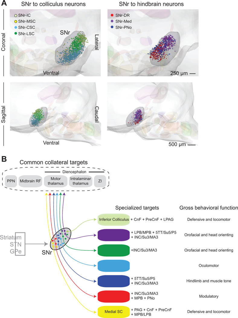
Figure 8. SNr subdivisions contain topographically organized projection populations.
(A) Three-dimensional reconstructions of SNr projection neurons labeled by retrograde viral tracers, aligned to a common brainstem atlas. The composite displays the relative position of SNr neurons projecting to midbrain (left) and hindbrain targets (right). Coronal (top) and sagittal (bottom) views of neurons projecting to the Inferior Colliculus (SNr-IC, light green), medial Superior Colliculus (SNr-MSC, yellow), central Superior Colliculus (SNr-CSC, light blue), lateral Superior Colliculus (SNr-LSC, green), Dorsal Raphe (SNr-DR, red), and PNo reticular formation (SNr-PNo, dark blue), and medullary reticular formation (SNr-Med, purple).
(B) Summary of SNr output pathways demonstrating unique and common targets of distinct SNr projection populations. Each SNr population projects to large, functionally-distinct brainstem regions and collateralizes to small brainstem nuclei and a set of common target regions. All projection populations demonstrate a distinct one-to-many projection pattern.

