Abstract
African swine fever (ASF) has severely influenced the swine industry of the whole world. Fast and accurate African swine fever virus (ASFV) antigen detection is very important for ASF prevention. This study aims to establish a new detection method for detection ASFV antigen using time-resolved fluorescence immunoassay (TRFIA) in the nose and mouth discharge. A double antibody sandwich TRFIA method was optimized and established. Recombinant P30 recombinant antigen was captured by its antibodies immobilized on 96-well plate, and then banded together with another detection antibodies labeled with Europium(III) (Eu3+) chelates, finally time-resolved analyzer measured the fluorescence intensity. The performance of this TRFIA (sensitivity, specificity and accuracy) was evaluated using the clinical samples and compared with the nucleic acid testing method. The sensitivity of this TRFIA was 0.015 ng/mL (dynamic range 0.24–500 ng/mL) with high specificity. The recovery ranged from 92.00 to 103.62 %, the inter-assay CVs ranged from 5.50 to 11.96 %, and the intra-assay CVs was between 5.20 and 10.53 %. Additionally, the cutoff value was 0.016. TRFIA took only 45 min to generate results, and its detection capability comparable to the nucleic acid detection. This study developed a TRFIA method that could be used for qualitative/quantitative detection of ASFV antigen in pigs nasal discharge, which has high sensitivity, specificity and accuracy. This TRFIA provides a new method for rapidly screening ASFV infection in pigs industry.
Keywords: African swine fever, African swine fever virus, Time-resolved fluorescence immunoassay, Double antibody sandwich
Introduction
African swine fever (ASF) is a viral hemorrhagic disease of pigs with mortality rates approaching 100 %, it is caused by a large DNA virus, African swine fever virus (ASFV) [1, 2]. ASF has caused major economic losses, limited pig industry and even threatened food security in affected countries. ASF has erupted and spread in Poland, Lithuania, China, Latvia, Belarus, USA and the Russian Federation etc.[3, 4]. Unfortunately, there is currently no effective antiviral drug or vaccine against ASFV [5]. Stamping out and movement control are the mainly control measures. Currently, the diagnostic methods for ASFV include nucleic acid detection, ASFV antigen detection and ASFV antibody detection (ELISA method), and establishment of new ASFV diagnostic technology is urgently needed [6, 7].
Time-resolved fluorescence immunoassay (TRFIA) is a novel detection technique using the unique lanthanide fluorescence properties, which can offer higher sensitivity, lower matrix interference and enlarge dynamic range than ELISA [8–10]. At present, TRFIA has been used in the diagnosis and screening of various viral infectious diseases of humans and animals [11, 12]. However, TRFIA has not yet been used in ASF screening. In this present study, we have optimized and established a double-antibody sandwich TRFIA method for detection of ASFV antigen. Compared with commercial ELISA kits, the detection performance of TRFIA is equivalent. This TRFIA provide a new method for sensitive, accurate and specific detection of ASFV.
Materials and Methods
Antigen, Antibody, Samples and Reagents
Recombinant P30 recombinant antigen (Escherichia coli), anti-P30 paired monoclonal antibodies of ASFV were prepared by Guangzhou Youdi Biotechnology Co., Ltd. 15 clinical nasal discharge samples with ASFV positive, and 110 healthy ral/nasopharyngeal swab samples came from South China Agricultural University. All samples were stored at -80℃. Europium(III) (Eu3+) labeling kit was purchased from PerkinElmer (Norwalk, USA). Sephadex G50 column was purchased from Amersham Pharmacia Biotech (Piscataway, NJ, USA). The buffers used in this study are all self-prepared using the routine laboratory reagents.
Optimized Coating Procedure
Anti-P30 coating monoclonal antibodies were diluted into different concentrations (1 µg/mL, 2 µg/mL, 5 µg/mL, 10 µg/mL) and added into the 96-well plates. Coated the plates at 37℃ for 2 h, and then blocked the plates with 200 µL/well blocking buffer (50 mmol/L, pH 7.4 PBS supplemented with 3 % BSA) at 37℃ for 1 h. Then, removed the blocking buffer and dried in vacuum, and finally stored the 96-well plates at -4℃. Using different concentrations of anti-P30 coating monoclonal antibodies to optimize antibody coating concentration.
Optimized Labeling Procedure
According to the protocol provided by the Eu3+ labeling kit, anti-P30 detection monoclonal antibodies was labeled with Eu3+-chelates. Briefly, washed the 1 mg detection antibodies with labeling buffer (2 mg/mL), and then added into 50, 100 or 200 µg Eu3+-chelates. Gently shaken the mixture for 24 h at -4℃, and then purified the Eu3+-labeled detection antibodies using a Sephadex G50 column. Finally, the Eu3+-labeled detection antibodies purified were stored at -20℃.
Optimized Assay Procedure
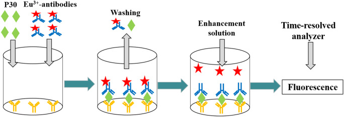
Optimized assay procedure using the one-step procedure, including the volume of Eu3+-labeled detection antibodies and enhancement solution, and the immunoreaction time. Briefly, standards, samples and Eu3+-labeled detection antibodies were added into the coated 96-well plates, and then incubated at room temperature. After washing the wells, added the enhancement solution into the wells, and shaken gently for a few minutes. Finally, time-resolved analyzer (PerkinElmer, Auto 1420, Waltham, MA, USA) measured the fluorescence. Scheme of the present TRFIA was shown in Fig. 1.
Fig. 1.
Scheme of the present TRFIA method
Standard Curves and Sensitivity Assay
The standard curve was established using serial dilutions of P30 antigen (0, 0.01, 0.1, 1, 10, 100 and 500 ng/mL). The Log function values of P30 antigen were plotted as X axis, and the Log function value of their fluorescence as the Y axis, performed a linear fit and draw the standard curve. The limit of the blank (LOB) was defined as the mean of the blank assayed in 20 independent measurements. The limit of detection (LOD) was determined by adding two standard deviations (SD) to the LOB, that means LOD = LOB + 2*SD [13].
Specificity Assay
High concentrations of transmissible gastroenteritis virus (TGEV), porcine respiratory coronavirus (PRCV), porcine hemagglutinating encephalomyelitis virus (PHEV), swine influenza H1N1 virus and porcine circovirus (PCV) were selected for specificity assay.
Accuracy and Recovery Assays
By adding the known concentrations of P30 antigen (high, medium, and low concentration respectively) into the nasal discharge samples of healthy pigs, we evaluated the accuracy and recovery of this TRFIA method. Performed six independent experiments and calculated the mean and their SD. The recovery, and the CVs of the intra-assay and interassay variations were obtained. The formula of recovery is: Recovery (%) = (measured concentration/theoretical concentration) ×100. CV(%) = SD/mean×100.
Qualitative Criteria
This TRFIA method was used to detect the 110 nasal discharge samples of the healthy pigs. Recorded the fluorescence values of swab samples (Fs) and blank control (background values, Fc), and calculate their antigen concentration according to the standard curves. SPSS 17.0 was used for data statistical analysis. If the values of antigen concentration belongs to the normal distribution, the normal distribution method is used to calculate the reference interval range, and the 95 % reference interval range is calculated by the following formula: cut off = mean + 1.96SD.
Comparison and Practical Application of TRFIA
15 clinical nasal discharge samples with ASFV positive and 110 nasal discharge samples of the healthy pigs were used for comparison and practical application. The clinical samples were simultaneous measured by the present TRFIA method and nucleic acid testing method (loop-mediated isothermal amplification, LAMP, Chinese Academy of Inspection and Quarantine, Beijing, China, No. 11 new veterinary drugs).
Statistical Analysis
SPSS 19.0 was used for the statistical analysis. Data were graphed using GraphPad Prism 5 (GraphPad Software, USA). The data are expressed as the mean ± SD. Correlations among different methods were calculated with χ2 test.
Results
Assay Procedure Optimization
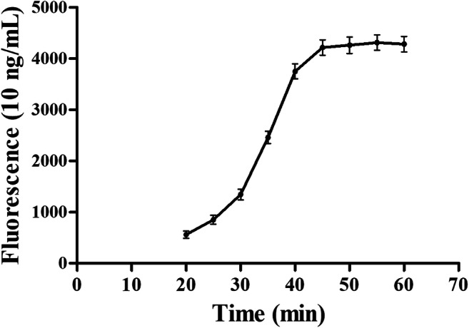
A double-antibody sandwich TRFIA method was developed under the following conditions: coating amount of anti-P30 coating antibodies: 2 µg/mL, 100µL/well; optimized labeling procedure: 1 mg detection antibodies (2 mg/mL) + 100 µg Eu3+-chelates; 100 µL/well Eu3+-labeled detection antibodies, 200µL/well enhancement solution, and the immunoreaction time was 45 min (Fig. 2). In a word, the optimized assay procedure is: 20 µL samples or standards, 100 µL/well Eu3+-labeled detection antibodies were added into the coated 96-well plates, and then incubated for 45 min at room temperature. After washed the wells 5 times, added 200 µL/well enhancement solution into the wells, and shaken gently for 2 min. Finally, time-resolved analyzer measured the fluorescence.
Fig. 2.

Optimization of immunoreaction time. The optimized TRFIA method detected the Fluorescence of the 10 ng/mL P30 antigen standards at the different immunoreaction time. The immunoreaction time were plotted as the X axis, and fluorescence values of the antigen standards as the Y axis, draw a curve
TRFIA Standard Curves
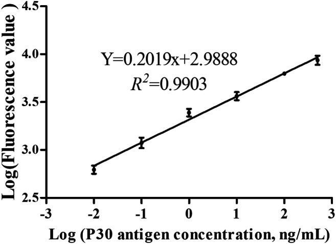
Standard curve determinations were carried out using linear regression and Log-Log regression. For the standard curve depicted in Fig. 3, The best-fit calibration was depicted in Fig. 3 by the following equation: LogY = 0.2019×LogX + 2.9888 (R2 = 0.9903). Sensitivity assay indicates the LOD of this TRFIA was 0.015 ng/mL, and the dynamic range ranged from 0.24 to 500 ng/mL.
Fig. 3.

Standard curves of ASFV antigen detection. The Log function values of P30 antigen were plotted as X axis, and the Log function value of their fluorescence as the Y axis, performed a linear fit and draw the standard curve
Specificity Results
This TRFIA detected the 5 interferents with high concentrations (TGEV, PRCV, PHEV, H1N1 and PCV), and their results are shown in Table 1. The cross-reactivity was very low, ranging from 0.18 to 0.23 %, which means this TRFIA method had high specificity for ASFV P30 antigen.
Table 1.
Specificity results of this TRFIA method
| Interferents | Nominal concentration (ng/mL) | Mean ± SD (ng/mL) | Cross-reactivity (%) |
|---|---|---|---|
| ASFV | 20 | 19.86 ± 0.34 | 99.30 |
| TGEV | 20 | 0.035 ± 0.0014 | 0.18 |
| PRCV | 20 | 0.044 ± 0.0016 | 0.22 |
| PHEV | 20 | 0.041 ± 0.0015 | 0.21 |
| H1N1 | 20 | 0.036 ± 0.0014 | 0.18 |
| PCV | 20 | 0.046 ± 0.0017 | 0.23 |
Note: TGEV: transmissible gastroenteritis virus, PRCV: porcine respiratory coronavirus, PHEV: porcine hemagglutinating encephalomyelitis virus, H1N1: swine influenza H1N1 virus, PCV: porcine circovirus
Accuracy and Recovery Results
As presented in Table 2, the inter-assay CVs ranged from 5.50 to 11.96 %, and the intra-assay CVs was between 5.20 and 10.53 %. All CVs were less than 15 %, which indicates that this TRFIA assay has high accuracy. Additionally, the recovery was between 92.00 and 103.62 %. The recovery results indicated that this TRFIA method was free from interferents in matrix samples.
Table 2.
Accuracy and recovery of the present TRFIA
| Theoretical (ng/mL) | mean ± SD (ng/mL) | CV (%) | Recovery (%) | |
|---|---|---|---|---|
| Inter-assay (n = 10) | 0.1 | 0.092 ± 0.011 | 11.96 | 92.00 |
| 10 | 9.46 ± 0.52 | 5.50 | 94.60 | |
| 100 | 103.62 ± 7.42 | 7.16 | 103.62 | |
| Intra-assay (n = 10) | 0.1 | 0.095 ± 0.010 | 10.53 | 95.00 |
| 10 | 9.61 ± 0.50 | 5.20 | 96.10 | |
| 100 | 102.56 ± 6.40 | 6.24 | 102.56 |
Qualitative Criteria
After performing a normality test with SPSS 17.0, the concentration values were normally distributed, so that the one-sided upper limit of the 95 % reference interval range was performed for the calculation. For 110 nasal discharge samples of the healthy pigs, the mean concentration value was 0.011, SD was 0.0026, the cutoff value was 0.016 (cut off = mean + 1.96SD), which meant that when antigen concentration was greater than 0.016, the sample may be ASFV positive.
Comparison and Practical Application of TRFIA
To confirm the diagnostic accuracy of our assay, we used it to synchronously test 15 clinical nasal discharge samples with ASFV positive and 110 nasal discharge samples of the healthy pigs. As shown in Table 3, the χ2 test showed that the P-value of the McNemar test was 1.0, and the value obtained with the kappa test was 0.924 (P < 0.001), which means there was no statistically significant difference between our developed TRFIA method and the nucleic acid testing method.
Table 3.
χ2 test for diagnostic results of the developed TRFIA assay and nucleic acid testing method
| Nucleic acid testing | Total | |||
|---|---|---|---|---|
| Positive | Negative | |||
| TRFIA | Positive | 14 | 1 | 15 |
| Negative | 1 | 109 | 110 | |
| Total | 15 | 110 | 125 | |
Discussion
ELISA specific for ASFV antigen/antibody are most commonly used in the clinical diagnosis of ASF infection [14–16]. But most of them are qualitative and with narrow detection ranges. Although the PCR method is the gold standard for various types of virus detection, only a few PCR-based ASF diagnostic assays have been previously described for detection and identification of ASFV [17, 18]. This may be because the PCR method requires relatively high technical personnel and equipment. At present, various new methods have been established and applied in ASF screening. LAMP is an alternative to traditional PCR, which requires a shorter reaction time, higher specificity and has a lower cost [19]. James et al. developed a loop-mediated isothermal amplification (LAMP) assay, and it is a simple-to-use and inexpensive molecular assay format for ASF diagnosis. There was good concordance between the LAMP assay and real-time PCR in clinical samples[20].
TRFIA is a sophisticated detection technique, and has been widely used in the detection of multiple viruses or their antigens [21, 22]. Compared with ELISA, TRFIA has higher sensitivity and wider measurement range. Schröder and Kuhlmann found that the use of a TRFIA significantly improved the quantitative detection of tetanus antitoxin over that of the ELISA technique, and the wide measurement range of TRFIA enabled fast examination of large numbers of serum samples without the need for repetition [23]. In this study, a novel TRFIA methodology was developed for the quantitative determination of ASFV antigen in pigs nasal discharge. The comparison experiment found its detection performance is equivalent with LAMP-based kits (Chinese Academy of Inspection and Quarantine, Beijing, China, No. 11 new veterinary drugs). The result shows that TRFIA is a promising detection method, which has better performance than ELISA and real-time PCR.
In this study, we developed a high sensitivity, specificity, and accuracy TRFIA using Eu3+ chelates. The assay was based on an double-antibody sandwich immunoassay format, and performed in 96-well plates. The sensitivity of this TRFIA is 0.015 ng/mL (dynamic range 0.24–500 ng/mL). There is very low cross-reaction with 5 interferents. The recovery ranged from 92.00 to 103.62 %, the inter-assay CVs ranged from 5.50 to 11.96 %, and the intra-assay CVs was between 5.20 and 10.53 %, which means this TRFIA has high accuracy. Additionally, we calculated the cutoff values using 110 nasal discharge samples of the healthy pigs. When antigen concentration ≥ 0.016 ng/mL, the sample (pig) may be ASFV positive. Compared with the nucleic acid method, χ2 test showed the detection performance of TRFIA is equivalent. Importantly, the main advantage of this TRFIA is in quantitative detection (not just qualitative detection). The cutoff values provide the basis for qualitative screening, and the drawn standard curve realizes quantitative detection for ASFV.
In conclusion, we developed a TRFIA method that could be used for qualitative/quantitative detection of ASFV antigen in pigs nasal discharge, which has high sensitivity, specificity and accuracy. TRFIA takes only 45 min to generate results, and its detection capability comparable to the nucleic acid detection. Therefore, the kit can be used in the veterinary services for effective monitoring of ASFV to contain, eliminate and prevent further spread of the disease.
Author Contributions
Cuicui Chen and Hongrui Lai: Methodology and Writing- Original draft preparation. Huankun Liang: Quality Control. Ying He: Samples detection. Guiling Guo: Software and Data analysis. Laiqing Li: Conceptualization.
Data Availability
The data used and/or analysed in the current study are given in the submitted article.
Code Availability
No applicable.
Declarations
Conflicts of Interest
The authors declared that they have no conflicts of interest.
Ethics Approval
No applicable.
Consent to Participate
No applicable.
Consent for Publication
No applicable.
Footnotes
Publisher’s Note
Springer Nature remains neutral with regard to jurisdictional claims in published maps and institutional affiliations.
References
- 1.Costard S, Wieland B, de Glanville W, Jori F, Rowlands R, Vosloo W, Roger F, Pfeiffer Dirk U, Dixon Linda K. African swine fever: how can global spread be prevented? Phil Trans R Soc B. 2009;364:2683–2696. doi: 10.1098/rstb.2009.0098. [DOI] [PMC free article] [PubMed] [Google Scholar]
- 2.Jouvenet N, Wileman T. African swine fever virus infection disrupts centrosome assembly and function. J Gen Virol. 2005;86:589–594. doi: 10.1099/vir.0.80623-0. [DOI] [PubMed] [Google Scholar]
- 3.Pejsak Z, Truszczyński M, Niemczuk K, Kozak E, Markowska-Daniel I. Epidemiology of African Swine Fever in Poland since the detection of the first case. Pol J Vet Sci. 2014;17(4):665–672. doi: 10.2478/pjvs-2014-0097. [DOI] [PubMed] [Google Scholar]
- 4.Ge S, Li J, Fan X, Liu F, Li L, Wang Q, Ren W, Bao J, Liu C, Wang H, Liu Y, Zhang Y, Xu T, Wu X, Wang Z. Molecular characterization of African swine fever virus, China, 2018. Emerg Infect Dis. 2018;24(11):2131–2133. doi: 10.3201/eid2411.181274. [DOI] [PMC free article] [PubMed] [Google Scholar]
- 5.Zhu Z, Fan Y, Liu Y, et al. Prediction of antiviral drugs against African swine fever viruses based on protein-protein interaction analysis. PeerJ. 2020;8(5):e8855. doi: 10.7717/peerj.8855. [DOI] [PMC free article] [PubMed] [Google Scholar]
- 6.Randriamparany T, Kouakou KV, Michaud V, Fernández-Pinero J, Gallardo C, Le Potier MF, Rabenarivahiny R, Couacy-Hymann E, Raherimandimby M, Albina E. African swine fever diagnosis adapted to tropical conditions by the use of dried-blood filter papers. Transbound Emerg Dis. 2016;63(4):379–388. doi: 10.1111/tbed.12295. [DOI] [PubMed] [Google Scholar]
- 7.Agüero M, Fernández J, Romero L, Sánchez Mascaraque C, Arias M, Sánchez-Vizcaíno JM. Highly sensitive PCR assay for routine diagnosis of African swine fever virus in clinical samples. J Clin Microbiol. 2003;41(9):4431–4434. doi: 10.1128/JCM.41.9.4431-4434.2003. [DOI] [PMC free article] [PubMed] [Google Scholar]
- 8.Yuan J, Wang G. Lanthanide complex-based fluorescence label for time-resolved fluorescence bioassay. J Fluoresc. 2005;15(4):559–68. doi: 10.1007/s10895-005-2829-3. [DOI] [PubMed] [Google Scholar]
- 9.De Pauw PE, Mackin RB, Goubert P, Van Schravendijk C, Gorus FK. Total error profiling of a proinsulin time-resolved fluorescence immunoassay. J Chromatogr B Analyt Technol Biomed Life Sci. 2009;877(23):2403–2406. doi: 10.1016/j.jchromb.2008.11.024. [DOI] [PubMed] [Google Scholar]
- 10.Chen JJ, Liu TC, Liang QN, Dong ZN, Wu YS, Li M. Development of a time-resolved fluorescence immunoassay for Epstein-Barr virus nuclear antigen 1-immunoglobulin A in human serum. J Med Virol. 2015;87(11):1940–1945. doi: 10.1002/jmv.24247. [DOI] [PubMed] [Google Scholar]
- 11.Maple PA, Gray J, Breuer J, Kafatos G, Parker S, Brown D. Performance of a time-resolved fluorescence immunoassay for measuring varicella-zoster virus immunoglobulin G levels in adults and comparison with commercial enzyme immunoassays and Merck glycoprotein enzyme immunoassay. Clin Vaccine Immunol. 2006;13(2):214–218. doi: 10.1128/CVI.13.2.214-218.2006. [DOI] [PMC free article] [PubMed] [Google Scholar]
- 12.Chen C, Liang H, Hu B, Ning B, Li L. Determination of parvovirus antigen in the vaccine using time-resolved fluorescence immunoassay. Biotechnol Appl Biochem. 2020 doi: 10.1002/bab.1967. [DOI] [PubMed] [Google Scholar]
- 13.Lin G, Chen S, Zhao H, et al. A time-resolved fluoroimmunoassay to assay the rabies virus glycoprotein: application for estimation of human rabies vaccine potency. Sci Rep. 2017;7(1):7288. doi: 10.1038/s41598-017-07687-7. [DOI] [PMC free article] [PubMed] [Google Scholar]
- 14.Hutchings GH, Ferris NP. Indirect sandwich ELISA for antigen detection of African swine fever virus: comparison of polyclonal and monoclonal antibodies. J Virol Methods. 2006;131(2):213–217. doi: 10.1016/j.jviromet.2005.08.009. [DOI] [PubMed] [Google Scholar]
- 15.Bergeron HC, Glas PS, Schumann KR. Diagnostic specificity of the African swine fever virus antibody detection enzyme-linked immunosorbent assay in feral and domestic pigs in the United States. Transbound Emerg Dis. 2017;64(6):1665–1668. doi: 10.1111/tbed.12717. [DOI] [PubMed] [Google Scholar]
- 16.Gallardo C, Reis AL, Kalema-Zikusoka G, Malta J, Soler A, Blanco E, Parkhouse RM, Leitão A. Recombinant antigen targets for serodiagnosis of African swine fever. Clin Vaccine Immunol. 2009;16(7):1012–1020. doi: 10.1128/CVI.00408-08. [DOI] [PMC free article] [PubMed] [Google Scholar]
- 17.Gonzague M, Plin C, Bakkali-Kassimi L, Boutrouille A, Cruciere C. Development of an internal control for the detection of the African swine fever virus by PCR. Mol Cell Probes. 2002;16(3):237–42. doi: 10.1006/mcpr.2002.0416. [DOI] [PubMed] [Google Scholar]
- 18.Fernández-Pinero J, Gallardo C, Elizalde M, Robles A, Gómez C, Bishop R, Heath L, Couacy-Hymann E, Fasina FO, Pelayo V, Soler A, Arias M. Molecular diagnosis of African Swine Fever by a new real-time PCR using universal probe library. Transbound Emerg Dis. 2013;60(1):48–58. doi: 10.1111/j.1865-1682.2012.01317.x. [DOI] [PubMed] [Google Scholar]
- 19.Pinhanelli V, Costa P, Fachin AL, Marins M. Evaluation and comparison of Loop-mediated Isothermal Amplification (LAMP) BMC Proc. 2014;8(Suppl 4):P140. doi: 10.1186/1753-6561-8-S4-P140. [DOI] [Google Scholar]
- 20.James HE, Ebert K, McGonigle R, Reid SM, Boonham N, Tomlinson JA, Hutchings GH, Denyer M, Oura CA, Dukes JP, King DP. Detection of African swine fever virus by loop-mediated isothermal amplification. J Virol Methods. 2010;164(1–2):68–74. doi: 10.1016/j.jviromet.2009.11.034. [DOI] [PubMed] [Google Scholar]
- 21.Hu Z, Li M, Huang B, Liu J, Yu L, Chen G. Detection of hepatitis B virus PreS1 antigen using a time-resolved fluoroimmunoassay. J Immunoassay Immunochem. 2012;33(2):156–165. doi: 10.1080/15321819.2011.609576. [DOI] [PubMed] [Google Scholar]
- 22.Liang RL, Yang YS, Zhou JW, et al. Dual-labeled time-resolved immunofluorometric assay for the simultaneous quantitative detection of hepatitis B virus antigens in human serum. J Fluoresc. 2017;27(1):309–316. doi: 10.1007/s10895-016-1959-0. [DOI] [PubMed] [Google Scholar]
- 23.Schröder JP, Kuhlmann WD. Detection of tetanus antitoxin using Eu(3+)-labeled anti-human immunoglobulin G monoclonal antibodies in a time-resolved fluorescence immunoassay. J Clin Microbiol. 1991;29(7):1504–1507. doi: 10.1128/JCM.29.7.1504-1507.1991. [DOI] [PMC free article] [PubMed] [Google Scholar]
Associated Data
This section collects any data citations, data availability statements, or supplementary materials included in this article.
Data Availability Statement
The data used and/or analysed in the current study are given in the submitted article.
No applicable.


