Abstract
Background
New-generation adjuvants for foot-and-mouth disease virus (FMDV) vaccines can improve the efficacy of existing vaccines. Chinese medicinal herb polysaccharide possesses better promoting effects.
Objectives
In this study, the aqueous extract from Artemisia rupestris L. (AEAR), an immunoregulatory crude polysaccharide, was utilized as the adjuvant of inactivated FMDV vaccine to explore their immune regulation roles.
Methods
The mice in each group were subcutaneously injected with different vaccine formulations containing inactivated FMDV antigen adjuvanted with three doses (low, medium, and high) of AEAR or AEAR with ISA-206 adjuvant for 2 times respectively in 1 and 14 days. The variations of antibody level, lymphocyte count, and cytokine secretion in 14 to 42 days after first vaccination were monitored. Then cytotoxic T lymphocyte (CTL) response and antibody duration were measured after the second vaccination.
Results
AEAR significantly induced FMDV-specific antibody titers and lymphocyte activation. AEAR at a medium dose stimulated Th1/Th2-type response through interleukin-4 and interferon-γ secreted by CD4+ T cells. Effective T lymphocyte counts were significantly elevated by AEAR. Importantly, the efficient CTL response was remarkably provoked by AEAR. Furthermore, AEAR at a low dose and ISA-206 adjuvant also synergistically promoted immune responses more significantly in immunized mice than those injected with only ISA-206 adjuvant and the stable antibody duration without body weight loss was 6 months.
Conclusions
These findings suggested that AEAR had potential utility as a polysaccharide adjuvant for FMDV vaccines.
Keywords: Polysaccharides, adjuvants, foot-and-mouth disease virus, vaccines, immune response
INTRODUCTION
Foot-and-mouth disease (FMD) is a highly infectious and rapidly spreading viral disease of cloven-hoofed animals. Inactivated FMD virus (FMDV) vaccines can largely control FMD in endemic regions [1,2]. However, FMDV vaccines have some defects, such as instable antigens, low immunogenicity, short antibody duration, and repetitive vaccination [3]. FMDV vaccines can be optimized with appropriate adjuvants to some degree. Adjuvants of FMDV vaccine formulations largely affect both strong humoral and cellular immunity, especially cytotoxic T lymphocyte (CTL) and long-lasting immunity. Vaccine adjuvants, such as saponins, mineral oil, liposomes, and cytokines, can enhance immune responses to FMDV vaccines [4,5]. Common FMDV vaccines are inactivated whole-virus preparations formulated with adjuvants, which often lead to carcinogenesis [6]. New-generation of adjuvants with low toxicity, high efficiency, and wide resource have been extensively explored and efficacious adjuvants might be a breakthrough in the development of FMDV vaccines.
Chinese medicinal herbs have been used as an immunopotentiator for thousands of years in China. Chinese medicinal herbs and their ingredients can boost immune responses. Many animal trials and clinical trials demonstrated that some natural polysaccharides could promote the better immune effects [7,8]. Many researchers utilized the polysaccharides from Chinese medicinal herbs as potent adjuvants for many vaccines of infectious diseases, such as Advax™ in influenza vaccine, West Nile virus vaccine and hepatitis B virus (HBV) vaccine, Astragalus polysaccharide in influenza vaccine, FMDV vaccine and HBV vaccine, Angelica polysaccharide in Newcastle disease virus vaccine, and panax ginseng polysaccharide in avian influenza vaccine [9,10,11,12]. Artemisia rupestris L. is one of the widely used Chinese medicinal herbs due to its anti-inflammatory, anti-virus and antioxidant activities [13,14]. A crude polysaccharide, the aqueous extract from Artemisia rupestris L. (AEAR) can work as the adjuvant to increase serum antibody titers, enhance secretion of cytokines, and stimulate T-cell-mediated immune responses to ovalbumin antigen and influenza vaccine, and the safety of AEAR has been tested in mice [15,16]. AEAR may be developed as a valuable adjuvant to improve the efficacy of FMDV vaccines.
Due to the high costs of large experimental animals such as swine, the immunologic analysis of AEAR combined FMDV vaccines is generally performed in mice to explore the immunological responses in natural hosts. Our previous study showed the immunogenicity of an FMD vaccine formulated with AEAR involved intra-muscular immunization [17]. The aim of the study focused on the vaccination subcutaneous route. The study attempted to experimentally investigate whether inactivated FMDV antigen adjuvanted with AEAR or AEAR and ISA-206 adjuvant after subcutaneous injection in mice could induce the better FMDV-specific antibodies and T-cell response. In addition, the antibody duration of AEAR combined FMDV vaccines was longer than FMDV vaccine with ISA-206 adjuvant. The study provides the basis for improving polysaccharide adjuvants for FMDV vaccines.
MATERIALS AND METHODS
Mice
Female Institute of Cancer Research (ICR) mice (5–6 weeks old; body weight of 18–22 g) from Xinjiang Medical University Animal Center (China) were maintained in polypropylene cages (24 ± 1°C, a light cycle of 12 h) and fed with pathogen-free foods and water. Animal experiments had been approved by the Committee on the Ethics of Animal Experiments of Xinjiang Key Laboratory of Biological Resources and Genetic Engineering (BRGE-AE001) in Xinjiang University.
Preparations of AEAR
AEAR was obtained according to the previously described method [18]. Briefly, after colored ingredients and lipids were extracted with petroleum ether from dried powder of Artemisia rupestris L., which was then extracted with boiling water. Then the filtrates were precipitated together with 95% ethanol. The obtained fractions were dissolved in distilled water and then proteins were removed with the Sevag method [19]. Then, the total sugar content was calculated as 32.39% based on the phenol-sulfuric acid analysis with glucose as a standard. Finally, the extracts were freeze-dried and stored until use.
Preparation of experimental vaccine and vaccination
FMDV OHM/02 strain, BHK-21 cell line, FMDV O-serotype inactivated 146S antigen and ISA-206 adjuvant were provided by Tecon Biology Co., Ltd. (China). According to the previously reported steps [17], the vaccine was prepared. Briefly, 0.3 µg (1 dose) of FMDV O-serotype inactivated 146S antigen for the induction of strong immunity was thoroughly mixed with three doses of aqueous AEAR (low, medium, and high) respectively or oil-based ISA-206 adjuvant according to the volumetric ratio of 1:1.1. All groups of mice were injected subcutaneously (S.C.) twice at 0 and 14 days with 125 µL of the antigen-adjuvant mixture at two sites. In a random way, ICR mice were divided into 7 groups (n = 5) and injected with different agents: control group (0.9% NaCl), FMDV group (FMDV O-serotype inactivated antigen), FMDV + AEAR-L group (FMDV O-serotype inactivated antigen and low dose of AEAR 13.3 mg/kg), FMDV + AEAR-M group (FMDV O-serotype inactivated antigen and medium dose of AEAR 26.6 mg/kg), FMDV + AEAR-H group (FMDV O-serotype inactivated antigen and high dose of AEAR 53.3 mg/kg), FMDV + oil emulsion group (FMDV O-serotype inactivated antigen and ISA-206 adjuvant), and FMDV + oil emulsion + AEAR-L group (FMDV O-serotype inactivated antigen, ISA-206 adjuvant and 13.3 mg/kg AEAR). Blood samples were obtained from immunized mice at 14, 28, 35, and 42 days after vaccination to analyze the antibodies. The same immunization strategy (n = 5) was performed to detect splenocyte proliferation, T-cell response, cytokine levels, CTL response, antibody duration.
Antibody test after immunization with the vaccine
To determine the effect of AEAR on antibody levels using enzyme-linked immunosorbent assay (ELISA), five serum samples obtained after the vaccination were pooled and serially diluted for analyzing FMDV-specific immunoglobulin G (IgG) titers, each serum samples were diluted 1:500 for detecting isotypes respectively. Briefly, mouse serum diluted was added to 96-well plates coated with FMDV antigen (0.75 μg/mL) for 12 h at 4°C in the carbonate-bicarbonate buffer for 60-min incubation at 37°C. Subsequently, goat anti-mouse IgG, IgG1 or IgG2a (Southern Biotech, USA) conjugated to horseradish peroxidase was added to each well for 60-min incubation. Then, TMB solution (Sangon Biotech, China) was added to develop the colorimetric detection reaction, which was terminated by the addition of 2M H2SO4 stop solution. The absorbance value of each well was analyzed in an ELISA reader (Bio-Rad, USA) at 450/655 nm.
Analysis of splenocyte proliferation
Mouse splenocyte proliferation in response to mitogen was evaluated by MTT method. Single-cell suspensions were obtained from the spleen of the immunized mice and the erythrocytes were lysed. After trypan blue exclusion staining, cell viability was measured. Cells (5 × 10⁶ cells/mL) were transferred into a 96-well plate (Nunc) in RPMI-1640 complete medium (Gibco, USA) and then Concanavalin A (Con A 5 μg/mL; Sigma, USA), lipopolysaccharide (LPS 10 μg/mL; Sigma) or RPMI-1640 medium was added to obtain a volume of 200 µL for 48 h at 37°C. All experiments were performed in triplicate. MTT (5 mg/mL; Sigma) solution was added into every well and incubated for another 4 h, then and the color development was stopped by adding dimethyl sulfoxide into each well. The optical density (OD) was determined in a microtiter plate reader at 570 nm. Stimulation index (SI) was calculated as: SI = (OD570nm of stimulated cells − OD570nm of the medium) divided by (OD570 nm of non-stimulated cells − OD570nm of the medium).
On day 21 after the primary vaccination, Flow cytometry was used to analyze T cell activation. Cell suspensions (5 × 10⁶ cells) of spleen from immunized mice were stained with phycoerythrin (PE)-CD3, allophycocyanin (APC)-CD4 or fluorescein isothiocyanate (FITC)-CD8 (BD Bioscience). Cells were blocked with Mouse BD FcBlock (BD Bioscience) before the addition of specific monoclonal antibodies for 30 min at 4°C. Non-labeled and single-labeled controls were used. After cells were washed and re-suspended in cold phosphate-buffered saline, the treated cells were detected with a FACS Calibur system (BD Bioscience). The results were analyzed in FlowJo (Treestar, USA) software.
Flow cytometry analysis of intracellular cytokine
For intracellular cytokine analysis, on day 7 after the second injection, splenocytes from the immunized mice were incubated with FMDV antigen (4.5 μg/mL) for 4 h in 96-well plates, followed by the addition of Golgi stop (BD Biosciences) for 12 h. After the collected cells (1 × 10⁶) were washed three times, cells were blocked for 30 min and stained with cell surface molecules for the analysis of APC-CD4 or FITC-CD8. After surface marker staining, the cells were fixed and permeabilized with Cytofix/Cytoperm (BD Bioscience) and stained with PE-interleukin (IL)-4 or PE-interferon (IFN)-γ (BD Bioscience) for 30 min at 4°C. The cells were detected with FACS system and the detection results were analyzed in FlowJo software.
Determination of FMDV-specific CTL response
To evaluate the antigen-specific CTL response, ICR mice were firstly divided into 6 groups (n = 5) and the same immunization strategy was performed as described above. Splenocytes of immunized mice on day 7 after the second immunization were used as the effector cells. Then, the splenocytes of naive mouse were used as the target cells and seeded in a 6-well plate in two aliquots according to the cell concentration of 1 × 10⁷ cells/mL. The first aliquot was stimulated with 50 μg FMDV antigen and the second aliquot was incubated in the medium. After 4-h incubation, pelleted cells from the first aliquot were washed with PBS for three times, resuspended in the complete medium as Ag-stimulated target cells according to the cell concentration of 1 × 10⁷ cells/mL, and incubated with 10 μM carboxyfluorescein succinimidyl ester (CFSE; CFSEhigh cells). The second aliquot was used as Ag-unstimulated target cells and incubated with 1 μM CFSE (CFSElow cells) at 1×10⁷ cells/mL. The two aliquots were mixed in the complete medium to realize the cell ratio of 1:1 (CFSEhigh/CFSElow) and then injected into the above immunized mice in 7 days after boost immunization. In 4 h after the injection, the splenocytes from the mice were isolated and effector cells and target cells were collected on FACS. The ratio of killed target cells was calculated as: Killing ratio = the percentage of CFSElow divided by the percentage of CFSEhigh; % lysis = [1 − (killing ratio unprimed/killing ratio primed) × 100].
Serum neutralization assay
On day 28 after the primary immunization, serum samples from mice were collected. and determined for neutralizing assay on BHK-21 cells. Briefly, after serum samples were inactivated, serum were serially diluted and mixed in a 96-well plates with a 100 TCID50 dose of FMDV OHM/02 at 37°C with 5% CO2 for 60 min. Next, BHK-21 cells (1.5 × 10⁶/mL) was added to each well of the microtiter plate and incubated at 37°C for 48 h. Neutralizing antibody titers of the sera were expressed as the highest dilutions that neutralized 100 TCID50 of FMDV in 50% of the wells. Results were calculated and analyzed using GraphPad Prism 5.0 software.
Statistical analysis
Experimental data are expressed as mean ± SE. The experimental data were analyzed with analysis of variance and Tukey's test. The p < 0.05 indicated the statistically significant differences.
RESULTS
Determination of specific humoral immune responses after vaccination
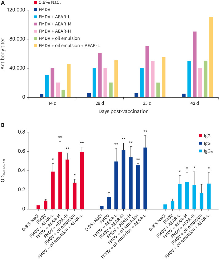
To explore the effect of AEAR on the induction of humoral immunity against FMDV, the mice were S.C. immunized twice with different vaccine formulations. Blood samples of mice (n = 5) were collected after the vaccination to determine FMDV-specific IgG titers and IgG isotypes levels by indirect ELISA. In two weeks after the first injection, a homogenous immunity was generated. FMDV-specific IgG titers in AEAR of medium-dose group were determined as 1:40,000. In four weeks after the second injection, IgG titers were determined as 1:90,000. IgG titers in AEAR of medium-dose group were almost 10 times higher than that in FMDV group and 3 times higher than that in FMDV + oil emulsion group. After the first injection, antibody titers in FMDV + oil emulsion + AEAR-L group were high (1:4,500–1:110,000) (Fig. 1A). In three weeks after the second injection, IgG1 level was significantly improved and IgG2a level was slightly induced, but no significant difference was observed between FMDV + AEAR group and FMDV + oil emulsion + AEAR-L group (Fig. 1B). The results revealed that the AEAR or the AEAR mixture with oil emulsion increased specific antibody levels in the FMDV-immunized ICR mice.
Fig. 1. Effects of AEAR on antibody levels. Sera were collected after the vaccination and FMDV-specific IgG, IgG1 and IgG2a antibodies were determined by enzyme-linked immunosorbent assay. (A) IgG titers of pooled serum from each group was assessed in 14–42 days after the first injection; (B) IgG isotypes were assessed in 35 days after the first injection. The values are presented as means ± SE (n = 5). Significant differences relative to FMDV group alone were designated.
FMDV, foot-and-mouth disease virus; AEAR, aqueous extract from Artemisia rupestris L.; IgG, immunoglobulin G; OD, optical density.
*p < 0.05, **p < 0.01.
Evaluation of cellular immune response
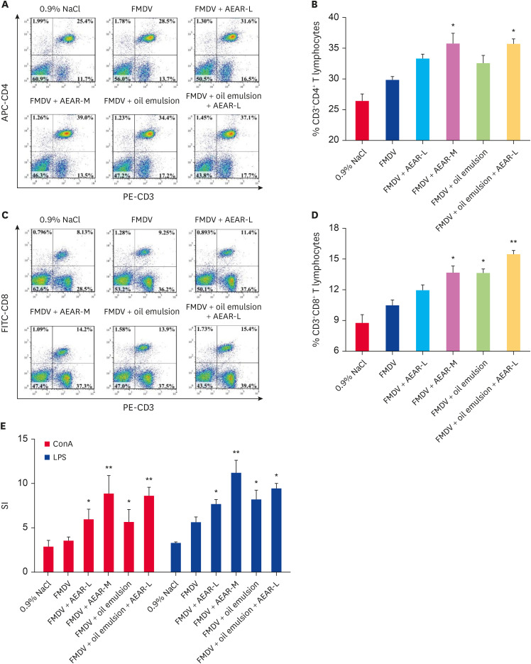
The immune control of FMDV involves a combination of cellular and humoral responses. T-cell responses largely affect the vaccination effect of FMDV vaccines. The cellular immune status could be roughly assessed with CD4+ helper and CD8+ CTLs. To evaluate the effect of AEAR on the cellular immunity, the counts of CD4+ and CD8+ T splenocytes of mice (n = 5) in 7 days after boost immunization were measured using surface marker staining. AEAR at a medium dose induced the more significant proliferative response than that in the mice immunized with FMDV alone or FMDV + oil emulsion in the population of CD4+ lymphocytes (Fig. 2A-D) and the proliferative response of CD8+ lymphocytes were the highest in the AEAR and ISA-206 adjuvant co-immunization group.
Fig. 2. Effects of AEAR on T cell subsets and splenocyte proliferation. With the cells isolated from the spleen of each group in 7 days after boost immunization, splenocyte proliferation was determined by MTT and T-cell activation was determined by FACS. (A, B) The double positive percentages of CD3+CD4+ T cells. (C, D) The double positive percentage CD3+CD8+ T cells. (E) ConA- and LPS-stimulated splenocyte proliferation was shown as a SI. Values are shown means ± SE (n = 5). Significant differences relative to FMDV group were designated.
APC, allophycocyanin; PE, phycoerythrin; FITC, fluorescein isothiocyanate; FMDV, foot-and-mouth disease virus; AEAR, aqueous extract from Artemisia rupestris L.; LPS, lipopolysaccharide; SI, stimulation index.
*p < 0.05, *p < 0.01.
Splenocyte proliferation was assessed by MTT assay. After splenocytes isolated of mice above (n = 5) were stimulated with ConA or LPS for 48 h, the effects of AEAR on splenocyte proliferation in the mice were explored (Fig. 2E), Con A- and LPS-stimulated splenocyte proliferation in the mice immunized with AEAR at a medium dose was higher than that in FMDV group and FMDV + oil emulsion group (p < 0.05). The differences between the FMDV + AEAR-L group and FMDV + oil emulsion + AEAR-L group were significant. These data indicated that cell-mediated immunity was strongly induced in the group vaccinated with various vaccines including AEAR or the mixture of AEAR and oil emulsion.
Effects of AEAR on cytokine levels in T cells
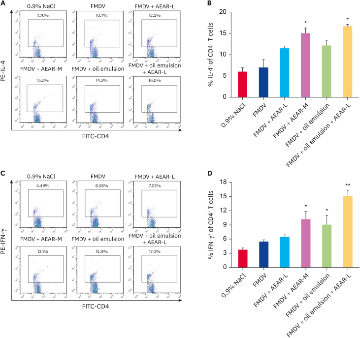
The cytokine profile of immune responses can indicate the T-helper-biased response. To explore the effects of AEAR on FMDV-specific Th cell response, the cytokines levels of IFN-γ and IL-4 in CD4+ T cells were explored after intracellular staining. Single splenocytes of mice (n = 5) were obtained in 7 days after final immunization and co-cultured with inactivated FMDV antigen. The expression of cytokines was detected by the FACS system with a gate set on CD4+ T cells (Fig. 3). The data showed that AEAR at a medium dose and AEAR with oil emulsion induced the highest IL-4 level in the antigen-specific CD4+ T cells. AEAR at medium dose induced the moderate IFN-γ level, whereas the mixture of AEAR and oil emulsion induced the highest IFN-γ level. Therefore, AEAR enhanced Th2 and Th1 immune responses. Especially, the mixture of AEAR and oil emulsion could promote IFN-γ level through activated T cells.
Fig. 3. Analysis of antigen-specific cytokine levels in T cells by FACS. Cells isolated from the spleen of mice post-vaccination were stimulated for 12 h with FMDV antigen in the presence of Golgi stop in vitro. These Cells were double stained with anti-CD4/anti-IL4, anti-CD4/anti-IFN-γ antibodies and determined by FACS. (A, B) The double positive percentage of IL-4 in total CD4+ T cells. (C, D) The double positive percentage of IFN-γ in total CD4+ T cells. The values are shown as means ± SE (n = 5). Significant differences relative to FMDV group were designated.
PE, phycoerythrin; IL, interleukin; FMDV, foot-and-mouth disease virus; AEAR, aqueous extract from Artemisia rupestris L.; FITC, fluorescein isothiocyanate; IFN, interferon.
*p < 0.05, *p < 0.01.
Effect of AEAR on effector T cells
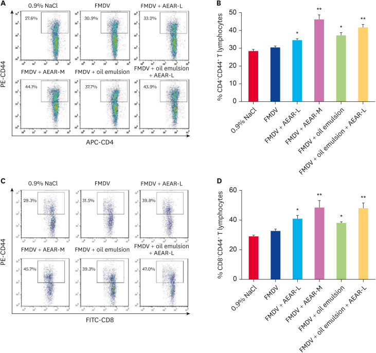
Effector T-cell-mediated responses may have important implications of vaccination against FMDV. To further evaluate T lymphocyte counts in spleen, the mice were immunized two times according to an interval of 2 weeks with FMDV antigen in different doses of AEAR or the mixture of AEAR and oil emulsion (Fig. 4). Splenocytes from individual mice (n = 5) were removed in 7 days after final immunization. The percentages of CD4+CD44+ and CD8+CD44+ of spleen cells were then measured. The percentages of CD4+CD44+ and CD8+CD44+ from the mice treated with the AEAR at medium dose were significantly higher than those in FMDV group. No significant difference in the percentages was observed between FMDV + AEAR group and FMDV + oil emulsion + AEAR-L group. The data indicated that AEAR could promote the activation of CD4+CD44+ and CD8+CD44+ T-lymphocyte subpopulations. AEAR and oil emulsion could synergistically promote the activation of T cells.
Fig. 4. Effects of AEAR on effective T cell activation. Cells were isolated from the spleen of each group after the vaccination. The population of T cells was detected by FACS. (A, B) The number of positive cells of CD44+ in total CD4+ T cells. (C, D) The number of positive cells of CD44+ in total CD8+ T cells. The values are shown as mean ± SE (n = 5). Significant differences relative to FMDV groups were designated.
PE, phycoerythrin; FMDV, foot-and-mouth disease virus; AEAR, aqueous extract from Artemisia rupestris L.; FITC, fluorescein isothiocyanate.
*p < 0.05, *p < 0.01.
Evaluation of CTL activity
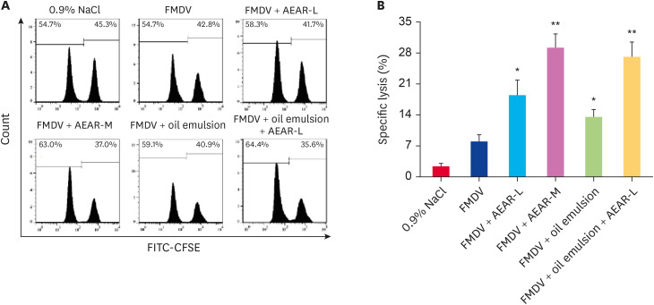
The effects of AEAR on CTL activity in the FMDV-immunized mice (n = 5) in 21 days after prime immunization were explored (Fig. 5). The lower CTL activity was induced in the mice immunized with FMDV alone. The addition of oil emulsion to FMDV antigen did not result in the further increase in FMDV-specific CTL activity above that in FMDV group. In contrast, AEAR at a medium dose mixed with FMDV significantly enhanced the killing activity of CTL in immunized mice, but no significant difference was observed between FMDV + AEAR-M group and FMDV + oil emulsion + AEAR-L group. Therefore, AEAR increased the specific CTL activity and AEAR and oil emulsion could synergistically elevate CTL response.
Fig. 5. Evaluation of AEAR on cytotoxic T lymphocyte responses. CFSE-labelled target cells from naive mice were injected into the immunized mice after the vaccination. In 4 h after the injection, splenocytes were analyzed to acquire the ratio changes by FACS. (A) Cell populations indicating the changes of CFSEhigh and CFSElow in CFSE-labelled cells. (B) The percentage of specific lysed cells was determined for each group. The values are shown as means ± SE (n = 5). Significant differences relative to FMDV groups were designated.
FMDV, foot-and-mouth disease virus; AEAR, aqueous extract from Artemisia rupestris L.; FITC, fluorescein isothiocyanate; CFSE, carboxyfluorescein succinimidyl ester.
*p < 0.05, *p < 0.01.
Neutralizing antibodies response
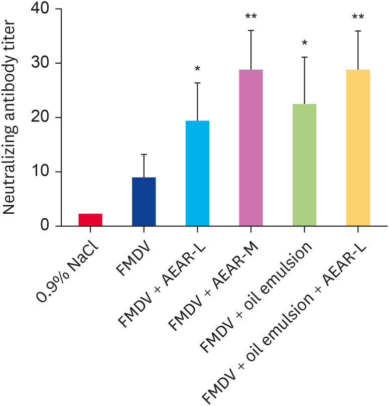
To further assess AEAR effects on FMDV, neutralizing antibody responses were performed at day 28 post immunization. The immunization schedule included a prime and a boost. As shown in Fig. 6, FMDV + AEAR-M group and FMDV + oil emulsion + AEAR-L group exhibited significantly higher FMDV-neutralizing activity than those for other groups. No significant difference in the titers was observed between FMDV + AEAR-L group and FMDV + oil emulsion group. The date showed AEAR or with oil emulsion promoted the induction of FMDV neutralizing antibodies.
Fig. 6. Effects of AEAR on serum neutralizing antibody response against the FMDV in mice. Institute of Cancer Research mice were subcutaneously immunized twice at a 2-week interval. Serum samples were collected on 28 day after the primary vaccination to detecting neutralizing antibody titers against the FMDV in BHK-21 cells. The values are shown as means ± SE (n = 5). Significant differences relative to FMDV groups were designated.
FMDV, foot-and-mouth disease virus; AEAR, aqueous extract from Artemisia rupestris L.
*p < 0.05, *p < 0.01.
Determination of the antibody duration and body weight
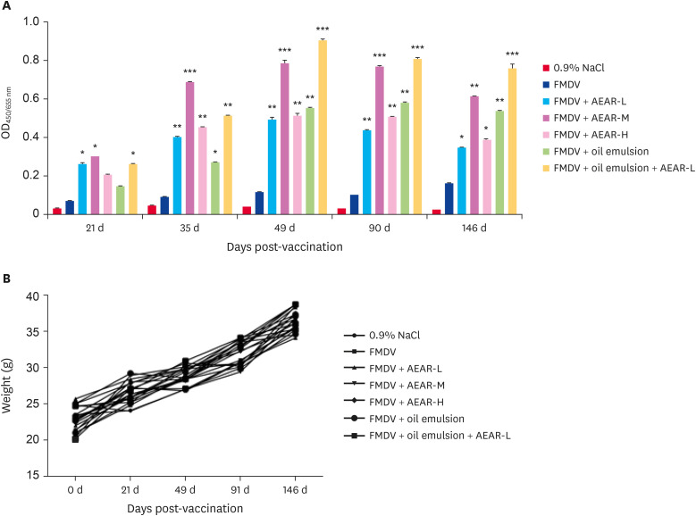
For the purpose of determining antibody duration, the serum from immunized mice (n = 5) was collected in 21, 35, 49, 90 and 146 days after primary immunization to determine the presence of FMDV antibodies (Fig. 7A). After the second vaccination, specific-FMDV antibodies were generated. After five weeks, the determined antibody levels in the mice immunized with AEAR at medium dose or with AEAR mixed oil emulsion were high and the high levels were kept for up to six months.
Fig. 7. Effects of AEAR on the duration of FMDV-specific IgG responses and weight loss. Institute of Cancer Research mice were subcutaneously immunized twice at a 2-week interval with FMDV antigen in different doses of AEAR or the mixture of AEAR and oil emulsion. (A) Blood samples were collected in 0, 21, 49, 91, and 146 days after the vaccination to measure FMDV-specific IgG. (B) The body weight was monitored after the vaccination. The values are shown as mean ± SE (n = 5). Significant differences relative to FMDV groups were designated.
FMDV, foot-and-mouth disease virus; AEAR, aqueous extract from Artemisia rupestris L.; OD, optical density; IgG, immunoglobulin G.
*p < 0.05, *p < 0.01, ***p < 0.001.
Mice (n = 5) were monitored to observe adverse post-vaccination reactions to obtain critical information regarding AEAR safety. Weight loss, abnormal behaviors and vaccination site reactions were monitored after the vaccination (Fig. 7B). No weight loss or severe adverse reactions after the vaccination with different vaccine formulations were observed during the experimental period.
DISCUSSION
It is important to optimize the vaccination effect of FMDV vaccine with a potent adjuvant [20,21]. Since the formation of granulomas at vaccination sites is problematic, the volume of FMDV vaccine is reduced under the premise that the same effect as existing vaccines is maintained for the purpose of improving the safety of vaccines [22,23]. Therefore, an adjuvant should be selected to establish all antibody levels while maintaining the efficacy of vaccines. Currently, only several vaccine adjuvants are commercially applied.
Polysaccharide adjuvants from Chinese medicinal herbs have attracted wide attention due to the characteristics of intrinsic immunomodulating, ease of availability and biocompatibility as well as lower toxicity in most of the cases. Adjuvants may contain various components with different functions and activities and crude extracts and isolated phytochemicals from Chinese medicinal herbs may be developed as promising adjuvants [24,25]. In recent years, we have explored AEAR (a complex of crude extracts) polysaccharide adjuvants enhancing adaptive immunity by screening new adjuvant formulations for different vaccines. Based on the results, it is necessary further to explore the contribution of AEAR to the efficacy of FMDV vaccines.
The vaccination responses of FMDV vaccine may be affected by the vaccination regime, including immune frequency, the optimal route, timing of administration as well as adjuvant vaccine-matching dose [26]. We previously demonstrated the adjuvant effects of the mixture of oil emulsion and AEAR in FMDV vaccine by intramuscular inoculation in mice [17]. Due to the limitation of using large experimental animals, in this study, we choose ICR mice mouse model for FMDV, we found that AEAR could induce elevated FMDV-specific antibodies and cytokine secretion to activate immune responses, improve CTL response through promoting T cell activation, and maintain a long-lasting antibody via subcutaneous routes. Our investigation of AEAR efficacy in the mouse model provided a theoretical basis for expanding its application in host animals.
In order to shed light on the determination of adjuvant vaccine-matching dose, the optimal combination of FMDV vaccine formulation and AEAR should be explored. Firstly, we realized an enhanced humoral response by AEAR at a medium dose with the increased IgG, and an enhanced immunity level in the AEAR at a low dose in the AEAR-oil emulsion group, indicating that AEAR maybe enhance the efficacy of the adjuvanted FMDV vaccine. IgG1 reached a similar level as that in the immunized group and AEAR and oil emulsion could synergistically promote humoral immune response.
A humoral immune response is the key factor in the protection against FMDV, but cell-mediated responses induced by FMDV vaccination are also important for viral clearance and recovery from infection [27,28]. In our previous results, AEAR enhanced Th1/ Th2 immune response, which was crucial for stimulating antiviral immune responses [15,16,19]. In line with the previous studies, AEAR or the mixture of AEAR and oil emulsion significantly enhanced splenocyte proliferation and promoted the proliferation of CD4+ and CD8+ T cells compared with FMDV group and adjuvanted FMDV vaccine group. Increasing evidences proved that effective T cells and CTL response to FMDV were important in virus resolution during natural infection of FMDV [29,30]. Some studies showed that the animals with low total antibody levels but high IgG1 or strong IFN-γ responses were well protected. New adjuvants together with FMDV vaccines contributed to the induction of stronger T-cell responses [31]. We confirmed that AEAR increased the levels of CD4+CD44+ and CD8+CD44+ effective T cells. AEAR could promote the activation of CD4+ IFN-γ cells. Besides, AEAR significantly increased CTLs response from splenocytes in the FMDV-immunized mice. AEAR and oil emulsion could synergistically improve cellular immune response.
As a hallmark of FMDV vaccine’s efficacy, the neutralizing antibody against the virus is very important and prevent it to enter the host cell. In this study, neutralizing antibody responses were assayed at day 28 post vaccination. The addition of AEAR elicited the production of FMDV-neutralizing antibody in serum in mice, which could show that a protective Th2 type immune response was induced. AEAR and oil emulsion had a synergistic effect.
In vivo results clearly showed that AEAR promoted antibody responses and T cell responses. It is necessary to explore the long-lasting immunity and safety in experimental animals for the development of new FMDV adjuvanted vaccines [32]. The long-lasting immunity may be developed after two rounds of vaccination. We confirmed that also AEAR prolonged the humoral response compared with a period of 5 months after the vaccination without a loss of body weight, abnormal behaviors and reactions in the experimental period. These data suggested that the AEAR-adjuvanted FMDV vaccine had the potential to induce a durable antibody response without adverse reactions. The durable humoral immunity might be related to T helper cells.
In summary, AEAR combined with FMDV vaccines significantly increased not only humoral immune response, but also the cellular immune response. Low dose of AEAR appeared to act synergistically with ISA-206 oil adjuvant to increase the immune response and stimulated long lasting immunity without severe adverse reactions. The results may help us for answering the immunological enhancement characteristics of AEAR for FMDV vaccines and provide foundation for new adjuvant discovery. In the future experiment, it should be conducted step by step to further confirmed whether the enhancing immune responses of AEAR to inactivated foot-and-mouth virus vaccine are available in host animals for improving the prevention and control of FMD.
ACKNOWLEDGMENTS
We thank Dr. Jiong Huang and Hui Cao from Tecon Biology Co., Ltd for experiment's help. We wish to thank Prof. J He (Urumqi, China) for kindly providing materials.
Footnotes
Funding: This work was supported by the National Natural Science Foundation of China (grant numbers 31660259, 31960164 and 31360224).
Conflict of Interest: The authors declare no conflicts of interest.
- Conceptualization: Zhang A.
- Data curation: Wang D, Yang Y, Li J.
- Formal analysis: Wang D, Li J.
- Funding acquisition: Zhang A.
- Investigation: Zhang A.
- Methodology: Zhang A, Wang B.
- Project administration: Zhang A.
- Resources: Zhang A.
- Software: Wang D.
- Supervision: Zhang A.
- Validation: Zhang A.
- Visualization: Zhang A.
- Writing - original draft: Zhang A.
- Writing - review & editing: Zhang A.
References
- 1.Grubman MJ, Baxt B. Foot-and-mouth disease. Clin Microbiol Rev. 2004;17(2):465–493. doi: 10.1128/CMR.17.2.465-493.2004. [DOI] [PMC free article] [PubMed] [Google Scholar]
- 2.Diaz-San Segundo F, Medina GN, Stenfeldt C, Arzt J, de Los Santos T. Foot-and-mouth disease vaccines. Vet Microbiol. 2017;206:102–112. doi: 10.1016/j.vetmic.2016.12.018. [DOI] [PubMed] [Google Scholar]
- 3.Park JH. Requirements for improved vaccines against foot-and-mouth disease epidemics. Clin Exp Vaccine Res. 2013;2(1):8–18. doi: 10.7774/cevr.2013.2.1.8. [DOI] [PMC free article] [PubMed] [Google Scholar]
- 4.Dar P, Kalaivanan R, Sied N, Mamo B, Kishore S, Suryanarayana VV, et al. Montanide ISA™ 201 adjuvanted FMD vaccine induces improved immune responses and protection in cattle. Vaccine. 2013;31(33):3327–3332. doi: 10.1016/j.vaccine.2013.05.078. [DOI] [PubMed] [Google Scholar]
- 5.Saravanan P, Sreenivasa BP, Selvan RP, Basagoudanavar SH, Hosamani M, Reddy ND, et al. Protective immune response to liposome adjuvanted high potency foot-and-mouth disease vaccine in Indian cattle. Vaccine. 2015;33(5):670–677. doi: 10.1016/j.vaccine.2014.12.008. [DOI] [PubMed] [Google Scholar]
- 6.Cao Y. Adjuvants for foot-and-mouth disease virus vaccines: recent progress. Expert Rev Vaccines. 2014;13(11):1377–1385. doi: 10.1586/14760584.2014.963562. [DOI] [PubMed] [Google Scholar]
- 7.Jiang MH, Zhu L, Jiang JG. Immunoregulatory actions of polysaccharides from Chinese herbal medicine. Expert Opin Ther Targets. 2010;14(12):1367–1402. doi: 10.1517/14728222.2010.531010. [DOI] [PubMed] [Google Scholar]
- 8.Li P, Wang F. Polysaccharides: candidates of promising vaccine adjuvants. Drug Discov Ther. 2015;9(2):88–93. doi: 10.5582/ddt.2015.01025. [DOI] [PubMed] [Google Scholar]
- 9.Xie F, Li Y, Su F, Hu S. Adjuvant effect of Atractylodis macrocephalae Koidz. polysaccharides on the immune response to foot-and-mouth disease vaccine. Carbohydr Polym. 2012;87(2):1713–1719. [Google Scholar]
- 10.Gordon D, Kelley P, Heinzel S, Cooper P, Petrovsky N. Immunogenicity and safety of Advax™, a novel polysaccharide adjuvant based on delta inulin, when formulated with hepatitis B surface antigen: a randomized controlled phase 1 study. Vaccine. 2014;32(48):6469–6477. doi: 10.1016/j.vaccine.2014.09.034. [DOI] [PMC free article] [PubMed] [Google Scholar]
- 11.Saade F, Honda-Okubo Y, Trec S, Petrovsky N. A novel hepatitis B vaccine containing Advax™, a polysaccharide adjuvant derived from delta inulin, induces robust humoral and cellular immunity with minimal reactogenicity in preclinical testing. Vaccine. 2013;31(15):1999–2007. doi: 10.1016/j.vaccine.2012.12.077. [DOI] [PMC free article] [PubMed] [Google Scholar]
- 12.Zhang N, Li J, Hu Y, Cheng G, Zhu X, Liu F, et al. Effects of astragalus polysaccharide on the immune response to foot-and-mouth disease vaccine in mice. Carbohydr Polym. 2010;82(3):680–686. [Google Scholar]
- 13.Juteau F, Masotti V, Bessière JM, Dherbomez M, Viano J. Antibacterial and antioxidant activities of Artemisia annua essential oil. Fitoterapia. 2002;73(6):532–535. doi: 10.1016/s0367-326x(02)00175-2. [DOI] [PubMed] [Google Scholar]
- 14.Li J, Li J, Zhang F. The immunoregulatory effects of Chinese herbal medicine on the maturation and function of dendritic cells. J Ethnopharmacol. 2015;171:184–195. doi: 10.1016/j.jep.2015.05.050. [DOI] [PubMed] [Google Scholar]
- 15.Zhang A, Yang Y, Wang Y, Zhao G, Yang X, Wang D, et al. Adjuvant-active aqueous extracts from Artemisia rupestris L. improve immune responses through TLR4 signaling pathway. Vaccine. 2017;35(7):1037–1045. doi: 10.1016/j.vaccine.2017.01.002. [DOI] [PubMed] [Google Scholar]
- 16.Zhang A, Wang D, Li J, Gao F, Fan X. The effect of aqueous extract of Xinjiang Artemisia rupestris L. (an influenza virus vaccine adjuvant) on enhancing immune responses and reducing antigen dose required for immunity. PLoS One. 2017;12(8):e0183720. doi: 10.1371/journal.pone.0183720. [DOI] [PMC free article] [PubMed] [Google Scholar]
- 17.Wang D, Cao H, Li J, Zhao B, Wang Y, Zhang A, et al. Adjuvanticity of aqueous extracts of Artemisia rupestris L. for inactivated foot-and-mouth disease vaccine in mice. Res Vet Sci. 2019;124:191–199. doi: 10.1016/j.rvsc.2019.03.016. [DOI] [PubMed] [Google Scholar]
- 18.Yang Y, Yang XM, Zhao G, Yu Y, Zhang HZ, Zhang AL. Immunoregulative action of polysaccharides in wild and cultivated Artemisia rupestris L. in Xinjiang on bone marrow dendritic cells. Shengwu Jishu Tongbao. 2016;32(7):217–226. [Google Scholar]
- 19.Guo XP, Tian CR, Gao CY, Meng YW. Study on removal process of proteins from crude Sphallerocarpus gracilis polysaccharides and its scavenging capability to nitrite. Sci Technol Food Ind. 2011;32:274–276. [Google Scholar]
- 20.Park ME, Lee SY, Kim RH, Ko MK, Lee KN, Kim SM, et al. Enhanced immune responses of foot-and-mouth disease vaccine using new oil/gel adjuvant mixtures in pigs and goats. Vaccine. 2014;32(40):5221–5227. doi: 10.1016/j.vaccine.2014.07.040. [DOI] [PubMed] [Google Scholar]
- 21.Rodriguez LL, Gay CG. Development of vaccines toward the global control and eradication of foot-and-mouth disease. Expert Rev Vaccines. 2011;10(3):377–387. doi: 10.1586/erv.11.4. [DOI] [PubMed] [Google Scholar]
- 22.Park ME, You SH, Lee SY, Lee KN, Ko MK, Choi JH, et al. Immune responses in pigs and cattle vaccinated with half-volume foot-and-mouth disease vaccine. J Vet Sci. 2017;18(S1):323–331. doi: 10.4142/jvs.2017.18.S1.323. [DOI] [PMC free article] [PubMed] [Google Scholar]
- 23.de Los Santos T, Diaz-San Segundo F, Rodriguez LL. The need for improved vaccines against foot-and-mouth disease. Curr Opin Virol. 2018;29:16–25. doi: 10.1016/j.coviro.2018.02.005. [DOI] [PubMed] [Google Scholar]
- 24.Licciardi PV, Underwood JR. Plant-derived medicines: a novel class of immunological adjuvants. Int Immunopharmacol. 2011;11(3):390–398. doi: 10.1016/j.intimp.2010.10.014. [DOI] [PubMed] [Google Scholar]
- 25.Rey-Ladino J, Ross AG, Cripps AW, McManus DP, Quinn R. Natural products and the search for novel vaccine adjuvants. Vaccine. 2011;29(38):6464–6471. doi: 10.1016/j.vaccine.2011.07.041. [DOI] [PubMed] [Google Scholar]
- 26.Zhang L, Zhang J, Chen HT, Zhou JH, Ma LN, Ding YZ, et al. Research in advance for FMD novel vaccines. Virol J. 2011;8(1):268. doi: 10.1186/1743-422X-8-268. [DOI] [PMC free article] [PubMed] [Google Scholar]
- 27.Capozzo AV, Periolo OH, Robiolo B, Seki C, La Torre JL, Grigera PR. Total and isotype humoral responses in cattle vaccinated with foot and mouth disease virus (FMDV) immunogen produced either in bovine tongue tissue or in BHK-21 cell suspension cultures. Vaccine. 1997;15(6-7):624–630. doi: 10.1016/s0264-410x(96)00284-8. [DOI] [PubMed] [Google Scholar]
- 28.Carr BV, Lefevre EA, Windsor MA, Inghese C, Gubbins S, Prentice H, et al. CD4+ T-cell responses to foot-and-mouth disease virus in vaccinated cattle. J Gen Virol. 2013;94(Pt 1):97–107. doi: 10.1099/vir.0.045732-0. [DOI] [PMC free article] [PubMed] [Google Scholar]
- 29.Shi XJ, Wang B, Zhang C, Wang M. Expressions of bovine IFN-gamma and foot-and-mouth disease VP1 antigen in P. pastoris and their effects on mouse immune response to FMD antigens. Vaccine. 2006;24(1):82–89. doi: 10.1016/j.vaccine.2005.07.051. [DOI] [PubMed] [Google Scholar]
- 30.Oh Y, Fleming L, Statham B, Hamblin P, Barnett P, Paton DJ, et al. Interferon-γ induced by in vitro re-stimulation of CD4+ T-cells correlates with in vivo FMD vaccine induced protection of cattle against disease and persistent infection. PLoS One. 2012;7(9):e44365. doi: 10.1371/journal.pone.0044365. [DOI] [PMC free article] [PubMed] [Google Scholar]
- 31.Guzman E, Taylor G, Charleston B, Skinner MA, Ellis SA. An MHC-restricted CD8+ T-cell response is induced in cattle by foot-and-mouth disease virus (FMDV) infection and also following vaccination with inactivated FMDV. J Gen Virol. 2008;89(Pt 3):667–675. doi: 10.1099/vir.0.83417-0. [DOI] [PubMed] [Google Scholar]
- 32.Cox SJ, Aggarwal N, Statham RJ, Barnett PV. Longevity of antibody and cytokine responses following vaccination with high potency emergency FMD vaccines. Vaccine. 2003;21(13-14):1336–1347. doi: 10.1016/s0264-410x(02)00691-6. [DOI] [PubMed] [Google Scholar]


