

Abstract
Purpose
The aim of this study was to evaluate the efficacy and vision-threatening complications of brachytherapy with ruthenium-106 (106Ru) plaque to treat uveal melanoma.
Material and methods
A literature review was performed based on results from searching PubMed, Embase, Web of Science, Scopus, and Cochrane databases, using the following key words: “choroidal melanoma”, “uveal melanoma”, “brachytherapy”, and “ruthenium-106”. We included studies performed on more than 30 patients since 1986, reporting on local control rate, complications rate, mean radiation dose, and mean tumor thickness. The cumulative analysis was performed using Metaprop command of Stata v.16, and meta-regression was conducted based on mean tumor thickness and mean radiation dose to tumor’s apex.
Results
Twenty-one retrospective studies were selected, involving 3,913 patients treated primarily with 106Ru plaque brachytherapy. The range of radiation dose to tumor apex was from 70 Gy to 250 Gy. The local control rate following brachytherapy ranged from 59% to 98%, and the overall weighted mean of local control was 84%. However, the heterogeneity between studies’ reports was remarkable (I2 = 95.40%). Meta-regression based on tumor thickness and mean dose of radiation to the apex showed that the studies’ heterogeneity was minimally related to the difference in mean tumor size (I2 = 92%). The correlation between larger tumor size and lower local control rate was statistically significant (p-value = 0.024). There was no significant correlation between the mean radiation dose and local control rate (p-value = 0.679). The most commonly reported complications were cataract and radiation-related retinopathy.
Conclusions
Although the studies’ heterogeneity was high, in a prescription dose ranging from 70 Gy to 250 Gy to the tumor apex, 106Ru brachytherapy seems to be successful in local control of uveal melanoma. The efficacy of 106Ru in controlling uveal melanomas decreased with the increase in tumor thickness. However, these outcomes should be verified in randomized comparative studies.
Keywords: uveal melanoma, choroidal melanoma, brachytherapy, complications, ruthenium-106
Purpose
Treatment of uveal melanoma has been revolutionized since the development of eye-preserving plaque brachytherapy. Different radioactive isotopes have been used as a brachytherapy source for treating these tumors. In 1930, Moore used radon active seeds to treat a case of choroidal melanotic sarcoma. In his report, radon seeds of 1 millicurie strength were embedded in the tumor’s thickest part [1]. Subsequent studies showed an evolution of ocular brachytherapy by introducing different radioisotopes and delivery devices. Nowadays, ocular brachytherapy plaques consist of gold, steel, silver, or titanium shells equipped with either low-energy photon emitter radioactive seeds or beta emitter isotopes. Iodine-125 (125I), palladium-103 (103Pd), and cesium-131 (131Cs) as low-energy photon emitter radioactive seeds, and ruthenium-106 (106Ru) as β emitter radioisotope, have been used as the radioactive plaque sources.
Using 106Ru plaques for ocular melanoma began from primary studies performed by Lommatzsch et al. [2]. The half-life of 106Ru can be six times as high as for 125I [3]. Moreover, the simplicity to implant over the sclera, lower theoretical complication rate, and cost-benefit transportation to distant hospitals from manufacturer country (due to longer half-life) can be considered as the advantages of using 106Ru rather than the other isotopes. On the other hand, lower penetration power into the tumor has created controversies over the use of beta emitter 106Ru in cases of large choroidal melanomas.
The present study aimed to meta-analyze the efficacy of 106Ru brachytherapy to treat choroidal melanoma, review complications, and if applicable, to evaluate the relationship between tumor thickness and local control after brachytherapy with these kinds of plaques.
Material and methods
Our study consisted of 21 peer-reviewed retrospective case series on the efficacy of 106Ru brachytherapy to treat uveal melanoma. PubMed, Embase, Web of Science, Scopus, and Cochrane databases were searched considering medical subject headings (MeSH) thesaurus for the literature published through July 31, 2020. Following key words were applied: “choroidal melanoma”, “uveal melanoma”, “brachytherapy”, and “ruthenium-106” for the title, abstract, and keywords. Following the initial search, 872 papers were selected. Finally, extracted search results were exported to EndNote (Clarivate Analytics, version X7), as a known reference management software, and duplicated records were merged, resulting in 298 indexed papers. Studies presented as reports in meetings and conferences were not included in the review. We applied the following inclusion criteria to extract relevant articles: 1) available English language text, 2) performed on malignant uveal melanoma with or without involving anterior uveal tumors, 3) containing reports about mean or median radiation dose to the apex of the tumor, 4) information about mean or median follow-up time, 5) containing reports on the percentage of local recurrence, 6) including reports about the percentage of vision-threatening complications, such as cataract and glaucoma following brachytherapy, 7) using 106Ru brachytherapy as a single primary treatment, 8) including more than 30 patients (eyes). Data were independently mined by three authors using a purpose-designed form. Following parameters were extracted from each study: number of patients, mean of tumor thickness (mm), follow-up time (months), radiation dose to tumor apex and sclera (Gy), local recurrence rate during follow-up (%), and rate of radiation-related cataract, glaucoma, papillopathy, and retinopathy (%). The present meta-analysis included the preferred reporting items for meta-analyses (PRISMA) checklist. Metaprop command Stata v.16 (StataCorp, Texas, USA) was used for data analysis. Estimates of the pooled proportion of local control and related confidence interval of 95% were combined using the inverse variance method. Between-study heterogeneity was assessed using Cochran Q and inconsistency index (I2). It was considered statistically significant when p-value was lower than 0.05, or I2 was higher than 50%. In case of a significant heterogeneity (I2 > 50%), a meta-regression analysis was used to evaluate the relationship between local control estimates and mean dose to the apex and mean tumor height.
Results
Twenty-one non-comparative observational studies were selected for this meta-analysis (Figure 1). Table 1 summarizes the studies included with their main descriptive characteristics [4-24]. Collectively, the studies involved 3,913 uveal melanomas with 106Ru brachytherapy plaques. The local control rate ranged from 59% to 98%, and the mean radiation dose to the apex of the tumor ranged from 70 Gy to 250 Gy.
Fig. 1.

Preferred reporting items for systematic reviews and meta-analyses (PRISMA) flowchart summary
Table 1.
Descriptive characteristics of included studies
| Study [ref.] | Year | No. of patients | Mean tumor thickness (mm) | Mean dose to tumor apex (Gy) | Mean dose to sclera (Gy) | Mean follow-up (months) | Local control (%) | Post-treatment lens opacity (%) | Post-treatment glaucoma (%) | Retinopathy (%) | Papillopathy (%) |
|---|---|---|---|---|---|---|---|---|---|---|---|
| Jiang et al. [4] | 2020 | 39 | 3.70 | 141.4 | 557.7 | 69.5 | 87.1 | 53.8 | N.R. | 28.2 | 10.3 |
| Espensen et al. [5] | 2019 | 226 | 3.90 | 100.0 | N.R. | 60.0 | 78.0 | 45.5 | 12.0 | 28.3 | 27.4 |
| Rospond-Kubiak et al. [6] | 2017 | 126 | 4.80 | 100.0 | 570.0 | 66.5 | 86.5 | 5.0 | 4.0 | 39.6 | N.R. |
| Pagliara et al. [7] | 2017 | 239 | 3.29 | 99.99 | 268.4 | 48.0 | 91.6 | 4.2 | N.R. | 25.5 | 5.4 |
| Naseripour et al. [8] | 2016 | 51 | 8.12 | 71.0 | 1269.0 | 36.1 | 82.4 | 37.2 | N.R. | 53.0 | 29.4 |
| Fili et al. [9] | 2015 | 952 | – | 100.0 | – | 37.9 | 72.0 | N.R. | N.R. | N.R. | N.R. |
| Tarmann et al. [10] | 2015 | 143 | 4.50 | 89.8 | 689.8 | 37.88 | 88.2 | 40.6 | 10.5 | 16.8 | 25.2 |
| Salkola et al. [11] | 2014 | 45 | 1.90 | 116.0 | 327.0 | 62.0 | 91.0 | 28.0 | 2.0 | – | 2.0 |
| Takiar et al. [12] | 2013 | 40 | 3.05 | 90.0 | – | 67.0 | 97.0 | 50.0 | 5.0 | 50.0 | 2.5 |
| Perri et al. [13] | 2012 | 133 | 3.26 | 100.0 | 800.0 | 92.4 | 82.7 | 15.0 | 0.0 | 24.0 | 2.2 |
| Papageorgiou et al. [14] | 2010 | 189 | 3.70 | 105.5 | N.R. | 33.0 | 85.7 | N.R. | N.R. | N.R. | N.R. |
| Kaiserman et al. [15] | 2009 | 63 | 9.29 | 69.9 | N.R. | 69.6 | 76.2 | N.R. | 8.0 | N.R. | N.R. |
| Frenkel et al. [16] | 2009 | 413 | 4.70 | – | – | 66.6 | 86.0 | N.R. | N.R. | – | N.R. |
| Mossbok et al. [17] | 2007 | 45 | 5.33 | 113.7 | 821.5 | 61.6 | 84.0 | N.R. | N.R. | 20.0 | N.R. |
| Damato et al. [18] | 2005 | 458 | 3.20 | 115.0 | 400.0 | 46.8 | 98.0 | N.R. | N.R. | – | N.R. |
| Novak-Andrejcic et al. [19] | 2003 | 65 | 4.79 | 100.0 | N.R. | 90.8 | 84.6 | N.R. | N.R. | N.R. | N.R. |
| Georgopoulos et al. [20] | 2003 | 41 | 5.00 | 137.0 | 977.0 | 55.0 | 98.0 | N.R. | N.R. | N.R. | N.R. |
| Stoffelns et al. [21] | 2002 | 52 | 3.20 | 105.0 | N.R. | 67.2 | 92.0 | N.R. | 2.0 | 40.0 | 20.0 |
| Kleineidam et al. [22] | 1993 | 184 | 3.50 | 250.0 | 747.0 | 73.2 | 82.0 | N.R. | 0.5 | N.R. | N.R. |
| Summanen et al. [23] | 1993 | 100 | 6.00 | 100.0 | 1000.0 | 33.6 | 59.0 | 2.6 | 10.0 | N.R. | 10.0 |
| Lommatzsch et al. [24] | 1986 | 309 | N.R. | 100.0 | N.R. | 80.4 | 69.9 | N.R. | N.R. | N.R. | N.R. |
N.R. – not reported
A random-effect model was used to analyze the efficacy of the treatment. The treatment’s overall efficacy in local control of the tumor was 84% (95% CI: 80-89%). The Cochrane Q analysis’s p-value was less than 0.05, emphasizing the heterogeneity of the studies’ results (Figure 2).
Fig. 2.

Forest plot of 21 studies included in the meta-analysis to assess the efficacy of 106Ru brachytherapy in the treatment of uveal melanoma
As the I2 was 95.40%, the inconsistency between 106Ru efficacy reports was concluded. To explore the reason, meta-regression analysis was performed based on the mean of tumor height and the radiation dose to apex. Following meta-regression, the I2 index decreased to 92.52%, which showed that the inconsistency was not related to the heterogeneity between dose and tumor size (Figures 3, 4, and Table 2); however, the local control rate of the tumors was correlated with the mean tumor size (p-value = 0.024). More meta-regression and sub-group analysis were not applicable.
Fig. 3.
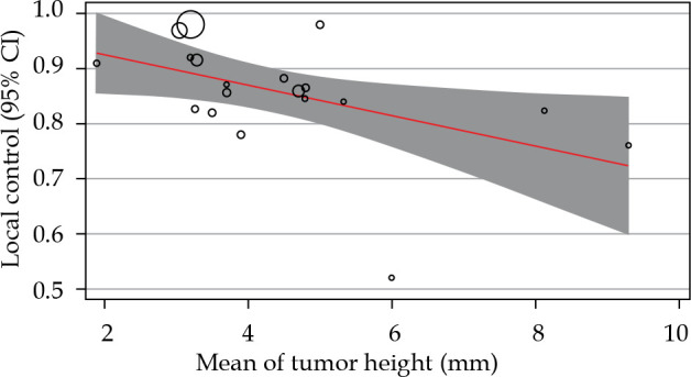
The relationship between local control rates and mean tumor thickness. Circles were the surrogate for each study sample size
Table 2.
The results of meta-regression based on tumor height and mean dose to the apex
| Variable of meta-regression | Number of included studies | Coefficients | P-value | 95% CI | Report of heterogeneity |
|---|---|---|---|---|---|
| Tumor height | 19 | –0.027 | 0.024 | –0.0518/–0.003 | Q = 154.34 P-value < 0.001 I2 = 92.52 |
| Radiation dose to the apex | 20 | 0.0002 | 0.679 | –0.0010/0.0016 | Q = 497.04 P-value < 0.001 I2 = 95.69 |
CI – confidence interval
Fig. 4.
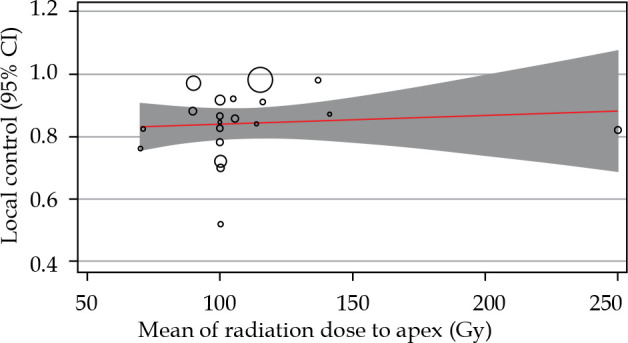
The relationship between local control rates and mean radiation dose to the apex of the tumor. Circles were the surrogate for each study sample size
The analysis of ocular complications was limited by authors’ poor reporting and was reflected in large and different results reported in the literature. According to the types of adverse effects, 14 studied described the rate of complications following brachytherapy [5, 6, 8-10, 14-16]. The rate of post-treatment retinopathy ranged from 20% to 53%. The rate of radiation-related crystalline lens opacity ranged from 4.2% to 53.8%. Radiation-related papillopathy and post-treatment ocular hypertension ranged from 2% to 29% and 2% to 12%, respectively.
Discussion
Our meta-analysis revealed a remarkable heterogeneity between the studies reporting the efficacy of 106Ru brachytherapy to treat uveal melanoma. However, 19 out of 21 reviewed studies reported a local control rate of more than 70%, and the weighted mean of this rate reached 84%.
Although the results are not conclusive, recent comparative studies have changed the primary concepts regarding the inferiority of 106Ru in local control of ocular melanoma. In a retrospective comparative case series with 2.5 years of follow-up, it has been reported that treatment with 106Ru is as effective as 125I [25]. Moreover, it has been suggested that 106Ru brachytherapy may be superior to 125I when the intended primary outcome is a reduction of thickness of melanoma. In another study comparing long-term efficacy and safety profiles of 106Ru and 125I brachytherapy, it was reported that both methods resulted in favorable control of the tumor, but 106Ru may provide additional benefit with reduced toxicity in tumors less than 5 mm of height [3].
The lower penetration power of 106Ru has limited its usage for melanomas thicker than five mm [3, 26]. Lack of reports on the effectiveness of 106Ru brachytherapy in treating large uveal melanoma makes it impossible to present a definite conclusion. In our study, 4 out of 21 reports have used 106Ru brachytherapy for tumors with a mean thickness of more than 5 mm, and reported rates of local control ranged from 59% to 84%. However, based on the negative slope of meta-regression, the thickness of uveal melanoma may be a predicting factor for the success of 106Ru brachytherapy, where the response of larger tumors could be lesser compared to smaller melanomas. Also, a minimal part of the heterogeneity in reported success rates was related to the tumors’ mean size.
Based on the previous reports and our analysis results, tumor location and radiation dose to the tumor’s apex seem to be additional determinants of therapeutic response to 106Ru plaque brachytherapy. It was suggested that the tumor location may be as important as the size of the treatment’s efficacy. In a study by Barker et al., tumors close to the edge of optic disc or the center of fovea, in addition to the cases with posterior tumor border near the posterior pole, were significantly associated with a higher rate of local recurrence. It may reflect the importance of tumor bulk coverage by the radiating plaque since, in the same study, smaller plaque diameter relative to the tumor’s largest base diameter was one of the predictors of 106Ru plaque brachytherapy failure [27].
In our meta-analysis, the correlation between the local control rates and the apex’s mean dose was positive but not statistically significant. Dose prescriptions for choroidal melanoma typically ranged from 70 Gy to 100 Gy to the tumor’s apex, with a treatment duration of 3 to 7 days. Although it may be interpreted as the presence of similar efficacy for different doses of apex radiation within the range of 70 Gy to more than 100 Gy, further studies should be conducted to evaluate this concept reliably. It would be logical to use lower doses to diminish the treatment’s short-term and long-term complications.
Predictably, the cumulative rate of local recurrence increases during the follow-up period. In a recent retrospective study, the local tumor recurrence rate increased from 3% at 12 months to nearly 15% at 48 months [10]. Similarly, uveal melanoma’s survival rates could decrease from 81.6% at five years to around 60% at ten years [10, 28]. After plaque brachytherapy, patients included in this review were followed for local control and complications with an interval of 3 to 6 months, and a follow-up duration of 1 to more than six years. The most common vision-threatening complications of 106Ru brachytherapy are retinopathy and cataract. However, ocular hypertension and optic neuropathy were also reported as complications of this treatment technique. According to the miscellaneous reports on the complications, plaque brachytherapy has also been associated with ocular surface disorders, sclera integrity, and ocular muscle functions [6, 10, 13, 22]. These complications must be treated through standard protocols to prevent loss of visual function and quality of life.
It seems that the preservation of visual acuity has been the primary goal of using beta-emitter isotopes in plaque brachytherapy. In preliminary non-comparative reports, assumed complications, such as neovascular glaucoma, are less common in patients treated with 106Ru [2]. According to previous reports, main risk factors associated with lower final visual acuity were older age, posterior and temporal tumor location, larger tumor, and posterior extension of the lesion [29]. Conservation of a visual acuity better than 20/200 and finger-counting were expected at long-term follow-up in 55% to 60% and over 80% of eyes treated with 106Ru, respectively [18]. Bergman et al. reported that up to 50% of patients treated with 106Ru were expected to have a visual acuity better than 0.1 in five years after the treatment [30].
Conclusions
The present study involves a systematic review of 21 retrospective studies. Our review’s main limitation was the retrospectivity of included studies, which made it impossible to perform a formal meta-analysis, since there were no randomized trials and prospective studies were rare. However, published studies suggest that plaque brachytherapy with 106Ru is successful in local control of uveal melanoma. Cataract and retinopathy are the main vision-threatening complication of radiation. Although pooled data analysis reveals a higher success rate in smaller tumors, this effect should be verified in randomized comparative studies.
Disclosure
The authors report no conflict of interest.
References
- 1.Moore RF. Choroidal sarcoma treated by the intraocular insertion of radon seeds. Br J Ophthalmol 1930; 14: 145-152. [DOI] [PMC free article] [PubMed] [Google Scholar]
- 2.Lommatzsch PK, Werschnik C, Schuster E. Long-term follow-up of Ru-106/Rh-106 brachytherapy for posterior uveal melanoma. Graefes Arch Clin Exp Ophthalmol 2000; 238: 129-137. [DOI] [PubMed] [Google Scholar]
- 3.Takiar V, Voong KR, Gombos DSet al. A choice of radionuclide: comparative outcomes and toxicity of ruthenium-106 and iodine-125 in the definitive treatment of uveal melanoma. Pract Radiat Oncol 2015; 5: e169-e176. [DOI] [PubMed] [Google Scholar]
- 4.Jiang P, Purtskhvanidze K, Kandzia Get al. (106)Ruthenium eye plaque brachytherapy in the management of medium sized uveal melanoma. Radiat Oncol 2020; 15: 183. [DOI] [PMC free article] [PubMed] [Google Scholar]
- 5.Espensen CA, Appelt AL, Fog LSet al. Predicting visual acuity deterioration and radiation-induced toxicities after brachytherapy for choroidal melanomas. Cancers (Basel) 2019; 11: 1124. [DOI] [PMC free article] [PubMed] [Google Scholar]
- 6.Rospond-Kubiak I, Wroblewska-Zierhoffer M, Twardosz-Pawlik H, Kociecki J. Ruthenium brachytherapy for uveal melanoma–single institution experience. J Contemp Brachytherapy 2017; 9: 548-552. [DOI] [PMC free article] [PubMed] [Google Scholar]
- 7.Pagliara MM, Tagliaferri L, Azario Let al. Ruthenium brachytherapy for uveal melanomas: Factors affecting the development of radiation complications. Brachytherapy 2018; 17: 432-438. [DOI] [PubMed] [Google Scholar]
- 8.Naseripour M, Jaberi R, Sedaghat Aet al. Ruthenium-106 brachytherapy for thick uveal melanoma: reappraisal of apex and base dose radiation and dose rate. J Contemp Brachytherapy 2016; 8: 66-73. [DOI] [PMC free article] [PubMed] [Google Scholar]
- 9.Fili M, Lundell G, Lundell M, Seregard S. High dose rate and low dose rate ruthenium brachytherapy for uveal melanoma. No association with ocular outcome. Br J Ophthalmol 2014; 98: 1349-1354. [DOI] [PubMed] [Google Scholar]
- 10.Tarmann L, Wackernagel W, Avian Aet al. Ruthenium-106 plaque brachytherapy for uveal melanoma. Br J Ophthalmol 2015; 99: 1644-1649. [DOI] [PubMed] [Google Scholar]
- 11.Salkola S, Heikkonen J, Eskelin S, Kivela T. Management of choroidal melanomas less than 10 mm in largest basal diameter with a 10-mm ruthenium plaque. Retina 2014; 34: 2110-2120. [DOI] [PubMed] [Google Scholar]
- 12.Takiar V, Gombos DS, Mourtada Fet al. Disease control and toxicity outcomes using ruthenium eye plaque brachytherapy in the treatment of uveal melanoma. Pract Radiat Oncol 2014; 4: e189-194. [DOI] [PubMed] [Google Scholar]
- 13.Perri P, Fiorica F, D’Angelo Set al. Ruthenium-106 eye plaque brachytherapy in the conservative treatment of uveal melanoma: a mono-institutional experience. Eur Rev Med Pharmacol Sci 2012; 16: 1919-1924. [PubMed] [Google Scholar]
- 14.Papageorgiou KI, Cohen VM, Bunce Cet al. Predicting local control of choroidal melanomas following 106Ru plaque brachytherapy. Br J Ophthalmol 2011; 95: 166-170. [DOI] [PubMed] [Google Scholar]
- 15.Kaiserman N, Kaiserman I, Hendler Ket al. Ruthenium-106 plaque brachytherapy for thick posterior uveal melanomas. Br J Ophthalmol 2009; 93: 1167-1171. [DOI] [PubMed] [Google Scholar]
- 16.Frenkel S, Hendler K, Pe’er J. Uveal melanoma in Israel in the last two decades: characterization, treatment and prognosis. Isr Med Assoc J 2009; 11: 280-285. [PubMed] [Google Scholar]
- 17.Mossbock G, Rauscher T, Winkler Pet al. Impact of dose rate on clinical course in uveal melanoma after brachytherapy with ruthenium-106. Strahlenther Onkol 2007; 183: 571-575. [DOI] [PubMed] [Google Scholar]
- 18.Damato B, Patel I, Campbell IRet al. Local tumor control after 106Ru brachytherapy of choroidal melanoma. Int J Radiat Oncol Biol Phys 2005; 63: 385-391. [DOI] [PubMed] [Google Scholar]
- 19.Novak-Andrejcic K, Jancar B, Hawlina M. Echographic follow-up of malignant melanoma of the choroid after brachytherapy with 106Ru. Klin Monbl Augenheilkd 2003; 220: 853-860. [DOI] [PubMed] [Google Scholar]
- 20.Georgopoulos M, Zehetmayer M, Ruhswurm Iet al. Tumour regression of uveal melanoma after ruthenium-106 brachytherapy or stereotactic radiotherapy with gamma knife or linear accelerator. Ophthalmologica 2003; 217: 315-319. [DOI] [PubMed] [Google Scholar]
- 21.Stoffelns BM, Kutzner J, Jochem T. Retrospective analysis of ruthenium-106 brachytherapy for small and medium-sized malignant melanoma of the posterior choroid. Klin Monbl Augenheilkd 2002; 219: 216-220. [DOI] [PubMed] [Google Scholar]
- 22.Kleineidam M, Guthoff R, Bentzen SM. Rates of local control, metastasis, and overall survival in patients with posterior uveal melanomas treated with ruthenium-106 plaques. Radiother Oncol 1993; 28: 148-156. [DOI] [PubMed] [Google Scholar]
- 23.Summanen P, Immonen I, Kivela Tet al. Radiation related complications after ruthenium plaque radiotherapy of uveal melanoma. Br J Ophthalmol 1996; 80: 732-739. [DOI] [PMC free article] [PubMed] [Google Scholar]
- 24.Lommatzsch PK. Results after beta-irradiation (106Ru/106Rh) of choroidal melanomas: 20 years’ experience. Br J Ophthalmol 1986; 70: 844-851. [DOI] [PMC free article] [PubMed] [Google Scholar]
- 25.Ghassemi F, Sheibani S, Arjmand Met al. Comparison of iodine-125 and ruthenium-106 brachytherapy in the treatment of choroidal melanomas. Clin Ophthalmol 2020; 14: 339-346. [DOI] [PMC free article] [PubMed] [Google Scholar]
- 26.Wilkinson DA, Kolar M, Fleming PA, Singh AD. Dosimetric comparison of 106Ru and 125I plaques for treatment of shallow (< or = 5 mm) choroidal melanoma lesions. Br J Radiol 2008; 81: 784-789. [DOI] [PubMed] [Google Scholar]
- 27.Barker CA, Francis JH, Cohen GNet al. (106)Ru plaque brachytherapy for uveal melanoma: factors associated with local tumor recurrence. Brachytherapy 2014; 13: 584-590. [DOI] [PMC free article] [PubMed] [Google Scholar]
- 28.Belaïd A, Nasr C, Jmour Oet al. Brachytherapy of uveal melanomas with ruthenium-106 plaques. Asian Pac J Cancer Prev 2016; 17: 5281-5285. [DOI] [PMC free article] [PubMed] [Google Scholar]
- 29.Damato B, Patel I, Campbell IRet al. Visual acuity after ruthenium(106) brachytherapy of choroidal melanomas. Int J Radiat Oncol Biol Phys 2005; 63: 392-400. [DOI] [PubMed] [Google Scholar]
- 30.Bergman L, Nilsson B, Lundell Get al. Ruthenium brachytherapy for uveal melanoma, 1979-2003: survival and functional outcomes in the Swedish population. Ophthalmology 2005; 112: 834-840. [DOI] [PubMed] [Google Scholar]