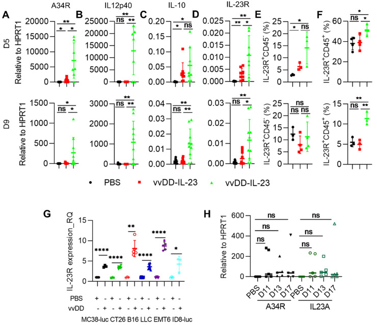
Figure 3.
vvDD-IL-23 treatment prolongs viral persistence. B6 mice were i.p. inoculated with 5×10⁵ MC38-luc cells and treated with PBS, vvDD, or vvDD-IL-23 at 2×10⁸ PFU/mouse five days after tumor inoculation. Tumor-bearing mice were sacrificed five or nine days post-treatment and primary tumors were collected and analyzed using RT-qPCR to determine the expression of A34R (A), IL12p40 (B), IL-10 (C) and IL-23R (D). The IL-23R expression on CD45- or CD45+ cells were determined by flow cytometry (E, F). MC38-luc (3×10⁵ cells), CT26 (3×10⁵ cells), B16 (2×10⁵ cells), LLC (3×10⁵ cells), EMT (3×10⁵ cells), or AB12-luc (3×10⁵ cells) tumor cells were mock-infected or infected with vvDD at an MOI of 1. The cell pellets were harvested to measure IL-23R expression at 24 h after infection using flow cytometry (G). MC38-luc-bearing mice treated as above were also sacrifice at D11, D13 and D17 to monitor viral persistence in tumors using RT-qPCR (H). *: P<0.05; **: P<0.01; and ****: P<0.0001. ns: not significant.

