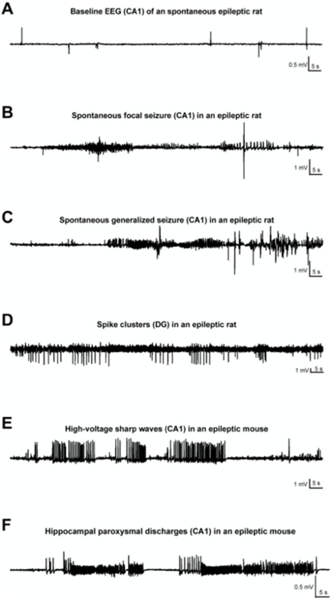
Figure 4.

Various patterns of EEG-activity during KA-induced chronic epilepsy. A, Baseline recording from CA1 of an epileptic rat. Note the occurrence of interictal spikes. B, Recording of a spontaneous focal seizure in CA1. C. A secondary generalized convulsive seizure in an epileptic rat. D, Spike clusters originating from the dentate gyrus 7 weeks post-SE. E, High-voltage sharp waves in the epileptic focus (CA1) of a mouse, several weeks post-SE. F, Hippocampal paroxysmal discharges (HPDs) in CA1 of an epileptic mouse. (Klee et al., 2017).
