PLOS ONE logoLink to PLOS ONE
. 2021 Jun 3;16(6):e0252628. doi: 10.1371/journal.pone.0252628

Serological profiles of pan-coronavirus-specific responses in COVID-19 patients using a multiplexed electro-chemiluminescence-based testing platform

Sidhartha Chaudhury 1, Jack Hutter 2, Jessica S Bolton 3,4, Shilpa Hakre 5, Evelyn Mose 6, Amy Wooten 6, William O’Connell 6, Joseph Hudak 6, Shelly J Krebs 5, Janice M Darden 4, Jason A Regules 3, Clinton K Murray 7, Kayvon Modjarrad 5, Paul Scott 5, Sheila Peel 8, Elke S Bergmann-Leitner 3,*
Editor: Etsuro Ito9
PMCID: PMC8174743  PMID: 34081747

Abstract

Serological assessment of SARS-CoV-2 specific responses are an essential tool for determining the prevalence of past SARS-CoV-2 infections in the population especially when testing occurs after symptoms have developed and limited contact tracing is in place. The goal of our study was to test a new 10-plex electro-chemiluminescence-based assay to measure IgM and IgG responses to the spike proteins from multiple human coronaviruses including SARS-CoV-2, assess the epitope specificity of the SARS-CoV-2 antibody response against full-length spike protein, receptor-binding domain and N-terminal domain of the spike protein, and the nucleocapsid protein. We carried out the assay on samples collected from three sample groups: subjects diagnosed with COVID-19 from the U.S. Army hospital at Camp Humphreys in Pyeongtaek, South Korea; healthcare administrators from the same hospital but with no reported diagnosis of COVID-19; and pre-pandemic samples. We found that the new CoV-specific multiplex assay was highly sensitive allowing plasma samples to be diluted 1:30,000 with a robust signal. The reactivity of IgG responses to SARS-CoV-2 nucleocapsid protein and IgM responses to SARS-CoV-2 spike protein could distinguish COVID-19 samples from non-COVID-19 and pre-pandemic samples. The data from the three sample groups also revealed a unique pattern of cross-reactivity between SARS-CoV-2 and SARS-CoV-1, MERS-CoV, and seasonal coronaviruses HKU1 and OC43. Our findings show that the CoV-2 IgM response is highly specific while the CoV-2 IgG response is more cross-reactive across a range of human CoVs and also showed that IgM and IgG responses show distinct patterns of epitope specificity. In summary, this multiplex assay was able to distinguish samples by COVID-19 status and characterize distinct trends in terms of cross-reactivity and fine-specificity in antibody responses, underscoring its potential value in diagnostic or serosurveillance efforts.

Introduction

The current SARS-CoV-2 pandemic is unparalleled in recent history, and countries have developed diverse approaches to combat and manage transmission. Most countries are attempting to curtail the infection rates through strict social distancing rules, rigorous testing, and contact tracing of individuals potentially exposed to infectious individuals. A population-wide serological assessment of SARS-CoV-2 immunity would have several applications: (a) conduct surveillance to determine exposure rates, (b) investigate the feasibility of using antibody titers as markers of immunity, (c) examine the durability of SARS-CoV-2 antibody responses and protection from reinfection, (d) establish the serological landscape of pan-CoV antibody responses and determine whether pre-existing immunity to common human CoVs affect COVID-19 disease course; (e) screen individuals for participation in COVID-19 vaccine trials or for prioritization for receiving FDA-approved or emergency use authorized COVID-19 vaccines; and (f) screen for antibody reactivity to newly emerging SARS-CoV-2 strains.

Several tests have been developed for measuring SARS-CoV-2-specific IgG and IgM responses are currently available [1] and the data obtained from these tests suggest that SARS-CoV-2-specific antibody responses are reliably measurable by 2–3 weeks after onset of symptoms [24]. The presence of antibodies was detected in a minority of COVID-19 patients within one week of onset and seroconversion ranges from 90 to 100% by 15 days after onset [5, 6]. IgM seroconversion is seen around 12 days from onset, and IgG seroconversion around 13 days from onset [7], with the caveat that studies on antibody response dynamics have so far largely focused on convenience samples that may fail to capture the earliest emergence of these responses. However, recent findings suggest the possibility of a lack of durable antibody responses, whereby some percentage of individuals previously infected with SARS-CoV-2 that were seropositive following infection and later became seronegative during early convalescence, especially in cases where the SARS-CoV-2 infection resulted in asymptomatic infection or low disease severity [8, 9]. This finding agrees with previous studies in humans with other coronaviruses in that antibody-responses to CoV infections can be short-lived [1012]. However, the durability of SARS-CoV-2 responses remains poorly characterized, and it is unclear how durability varies with respect to antibody fine-specificity, isotype profile, cross-reactivity to other coronaviruses, and how these aspects of the humoral immunity contribute to durable protection from reinfection.

Although numerous SARS-CoV-2-specific serological assays have been developed [13, 14], additional challenges remain in using these assays for surveillance or clinical management [15] including: 1) assay throughput, 2) the specificity of assay readouts to SARS-CoV-2 as compared to other related coronaviruses 3) and the need to perform sample testing at multiple dilutions due to the narrow linear range of the respective assay platform. Recently, we compared a newly developed multiplexed serological assay based on electro-chemiluminescence (ECLIA) customized for malaria serosurveillance with a qualified standard ELISA [16]. The results demonstrated superiority of the ECLIA-based assay in several aspects including a wide linear range eliminating the need for serial dilutions of test samples, low variability, robust reproducibility of the assay, and no signal reduction due to antigenic competition when testing closely related antigens. Given that only 0.1 μL of sample is needed for the assay, and with the use of the pre-manufactured CoV antigen plates described here, five antigen plates (450 samples) could be run by a single operator in five hours, and with substantially higher throughput if using a liquid handler, this platform would be appropriate for use in high-throughput serosurveillance applications.

The objective of the current study was to test the performance of a multiplexed pan-CoV ECLIA-based assay regarding its ability to establish a serological profile of responses to common beta CoVs (HKU-1, OC43), MERS-CoV, SARS-CoV-1 and SARS-CoV-2. For this study, three sample groups were compiled subjects diagnosed with COVID-19 from the Brian D. Allgood Army Community Hospital at Camp Humphreys in Pyeongtaek, South Korea from March 13 to April 18, 2020; healthcare administrators from the same hospital during the same time period but with no diagnosis of COVID-19; and pre-pandemic samples from several sources in the United States in 2019. The comparison of the COVID-19 and non-COVID-19 samples from the same site will enable us to compare antibody responses from COVID-19 diagnosed individuals from other individuals the same location and pandemic time period, who may have been exposed, but were not symptomatic or diagnosed with COVID-19. Comparison with pre-pandemic samples will determine whether there are differences in CoV exposure between pre-pandemic samples and pandemic samples, even in cases where there was no COVID-19 diagnosis. Our findings demonstrate the utility of this assay for SARS-CoV-2-serosurveillance based on its high sensitivity and specificity as well as its ability to discern between pre-existing immunity to common human CoVs and SARS-CoV-2.

Materials and methods

Sample collection

For this analysis, plasma samples were obtained from a public health investigation of COVID-19 patients at the Brian D. Allgood Army Community Hospital (BDAACH) at Camp Humphreys in Pyeongtaek, South Korea from March 13 to April 18, 2020 (WRAIR#2755). Three sample groups were compiled: patients diagnosed with COVID-19; health care personnel assigned to the same hospital during the same time period whose duties did not include regular interaction with patients and who did not have a diagnosis of COVID-19 (these individuals were assumed to be unexposed or minimally exposed from living and working in an outbreak setting with a very low prevalence of COVID-19 at the time of collection); and pre-pandemic samples from several sources in the United States in 2019. COVID-19 and Control subjects were drawn from the same overall population: the U.S. Department of Defense military, civilian, and contractor population working at Camp Humphreys. The comparison of the COVID-19 and Control samples from the same site enabled us to measure antibody responses from COVID-19 diagnosed individuals from those in the same location and pandemic time period, who may have been exposed, but were not symptomatic or diagnosed with COVID-19. Samples from ten COVID-19 subjects and eight control subjects, matched by study location and population, were obtained and compared to a similar number of pre-pandemic samples (ten) using the multiplex ECLIA assay. As this was a retrospective analysis of COVID-19 samples collected during a public health investigation of a local outbreak, no a priori power calculation was carried out.

All COVID-19 diagnoses were confirmed using a nasopharyngeal swab and RT-PCR-based diagnostic assay (Centers for Disease Control 2019-nCoV RT-PCR diagnostic panel run on the Applied Biosystems 7500 platform). All Control subjects were also tested via nasopharyngeal swab and RT-PCR and confirmed to be negative for COVID-19 at the time of sample collection. COVID-19 disease severity was assessed as asymptomatic, mild (symptomatic but not interfering with daily activity), moderate (interfering with daily activity but not requiring hospitalization), and severe (preventing daily activity and requiring hospitalization). All samples collected at BDAACH were sent to Walter Reed Army Institute of Research (WRAIR) for analysis. Pre-pandemic samples were obtained from a WRAIR blood collection protocol (WRAIR#2567) based on sample availability from August 2019 conducted in Silver Spring, Maryland. Finally, two pre-pandemic samples were commercially available as pooled plasma samples from GeminiBio (GemCell™ U.S. Origin Human Serum AB, Cat.No 100–512) that were delivered to WRAIR in 2018.

Ethics approval and consent to participate

The plasma sample use was reviewed by the WRAIR Human Subjects’ Protection Branch which determined that the research does not involve human subjects (NHSR protocol WRAIR #2567, WRAIR#2755, #EID-029) as the samples used were de-identified and no link between samples and subjects exists.

Antigens

Antigens for this study were manufactured by MSD in a mammalian expression system (Expi 293 F) and printed onto the 10-plex plates by Meso Scale Diagnostics, LLC (Cat No K15362U (IgG), and K15363U (IgM), MSD, Rockville, Maryland). The antigens used were: HA-trimer Influenza A (Hong Kong H3), spike (soluble ectodomain with T4 trimerization domain) trimers for SARS-CoV-2, SARS-CoV-1, MERS-CoV, and betacoronaviruses HKU-1 and OC43, as well as the spike N-terminal domain (NTD, Q14-L303 of the SARS-CoV-2 spike sequence), receptor binding domain (RBD, R319-F541 of the SARS-CoV-2 spike sequence), and nucleocapsid protein (N; full length) for SARS-CoV-2, and bovine serum albumin (BSA).

ECLIA

The MSD V-PLEX platform was used as 10-plex assays utilizing the pre-printed antigens described above with each printed on its own spot. Blocker A Solution (Cat.No R93BA, MSD) was added to the plates at 150 μl/well. The plates were sealed and incubated at room temperature (RT) for 1h on a plate shaker, shaking at 700 rpm. The plates were washed three times with 1x MSD Wash Buffer (Cat.No R61AA, 150 μl/well). Sera were diluted to 1:1000 dilution with Diluent 100 (Cat. No R50AA, MSD) and added to each well (50 μl/well). The same dilution was used for both IgM and IgG measurements. Plates were sealed and incubated at RT for 2h on a plate shaker, shaking at 700 rpm, then washed three times with 1x MSD Wash Buffer (150 μl/well). The detection antibody, SULFO-TAG either with anti-human IgG (Cat.No D20JL, MSD) antibody or anti-human IgM (Cat.No D20JP, MSD) was diluted to 2 μg/ml in Diluent 100 (MSD) and added to the wells (50 μl/well). The plates were sealed and incubated at RT for 1h on a plate shaker, shaking at 700 rpm. After washing, 150 μl a working solution of MSD GOLD Read Buffer B (Cat.No R60AM, MSD) was added to each well and immediately the plates were read on the MESO QuickPlex SQ 120 (MSD), per manufacturer’s instructions. We assessed the dynamic range of the MSD V-PLEX platform using this antigen panel across a serial dilution range from 1:1000 to 1:30,000 and found high signal-to-noise ratio and a linear response across that entire span of concentrations (S1 Fig).

Statistical analysis

The MSD assay provides a readout in units of mean luminescence intensity and all readouts were directly log-transformed prior to analysis without any normalization or subtraction of background. Univariate analysis comparisons between groups (COVID-19, Control, and pre-COVID) were made using a Shapiro-Wilk Normality Test followed by a student’s t test or a Wilcoxon signed rank test. We applied a multiple test correction using the Benjamin-Hochberg method; p-values were considered significant if their adjusted p-value was < 0.05. Principal Component Analysis (PCA) was carried out by normalizing and scaling the log-transformed values. Data points were colored by group, and ellipses were generated corresponding to 50% confidence intervals for each group, to identify general trends in the data set. Seropositivity for each CoV spike antigen for a given subject was assessed based on whether the readout for that antigen exceeded cutoff defined by the upper limit of the 99.9% confidence interval of the BSA (negative control) response, as determined from pooling the BSA response across all subjects in the study. This cutoff value was determined to be 8.85 for IgM and 8.96 for IgG in log-transformed units of mean luminescence intensity. Correlation plots were generated using pairwise Pearson correlation coefficients calculated from the log-transformed data. All statistical analysis was carried out in R using the stats, ggplot2, and corrplot,.

Results

Table 1 provides a summary of the COVID-19 subjects, date of the first positive COVID-19 test, date of sample collection, disease severity and clinical indications as well as demographic information (age range and sex). All samples were collected within three weeks of the first positive COVID-19 test. One subject had severe COVID-19, six subjects had mild symptoms, and three subjects were asymptomatic. Demographic information on Control subjects is shown in S1 Table.

Table 1. Summary of COVID-19 subjects.

Subject ID Age range (y.o.) [Sex] Day of Symptom Onset* Day of Sample Collection* Disease Severity Notes
i-0001 21–30 [M] -1 +17 Mild Chills, cough, from 18 to 15 days prior to participation (Day +17)
+33
+37
i-0002 21–30 [F] 0 +18 Mild Mild cough from 18 to 16 days prior to participation (Day +18)
i-0003 21–30 [F] 0 +1 Mild Anosmia, sore throat, cough and malaise 1 day prior to participation (Day +1), improved 9 days later
i-0004 51–60 [M] 0 +2 Severe Severe illness 2 days prior to participation (Day +2), hospitalized with hypoxia from -1 to 7 days after participation in the investigation; started improving by day 5; symptoms resolved by day 8
+7
+10
i-0005 41–50 [M] 0 +3 Mild Mild cough 3 days prior to participation (Day +3), persisting through admission
i-0006 51–60 [M] -12 +4 Mild Chills, cough, runny nose, loss of appetite 16 days prior to participation (Day +4), persisted for weeks
i-0007 41–50 [M] NA +3 Asymptomatic never symptomatic
i-0008 41–50 [M] NA +2 Asymptomatic never symptomatic
i-0009 51–60 [F] 0 +2 Mild Mild cough 2 days prior to participation (Day +2), and persisted
i-0010 21–30 [F] NA +2 Asymptomatic never symptomatic

* Day 0 defined as day of initial test positivity by nasopharyngeal COVID-19 test

SARS-CoV-2 specific IgG and IgM responses

Samples for all three groups were assayed on the MSD platform to determine IgM and IgG -specific responses to influenza H3, spike proteins for SARS-CoV-2, SARS-CoV-1, MERS-CoV, beta-coronaviruses OC43 and HKU1 relative to BSA (negative control) (summarized in Fig 1). In terms of assay sensitivity, we found that COVID-19 samples had a roughly 300-fold higher IgM signal to SARS-CoV-2 spike protein and a 1000-fold higher IgG antibodies binding to SARS-CoV-2 spike antigen compared to BSA. In Control and pre-pandemic samples, there was no significant difference in IgM responses to SARS-CoV-2 spike protein and BSA, but the IgG response to the SARS-CoV-2 spike was approximately 10-fold higher (p < 10−4) than to BSA, suggesting either some degree of cross-reactivity of pre-existing IgG antibodies to SARS-CoV-2 spike antigen in these samples or higher non-specific IgG binding to this antigen. We carried out serial dilutions to further assess assay sensitivity (S1 Fig).

Fig 1. Antibody responses to pan-CoV antigens.
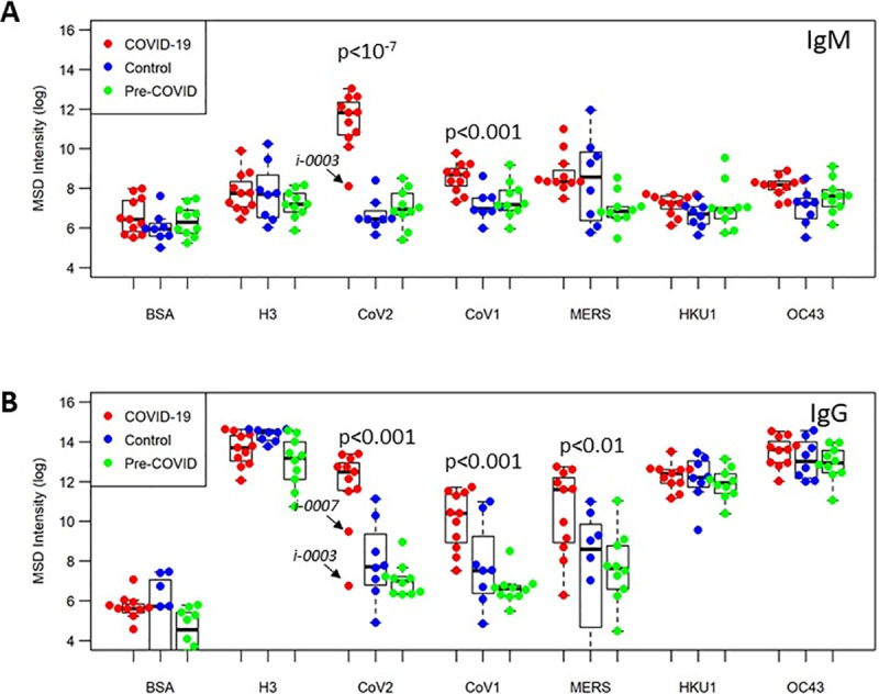
Fig 1

Plasma levels of IgM (A) or IgG (B) from COVID-19 samples (n = 10, red), individuals with no known history of SARS-CoV-2 infection (Control group, n = 8, blue) and samples from US health donors (pre-COVID-19 group, n = 10, green) were tested at 1:1000 dilution; plate antigens shown are: BSA (negative contol); influenza H3 trimer (reference antigen), along with full-length spike proteins for CoVs: SARS-CoV-2, SARS-CoV-1, MERS-CoV, OC43 and HKU1.

We found that COVID-19 samples showed significant higher IgM responses to the SARS-COV-2 spike protein (p<10−7) than the control sample group and pre-pandemic group, while no differences between the three groups were noted for BSA or influenza. On average, COVID-19 samples showed 100- to 200-fold higher IgM responses to SARS-CoV-2 spike antigen than the control or pre-pandemic samples. For IgG responses, the COVID-19 samples also showed significantly higher responses than either the control or pre-pandemic samples (p < 0.001), while no differences were seen between the sample groups in terms of their reactivity to BSA or H3. Like the IgM responses, the IgG responses in the COVID-19 samples to SARS-CoV-2 spike protein were approximately 100- to 200-fold higher than in the control and pre-pandemic samples. In both IgM and IgG responses to CoV-2 spike protein, there was a single subject in the COVID-19 group, Subject i-0003, that was a low outlier, showing similar responses to Control and pre-pandemic samples. This subject had mild symptoms and their plasma sample was obtained only one day following initial test positivity; it is possible this was early in the infection course and the subject had not yet seroconverted.

Cross-reactivity of SARS-CoV-2 antibody responses

Next, we analyzed differences in antibody binding response to the other CoV antigens between the three groups. COVID-19 subjects, in addition to showing significantly higher IgM binding antibodies to SARS-CoV-2 spike compared to the Control and pre-COVID subjects, also showed significantly higher IgM binding antibodies to SARS-CoV-1 (p < 0.001), HKU1 (p < 0.05), and OC43 (p < 0.01) spike proteins (Fig 1A). This data suggests significant IgM cross-reactivity between SARS-CoV-2 and SARS CoV-1, HKU-1, and OC43. Likewise, we found that COVID-19 subjects, in addition to showing significantly higher IgG binding antibodies to SARS-CoV-2, also show significantly higher IgG binding antibodies to SARS-CoV-1 (p < 0.001) and MERS-CoV (p < 0.01) spike proteins, compared with Control and pre-COVID-19 subjects, suggesting that SARS-CoV-2 IgG antibodies may be cross-reactive with SARS-CoV-1 and MERS-CoV (Fig 1B).

To determine the distinguishing features between COVID-19 and non-COVID-19 samples, we generated a principal component analysis (PCA) plot of the IgM and IgG responses to these five CoV spike antigens (Fig 2). The PCA using IgM data demonstrates that samples display antibody responses largely along two major axes: SARS-CoV-1/SARS-CoV-2 vs. HKU1/MERS-CoV. COVID-19 samples have high SARS-CoV-1/SARS-CoV-2 binding antibodies while Control and Pre-COVID-19 samples do not; HKU-1/OC43/MERS CoV responses appear to be independent of COVID-19 status. Analyzing the IgG responses revealed that the profile of cross-reactivity is different from that of IgM: samples show responses along two major axes: SARS-CoV-1/SARS-CoV-2/MERS-CoV and HKU-1/OC43. COVID-19 samples show high SARS-CoV-1/SARS-CoV-2/MERS-CoV responses, and responses along the HKU1/OC43 axis appear to be independent of COVID-19 status.

Fig 2. PCA plot of IgM and IgG pan-CoV responses.
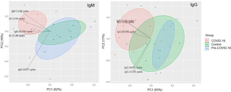
Fig 2

PCA plot showing IgM (left) and IgG (right) responses to spike proteins for SARS-CoV-2, SARS-CoV-1, MERS-CoV, OC43, and HKU1 for COVID-19 (red), Control (green), and pre-COVID (blue) samples. Each data point reflects a single sample colored by group; loading vectors reflecting the direction of the contribution of each parameter to the PCA plot is shown. Ellipses denote the 50% confidence interval for each group.

Seropositivity across CoV antigens

We next sought to determine the seropositivity of these samples across the five CoV spike antigens in the panel. We defined a cutoff above which a sample would be considered seropositive based on the IgG and IgM signal to BSA (noise). We set the cutoff at the upper limit of the 99.9% confidence interval calculated by pooling all the samples in the data set. This cutoff was determined to be 8.46 for IgM seropositivity and 8.96 for IgG seropositivity in log transformed units of luminescence intensity.

Overall, we found that 90% of the COVID-19 samples were seropositive for IgM binding antibodies to SARS-CoV-2, suggesting most COVID-19 patients tested here had seroconverted by the time the sample was taken (Fig 3, top). Additionally, 33% of CoV-2-seropositive COVID-19 samples were also seropositive for MERS-CoV, 33% were seropositive for both SARS-CoV-1, and 22% to both. By contrast, none of the Control and pre-pandemic subjects were seropositive for SARS-CoV-2, and 50% of the Control and 90% of the pre-pandemic samples were seropositive to none of the CoV antigens, including OC43 and HKU1, again reflecting the lack of IgM response detected against these seasonal CoVs. Interestingly, 50% of Control samples were seropositive for MERS-CoV, compared to none of the pre-pandemic U.S. samples.

Fig 3. Seropositivity Pan-CoV in IgM and IgG responses.
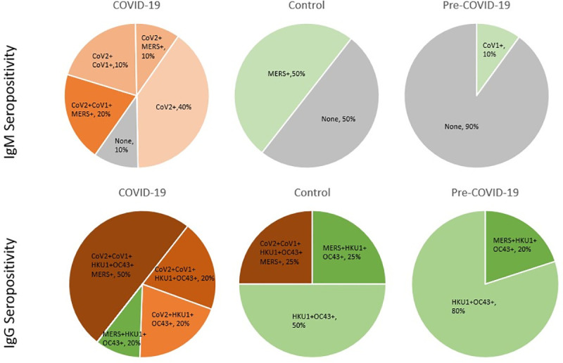
Fig 3

Pie charts showing percentage of subjects in the COVID-19 (left), Control (center), and Pre-COVID-19 (right) groups that are seropositive to each combination of CoV spike antigens for SARS-CoV-1, SARS-CoV-2, MERS-CoV, HKU1, and OC43, with ‘+’ denoting seropositivity for the IgM (top) and IgG (bottom) responses. SARS-CoV-2 seropositivity is reflected by shades of orange, non-SARS-CoV-2 seropositivity is shown in shades of green, and no seropositivity to any CoV antigen is shown in gray.

For the IgG responses, 90% of COVID-19 samples were seropositive for SARS-CoV-2, suggesting that one (of the 10) COVID-19 patients did not seroconvert at the time the sample was taken (Fig 3, bottom). Almost 60% of COVID-19 subjects that had seroconverted by IgG to SARS-CoV-2 were seropositive for all four other CoVs, suggesting substantial cross-reactivity in IgG SARS-CoV-2 binding antibodies. Interestingly, 25% of the subjects in the Control group showed seropositivity to not only SARS-CoV-2 but also to all five of the human CoVs tested, similar to what was observed in the COVID-19 group. A single subject in the COVID-19 group, aforementioned Subject i-0003, was seronegative to SARS-CoV-2 in both IgM and IgG responses. No clear association between seropositivity and symptom severity was observed in this data set.

None of the subjects in the pre-COVID-19 group showed seropositivity to SARS-CoV-2, but all showed seropositivity to the seasonal betacoronaviruses HKU1 and OC43. Overall two broad observations can be made from this seropositivity data: 1) IgM CoV binding antibodies likely reflect acute or recent infection while IgG CoV binding antibodies reflect both acute infection (in the case of SARS-CoV-2) or long-term memory responses (in the case of the seasonal CoVs) and 2) the IgG SARS-CoV-2 binding antibodies appear to be more cross-reactive than the IgM SARS-CoV-2 binding antibodies.

Fine specificity of SARS-CoV-2 antibody responses

The multiplex assay contains additional antigenic targets of SARS-CoV-2, i.e., RBD, NTD, and N (Fig 4). COVID-19 patients had significantly higher IgM levels directed at these antigens compared to the control groups. While all samples in the three groups had significant antibody responses to the seasonal CoVs (HKU-1, OC43), there was no significant recognition of the SARS-CoV-2 antigen fragments in the Control and pre-COVID-19 samples. The hierarchy of IgM binding to SARS-CoV-2 antigens reveals highest reactivity to spike and RBD, followed by binding to the nucleocapsid and the least reactivity to NTD. The antibody profile of SARS-CoV-2 specific IgG responses was different from the IgM profile. While COVID-19 patients had significantly higher IgG binding antibodies targeting the spike, RBD and nucleoprotein, the SARS-CoV-2-specific IgG responses to NTD were not significantly different between the three sample groups. As before, one subject in the COVID-19 group, aforementioned Subject i-0003, was a low outlier in responses to SARS-CoV-2 spike and RBD for both IgG and IgM responses.

Fig 4. Fine-specificity of SARS-CoV-2 specific antibody responses.
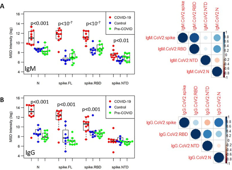
Fig 4

The IgM (A) and IgG (B) responses in samples from three groups (COVID-19, red; Control, blue; Pre-COVID, green) was assessed in the multiplex ECLIA platform against SARS-CoV-2 full-length spike protein, spike RBD, spike NTD, and nucleocapsid. Correlation matrices are shown on the right, with the color and size of the circles corresponding to pairwise Pearson correlation coefficient.

To identify the relationship between the different antibody specificities, correlation matrices were generated for IgM and IgG responses (Fig 4) demonstrating that the magnitude of IgM SARS-CoV-2 spike binding antibodies correlated strongly with RBD responses. To a lesser extent, there was also a positive correlation between the nucleocapsid and RBD specific antibodies. The antibody profile of SARS-CoV-2-specific IgG was distinct from the IgM profiles as there was a weak correlation between nucleocapid and spike-specific responses and NTD with RBD specific antibodies, suggesting that the fine specificity between the IgM and IgG SARS-CoV-2 spike responses may differ, specifically that the IgM spike binding antibodies target epitopes largely to the RBD, while the IgG spike binding antibodies may be more focused on epitopes that include regions outside of the RBD itself, or target RBD epitopes unique to the whole-spike structure that are not recapitulated in the recombinant protein.

Combining IgM and IgG CoV-2 responses to identify COVID-19 samples

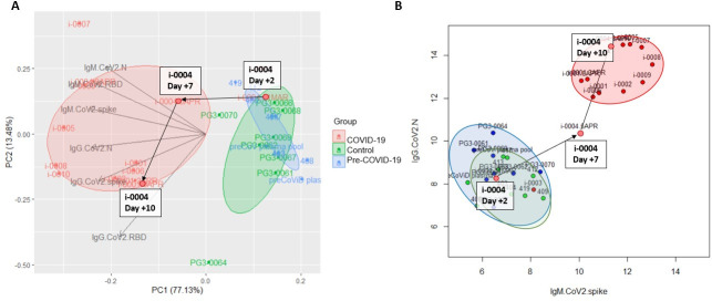
We combined all the SARS-CoV-2 specific antigen readouts (full-length spike, RBD, NTD, and N) for IgM and IgG to determine if they could clearly distinguish COVID-19 samples from Control or Pre-COVID-19 samples. Using an unsupervised PCA approach, we show that these groups can be readily distinguished (Fig 5A), and that even a reduction from these 12 parameters to two, IgG response to N and IgM response to the spike protein (Fig 5B), was sufficient to identify COVID-19 samples, for all subjects except the aforementioned Subject i-0003. Furthermore, we had longitudinal data for a COVID-19 subject (Subject i-0004) who seroconverted by IgG over the course of eight days. Mapping the data from this subject shows that the longitudinal course of this subject’s antibody response could clearly be mapped going from the pre-COVID-19 or non-infected region to the COVID-19 region.

Fig 5. Combining IgG and IgM responses to distinguish COVID-19 samples.

Fig 5

PCA plot of IgM and IgG responses to SARS-CoV-2 antigens spike, RBD, NTD, and Ncap (A) for COVID-19 (red), Control (green), and pre-COVID (blue) subjects. Loading vectors showing the direction of the contribution of each parameter to the PCA are shown. Ellipses correspond to 50% confidence intervals for each group. Scatterplot of IgM SARS-CoV-2 spike responses and IgG SARS-CoV-2 Ncap responses for all three groups. Longitudinal data for COVID-19 subject i-0004 collected on Day +2, Day +7, and Day +10, relative to day of initial test positivity, is highlighted as the subject seroconverted over this time span.

Analysis of IgM and IgG seropositivity in COVID-19 subjects, with respect to time from first positive test and onset of symptoms (S2 Fig) showed that (1) seronegative results were only found in two cases (i-0003 and i-0004) where the sample was collected within two days of the onset of symptoms and (2) that all samples that were seropositive by IgM were also seropositive by IgG, as measured by response to the SARS-CoV-2 spike protein. This apparent simultaneous seroconversion was seen as early as two and three days after onset of symptoms (i-0009 and i-0005, respectively).

Discussion

In the current study, we evaluated a new multiplex coronavirus antigen panel using an electro-chemiluminescence assay platform to conduct serological high-throughput testing of sera/plasma. The study had two objectives: (a) determine whether the methodology is useful for sero-surveillance and (b) to gain insights into serological cross-reactivity between five human coronaviruses. Analysis of the high-dimensional serological data (20 parameters per sample collected) revealed clear differences between pre-existing immunity and SARS-CoV-2 induced antibody responses and distinct patterns of cross-reactivity in IgM and IgG responses, demonstrating the value of this multiplex approach for SARS-CoV-2 serology studies.

The ECLIA-based MSD platform was chosen based on its superiority in previous studies using malarial antigens [16, 17]. In the present study, we evaluated a pan-CoV panel of recombinant proteins generated in a mammalian expression system to ensure proper glycosylation. We found that the linear range of the MSD ranged from 1:1,000 to 1:30,000, which eliminates the need to test serial dilutions for individual samples. An assay with such high sensitivity and specificity requires only very small sample volume (facilitating longitudinal studies), and is also more likely to detect SARS-CoV-2-specific antibodies for longer periods of time after recovery. This is critical for serosurveillance approaches, particularly in light of recent studies which report sero-reversion within weeks to months of infection [18, 19]. Multiplexing the various antigens and testing only at one dilution provides significant sample sparing and increases the throughput of the assay. Another advantage of the MSD platform is the lack of apparent competition for antibody binding between related test antigens [16] due to their physical separation within the assay wells. Such competition has the potential to introduce significant artifacts when antigen-antibody binding occurs in a liquid phase as is the case in fluorescent bead-based flow cytometry (e.g. Luminex) [16]. This aspect is critical to the current study where the spike proteins of five CoVs are simultaneously being tested.

While the ECLIA assay tests reactivity to multiple antigens simultaneously, the assay must ultimately be validated against samples from individuals with known exposure to each antigen in the panel to determine thresholds for seropositivity and assess the specificity. Here we provide this validation for SARS-CoV-2 antigens using samples known to be exposed to SARS-CoV-2 and utilized a single threshold for defining seropositivity based on negative controls, but cross-reactivity in antibody responses between the CoV antigens necessitates individual validation of responses to each antigen to maximize specificity. This limitation is highlighted in the apparent 50% IgM seropositivity for MERS-CoV in the Control Group. While there was a MERS outbreak in South Korea in 2015, there were only 186 confirmed cases in that outbreak [20] and a more likely explanation is that this reflects a cross-reactivity from immunity to a related beta coronavirus. On a similar note, we were surprised to find 25% IgG seropositivity for SARS-CoV-2 in the Control group. Given that they were all seronegative to SARS-CoV-2 by IgM, this suggests the possibility of either a prior asymptomatic SARS-CoV-2 infection or cross-reactivity from immunity to another coronavirus.

Assessing the serological landscape of CoV-specific IgM and IgG responses resulted in several key observations: 1) IgG seropositivity to seasonal OC43 and HKU1 as well as influenza H3 was high, while IgM seropositivity to these antigens was low; 2) IgM seropositivity to SARS-CoV-2 was highly specific, with 90% seropositivity in COVID-19 samples and 0% seropositivity in Control or Pre-COVID samples; 3) SARS-CoV-2 IgG responses were highly cross-reactive with almost 60% of SARS-CoV-2 IgG seropositive samples being seropositive for all five CoV spike antigens; and 4) IgM and IgG SARS-CoV-2 spike responses appear to show different fine specificities, with IgM spike responses being largely recapitulated by the SARS-CoV-2 RBD antigen, while IgG spike responses were not. Taken together these observations suggest a few explanations. First, that the IgM response measured here largely reflect short-term antibody responses to acute or recent infections, while the IgG response here reflects long-term memory responses (in the case of the seasonal influenza H3, OC43, and HKU1) and/or later-stage, possibly affinity-matured, responses (in the case of COVID-19 samples). Accordingly, the early IgM response is highly specific to SARS-CoV-2 and focused on the RBD, while the late IgG response is broadly cross-reactive to many CoVs and includes non-RBD or RBD-adjacent epitopes. One immunological explanation for this pattern of responses is that the SARS-CoV-2 IgM response is naïve-derived and thus highly specific to SARS-CoV-2, while the IgG response is largely memory-derived, from cross-reactive B cells from prior CoV infections, and thus biased towards conserved or broadly cross-reactive SARS-CoV-2 epitopes.

Different origins of the CoV-2 IgM and IgG response (naïve vs. memory derived) could explain the apparent near-simultaneous emergence of IgM and IgG responses [21], lacking the interval period thought to be associated isotype-class switching in a primary infection, that has also been observed in SARS in 2003 [22]. Wec et al [23] showed that the memory B cell repertoire from an individual that survived SARS-CoV-1 infection in 2003 contained hundreds of B cells that were broadly neutralizing across multiple human CoVs, including CoV-2, suggesting they derived from memory B cells to prior CoV infections. Further corroborating evidence is found by Ng et al. [24] who found that approximately 10–20% of pre-pandemic or non-CoV-2 infected samples showed CoV-2-reactive IgG responses while none showed CoV-2-specific IgM responses, very similar to our findings. They found that these cross-reactive CoV-2-reactive IgG antibodies largely target the more highly conserved spike S2 domain, not the S1 domain that contains the RBD, while CoV-2 infection induced IgG and IgM antibodies target both S1 and S2 domains, supporting the theory that pre-existing CoV immunity is largely biased towards conserved S2 epitopes. Finally, our findings suggest that for antibody-based diagnostics and serosurveillance, IgM and CoV-2-N-specific responses may have higher specificity than IgG and CoV-2-spike-responses, and that RBD-specific IgG responses in particular might have poor sensitivity in individuals with COVID-19 whose IgG response is largely derived from pre-existing CoV immunity focused on conserved S2 epitopes.

In this study, we demonstrate that by combining IgM and IgG responses to spike and N proteins, the ECLIA assay platform is able to reliably distinguish COVID-19 samples from Control or Pre-COVID-19 samples. IgM responses alone were found to be highly specific, but may have limited durability, while IgG responses were less specific, but potentially more long-lived–possibly distinguishing acute infection from convalescence or prior exposure. Furthermore, IgG responses of COVID-19 patients were more cross-reactive with spike proteins of other CoVs. This is an important finding since most reports on SARS-CoV-2 serology focus on assessing the level of SARS-CoV-2 specific IgG. Our findings support the strategy of some point of care antibody testing kits that assess IgM and IgG to identify ongoing/recent infection or previous exposure [7]. The fact that our unsupervised approach to combining IgM and IgG responses was able to distinguish COVID-19 subjects suggests that a machine-learning approach using a larger data set would have high potential for detecting acute infection status and prior exposure of an individual from their serological data.

There were several limitations to the present study. First, the sample size is relatively small and as such the study is intended primarily to demonstrate feasibility of the multiplex ECLIA assay. Second, the samples were obtained through a public health investigation of a local outbreak in Camp Humphreys, and thus largely consists of ‘convenience’ samples. While we matched Control subjects to the same location and study population, a rigorous case-control study was infeasible in the midst of an emergency outbreak response. Still, the samples reflect diversity in disease onset and severity that parallels samples collected in real-world serosurveillance efforts. Third, with some exceptions, the study did not include longitudinal sample collection which limits its findings with respect to disease progression. Fourth, while COVID-19 and Control groups were matched by site and population, pre-pandemic samples were obtained from a sample collection protocol carried out domestically, in Maryland, and thus provides an imperfect pre-pandemic comparison to the pandemic samples.

In summary, the new multiplex assay demonstrated the power of assessing both, IgM and IgG specific for pan-CoVs—and SARS-CoV-2 in particular—and showed the power of this readout to establish serological landscapes that contribute to our understanding of the role of cross-reactivities between the various CoV and the impact on immunity and protection. Furthermore, the present study also demonstrates the power of the MSD multiplex platform in quickly establishing serological profiles of specific populations and cohorts to guide vaccine design and optimization and identify biomarkers of immunity or disease.

Supporting information

S1 Fig. Sensitivity and specificity of assay to detect SARS-CoV-2-specific antibodies.

(PDF)

S2 Fig. IgM and IgG seropositivity with respect to disease progression.

(PDF)

S1 Table. Age and sex of control subjects.

(PDF)

S2 Table. Serological dataset.

(XLSX)

Acknowledgments

The authors would like to thank Ms. Elizabeth Duncan for technical assistance at various levels of the project. This study could not have been completed without the help of an excellent team of laboratory support personnel and other project staff working overseas who helped obtain and transport samples. Laboratory and project staff include: MAJ Ashley Torrence, SPC Bryanna Harris, SPC Christian Stevens, SPC Jacob Lacourse, SSG Jessie Rodriguez, CAPT Robert Pilla, CPT Scott Kim, SGT Taylor Wolik Finally, we express our sincere appreciation to all those volunteers that provided samples for this project.

Declarations

ESB-L is a government employee. Title 17 U.S.C. § 105 provides that “Copyright protection under this title is not available for any work of the United States Government, but the United States Government”. Title 17 U.S.C. § 101 defines US Government work as “work prepared by a military service member or employee of the US Government as part of that person’s official duties”.

Disclaimer

Material has been reviewed by the Walter Reed Army Institute of Research. There is no objection to its presentation and/or publication. The opinions or assertions contained herein are the private views of the authors, and are not to be construed as official, or as reflecting the views of the Department of the Army or the Department of Defense. The investigators have adhered to the policies for protection of human subjects as prescribed in AR 70–25. This paper has been approved for public release with unlimited distribution.

Data Availability

The serological data (expressed as Luminescence signal) are provided in S2 Table. These data are raw data and were log transformed for the downstream analysis described in the manuscript.

Funding Statement

The work was funded by the Military Infectious Disease Research program (MIDRP), which was not in the online database of funders. The funders did not have any influence on this study and the experimental plan.

References

  • 1.Whitman J.D., et al., Test performance evaluation of SARS-CoV-2 serological assays. medRxiv, 2020. doi: 10.1101/2020.04.25.20074856 [DOI] [PMC free article] [PubMed] [Google Scholar]
  • 2.To K.K., et al., Temporal profiles of viral load in posterior oropharyngeal saliva samples and serum antibody responses during infection by SARS-CoV-2: an observational cohort study. Lancet Infect Dis, 2020. 20(5): p. 565–574. doi: 10.1016/S1473-3099(20)30196-1 [DOI] [PMC free article] [PubMed] [Google Scholar]
  • 3.Xiang F., et al., Antibody Detection and Dynamic Characteristics in Patients with COVID-19. Clin Infect Dis, 2020. [DOI] [PMC free article] [PubMed] [Google Scholar]
  • 4.Liu T., et al., Prevalence of IgG antibodies to SARS-CoV-2 in Wuhan—implications for the ability to produce long-lasting protective antibodies against SARS-CoV-2. medRxiv, 2020: p. 2020.06.13.20130252. [Google Scholar]
  • 5.Long Q.X., et al., Antibody responses to SARS-CoV-2 in patients with COVID-19. Nat Med, 2020. 26(6): p. 845–848. doi: 10.1038/s41591-020-0897-1 [DOI] [PubMed] [Google Scholar]
  • 6.Wajnberg A., et al., Humoral immune response and prolonged PCR positivity in a cohort of 1343 SARS-CoV 2 patients in the New York City region. medRxiv, 2020: p. 2020.04.30.20085613. [Google Scholar]
  • 7.Jacofsky D., Jacofsky E.M., and Jacofsky M., Understanding Antibody Testing for COVID-19. J Arthroplasty, 2020. 35(7s): p. S74–s81. doi: 10.1016/j.arth.2020.04.055 [DOI] [PMC free article] [PubMed] [Google Scholar]
  • 8.Long Q.-X., et al., Clinical and immunological assessment of asymptomatic SARS-CoV-2 infections. Nature Medicine, 2020. doi: 10.1038/s41591-020-0965-6 [DOI] [PubMed] [Google Scholar]
  • 9.Seow J., et al., Longitudinal evaluation and decline of antibody responses in SARS-CoV-2 infection. medRxiv, 2020: p. 2020.07.09.20148429. [Google Scholar]
  • 10.Callow K.A., et al., The time course of the immune response to experimental coronavirus infection of man. Epidemiol Infect, 1990. 105(2): p. 435–46. doi: 10.1017/s0950268800048019 [DOI] [PMC free article] [PubMed] [Google Scholar]
  • 11.Cao W.C., et al., Disappearance of antibodies to SARS-associated coronavirus after recovery. N Engl J Med, 2007. 357(11): p. 1162–3. doi: 10.1056/NEJMc070348 [DOI] [PubMed] [Google Scholar]
  • 12.Mo H., et al., Longitudinal profile of antibodies against SARS-coronavirus in SARS patients and their clinical significance. Respirology, 2006. 11(1): p. 49–53. doi: 10.1111/j.1440-1843.2006.00783.x [DOI] [PMC free article] [PubMed] [Google Scholar]
  • 13.Amanat F., et al., A serological assay to detect SARS-CoV-2 seroconversion in humans. Nat Med, 2020. [DOI] [PMC free article] [PubMed] [Google Scholar]
  • 14.Weiss S., et al., A High-Throughput Assay for Circulating Antibodies Directed Against the S Protein of Severe Acute Respiratory Syndrome Coronavirus 2. J Infect Dis, 2020. 222(10): p. 1629–1634. doi: 10.1093/infdis/jiaa531 [DOI] [PMC free article] [PubMed] [Google Scholar]
  • 15.Krammer F. and Simon V., Serology assays to manage COVID-19. Science, 2020. 368(6495): p. 1060–1061. doi: 10.1126/science.abc1227 [DOI] [PubMed] [Google Scholar]
  • 16.Bolton J.S., et al., Comparison of ELISA with electro-chemiluminescence technology for the qualitative and quantitative assessment of serological responses to vaccination. Malar J, 2020. 19(1): p. 159. doi: 10.1186/s12936-020-03225-5 [DOI] [PMC free article] [PubMed] [Google Scholar]
  • 17.Chaudhury S., et al., Breadth of humoral immune responses to the C-terminus of the circumsporozoite protein is associated with protective efficacy induced by the RTS, S malaria vaccine. Vaccine, 2020. In Press. [DOI] [PubMed] [Google Scholar]
  • 18.Bölke E., Matuschek C., and Fischer J.C., Loss of Anti–SARS-CoV-2 Antibodies in Mild Covid-19. New England Journal of Medicine, 2020. doi: 10.1056/NEJMc2027051 [DOI] [PubMed] [Google Scholar]
  • 19.Ibarrondo F.J., et al., Rapid Decay of Anti–SARS-CoV-2 Antibodies in Persons with Mild Covid-19. New England Journal of Medicine, 2020. 383(11): p. 1085–1087. doi: 10.1056/NEJMc2025179 [DOI] [PMC free article] [PubMed] [Google Scholar]
  • 20.Oh M.D., et al., Middle East respiratory syndrome: what we learned from the 2015 outbreak in the Republic of Korea. Korean J Intern Med, 2018. 33(2): p. 233–246. doi: 10.3904/kjim.2018.031 [DOI] [PMC free article] [PubMed] [Google Scholar]
  • 21.Weisberg S.P., et al., Distinct antibody responses to SARS-CoV-2 in children and adults across the COVID-19 clinical spectrum. Nat Immunol, 2021. 22(1): p. 25–31. doi: 10.1038/s41590-020-00826-9 [DOI] [PMC free article] [PubMed] [Google Scholar]
  • 22.Hsueh P.R., et al., Chronological evolution of IgM, IgA, IgG and neutralisation antibodies after infection with SARS-associated coronavirus. Clin Microbiol Infect, 2004. 10(12): p. 1062–6. doi: 10.1111/j.1469-0691.2004.01009.x [DOI] [PMC free article] [PubMed] [Google Scholar]
  • 23.Wec A.Z., et al., Broad neutralization of SARS-related viruses by human monoclonal antibodies. Science, 2020. 369(6504): p. 731–736. doi: 10.1126/science.abc7424 [DOI] [PMC free article] [PubMed] [Google Scholar]
  • 24.Ng K.W., et al., Preexisting and de novo humoral immunity to SARS-CoV-2 in humans. Science, 2020. 370(6522): p. 1339–1343. doi: 10.1126/science.abe1107 [DOI] [PMC free article] [PubMed] [Google Scholar]

Decision Letter 0

Pierre Roques

3 Feb 2021

PONE-D-21-00516

Serological Profiles of Pan-CoV Responses in COVID-19 Patients Using a Multiplexed Electro-chemiluminescence-based Testing Platform

PLOS ONE

Dear Dr. Bergmann-Leitner,

Thank you for submitting your manuscript to PLOS ONE. After careful consideration, we feel that it has merit but does not fully meet PLOS ONE’s publication criteria as it currently stands. Therefore, we invite you to submit a revised version of the manuscript that addresses the points raised during the review process.

Both reviewer find the article of interest, even if many improvements are suggested by the reviewer 1, the results remained important even if a full description of the study limitations deserved to be added.

The main concern of the two reviewers, however, is the choice of the naïve population from a very different epidemiologic context: US population versus a Korean military group). This deserved to be adressed carefully and I suggest to add some data from a pre-epidemic Korean population or even from people previously exposed to other corona viruses as it is expected to be found in Korea.

Please submit your revised manuscript by Mar 20 2021 11:59PM. If you will need more time than this to complete your revisions, please reply to this message or contact the journal office at plosone@plos.org. When you're ready to submit your revision, log on to https://www.editorialmanager.com/pone/ and select the 'Submissions Needing Revision' folder to locate your manuscript file.

Please include the following items when submitting your revised manuscript:

  • A rebuttal letter that responds to each point raised by the academic editor and reviewer(s). You should upload this letter as a separate file labeled 'Response to Reviewers'.

  • A marked-up copy of your manuscript that highlights changes made to the original version. You should upload this as a separate file labeled 'Revised Manuscript with Track Changes'.

  • An unmarked version of your revised paper without tracked changes. You should upload this as a separate file labeled 'Manuscript'.

If you would like to make changes to your financial disclosure, please include your updated statement in your cover letter. Guidelines for resubmitting your figure files are available below the reviewer comments at the end of this letter.

If applicable, we recommend that you deposit your laboratory protocols in protocols.io to enhance the reproducibility of your results. Protocols.io assigns your protocol its own identifier (DOI) so that it can be cited independently in the future. For instructions see: http://journals.plos.org/plosone/s/submission-guidelines#loc-laboratory-protocols

We look forward to receiving your revised manuscript.

Kind regards,

Pierre Roques, Ph.D.

Academic Editor

PLOS ONE

Journal Requirements:

When submitting your revision, we need you to address these additional requirements.

1.) Please ensure that your manuscript meets PLOS ONE's style requirements, including those for file naming. The PLOS ONE style templates can be found at

https://journals.plos.org/plosone/s/file?id=wjVg/PLOSOne_formatting_sample_main_body.pdf and

https://journals.plos.org/plosone/s/file?id=ba62/PLOSOne_formatting_sample_title_authors_affiliations.pdf

2.) Please provide a sample size and power calculation in the Methods, or discuss the reasons for not performing one before study initiation.

3.) Supplementary materials are referenced in your manuscript but appear to be missing. Please upload these as supplementary files.

4.) PLOS ONE requires experimental methods to be described in enough detail to allow suitably skilled investigators to fully replicate and evaluate your study.

See https://journals.plos.org/plosone/s/submission-guidelines#loc-materials-and-methods for more information.

To comply with PLOS ONE submission guidelines, in your Methods section, please provide:

a) the sources, catalog numbers, and dilutions of the SULFO-TAG antibodies used in your study

b) the catalog/identifying numbers for the two commercially available pooled plasma samples

c)  the sequences or accession numbers of the antigens used in your study.

5.) Thank you for stating the following in the Funding Section of your manuscript:

'This work was supported by the Military Infectious Disease Research Program (MIDRP).'

We note that you have provided funding information that is not currently declared in your Funding Statement. However, funding information should not appear in the Acknowledgments section or other areas of your manuscript. We will only publish funding information present in the Funding Statement section of the online submission form.

a. Please remove any funding-related text from the manuscript and let us know how you would like to update your Funding Statement. Currently, your Funding Statement reads as follows:

'The funders had no role in study design, data collection and analysis, decision to publish, or preparation of the manuscript.'

b. Please include your amended statements within your cover letter; we will change the online submission form on your behalf.

6.) We note that you have indicated that data from this study are available upon request. PLOS only allows data to be available upon request if there are legal or ethical restrictions on sharing data publicly. For more information on unacceptable data access restrictions, please see http://journals.plos.org/plosone/s/data-availability#loc-unacceptable-data-access-restrictions.

In your revised cover letter, please address the following prompts:

a) If there are ethical or legal restrictions on sharing a de-identified data set, please explain them in detail (e.g., data contain potentially sensitive information, data are owned by a third-party organization, etc.) and who has imposed them (e.g., an ethics committee). Please also provide contact information for a data access committee, ethics committee, or other institutional body to which data requests may be sent.

b) If there are no restrictions, please upload the minimal anonymized data set necessary to replicate your study findings as either Supporting Information files or to a stable, public repository and provide us with the relevant URLs, DOIs, or accession numbers. For a list of acceptable repositories, please see http://journals.plos.org/plosone/s/data-availability#loc-recommended-repositories.

We will update your Data Availability statement on your behalf to reflect the information you provide.

7.) Your ethics statement should only appear in the Methods section of your manuscript. If your ethics statement is written in any section besides the Methods, please move it to the Methods section and delete it from any other section. Please ensure that your ethics statement is included in your manuscript, as the ethics statement entered into the online submission form will not be published alongside your manuscript.

8.) Please include captions for your Supporting Information files at the end of your manuscript, and update any in-text citations to match accordingly. Please see our Supporting Information guidelines for more information: http://journals.plos.org/plosone/s/supporting-information

9.) Please amend either the title on the online submission form (via Edit Submission) or the title in the manuscript so that they are identical.

[Note: HTML markup is below. Please do not edit.]

Reviewers' comments:

Reviewer's Responses to Questions

Comments to the Author

1. Is the manuscript technically sound, and do the data support the conclusions?

The manuscript must describe a technically sound piece of scientific research with data that supports the conclusions. Experiments must have been conducted rigorously, with appropriate controls, replication, and sample sizes. The conclusions must be drawn appropriately based on the data presented.

Reviewer #1: No

Reviewer #2: Yes

**********

2. Has the statistical analysis been performed appropriately and rigorously?

Reviewer #1: I Don't Know

Reviewer #2: Yes

**********

3. Have the authors made all data underlying the findings in their manuscript fully available?

The PLOS Data policy requires authors to make all data underlying the findings described in their manuscript fully available without restriction, with rare exception (please refer to the Data Availability Statement in the manuscript PDF file). The data should be provided as part of the manuscript or its supporting information, or deposited to a public repository. For example, in addition to summary statistics, the data points behind means, medians and variance measures should be available. If there are restrictions on publicly sharing data—e.g. participant privacy or use of data from a third party—those must be specified.

Reviewer #1: Yes

Reviewer #2: Yes

**********

4. Is the manuscript presented in an intelligible fashion and written in standard English?

PLOS ONE does not copyedit accepted manuscripts, so the language in submitted articles must be clear, correct, and unambiguous. Any typographical or grammatical errors should be corrected at revision, so please note any specific errors here.

Reviewer #1: Yes

Reviewer #2: Yes

**********

5. Review Comments to the Author

Please use the space provided to explain your answers to the questions above. You may also include additional comments for the author, including concerns about dual publication, research ethics, or publication ethics. (Please upload your review as an attachment if it exceeds 20,000 characters)

Reviewer #1: *Summary of the research

The clinical presentation of covid-19 (+) is wide, inconstant ad emerging, making the diagnostic, management and any implementation difficult. In response to that, the manuscript claims to value a new 10-plex electro-chemiluminescence assay as a multitasking Covid-19 screening platform. The dual IgM/IgG detection, respectively targeting Covid-19 Spike Covid-19 NP proteins, allows to achieved high specificity and sensitivity levels with a low samples-consuming. The cross reactivity is also considered in diagnosis to distinguish the covid-19 (+) persons (ill or cured) from the healthy population and/or covid-19 (-) persons and be a high-throughput sero-surveillance tool of quality.

*Overview of the manuscript’s

The multiplex approach is a commonly tool used in serological survey, including SARS-Cov2 as here : 1) The IgG/IgM detection allows to determine host responses profile independently to clinical signs and compared to control group, 2) The SARS-Cov-2 10-plex raises on the cross reactivity trouble. Nevertheless, this lacks of creativity " with a feeling of "déjà vu". The choice of a ECLIA approach remains unclear in implementation: diagnostic (combined with RT-qPR), management or survey (prevalence)?

Several papers already demonstrated the multiplex tools accuracy (%specificity and % sensitivity) to IgM and/or IgG detection in various population groups sera. Nevertheless, at the end of the discussion, the goal seems not to be fully achieved. Despite adequate statistical analysis, the data seems not enough exploited to draw the supported conclusions.

As reporting in others similar studies, the cross-reactivity concern between coronavirus is also discussed here. Its impact in the tools performance and the developing some plateforms depends on targeted population by covid-19. The sampling selection process and the choice of population categories are not enough justified. The groups detailed description lack and reducing the interpreted accurate. The authors forgot to address some possible limitations of the research such as a military environment choice: healthy and male person in majority.

To sum up, the improvement involves in giving details of the sampling collection process and providing more patients information. Analyses and discussion need to enrich in order to link the findings of this study to the supported conclusions (confer below recommendations).

*Recommended course of action (major)

1-Population study and selection criteria, and sampling:

-The choice of population groups and selection criteria support the study quality. The covid-19 and non-COVID-19 patients are enlisted at the same site and in the pandemic time period. However, the sampling process needs to be precise. The selection criteria has to be the same between these both populations in order to perform comparative study.

-Moreover, the negative status of non-Covid-19 patient (working in health center) seems only based on the observation (clincal forms) (evaluation bias) and/or the paitent declaration during the interview (no risk of exposition?) (= memory bias). While the prevalence of COVID-19 in Health Care Worker is low, providing evidence of (RT-qPCR) tests for non-covid-19 group is recommended.

-The military (covid-19 group) as well as the health care persons (non covid-19 group) are often considering as healthy and volunteer persons in participating in study compared to the general population. This behavior may impact on serological results and needs to be taking into consideration (analyses and discussion).

-The selection of pre-pandemic samples from several sources in the United States in 2019 is also questioned (Random or voluntary strategies?). In addition, epidemic information about coronavirus circulation during this 2019 summer would allow to determine whether the exposition to others CoV species differs between pre-pandemic samples and pandemic samples (Covid-19 (+) and Non-Covid-19 (-)) and enrich the cross-reactivity analyses.

-The lack of homogeneity in the inclusion criteria between 3 groups making difficult the value of antibody responses and their interpretation.

2-Sex and ages:

As influencing the antibodies response (i.e.= kinetic, duration, level), the sex and ages need to be noticed in groups.

3-Results graphic:

In addition to table 1, the illustration of patient features through a timeline-scale would better link the diseases progression, onset with the antibody responses and potentially other determinants factors.

*Recommended course of action (minor)

1-Severity level determination:

The illness definition and the levels severity remain uncleared for the covid-19 group: is it related to a score scale and performed by a same medical person (table 1: page 186)?

2-Chapter reorganisation:

The 334-347 discussion paraph, including particular objectives, should be moved in the introduction than in the conclusion to facilitate the results analyses and authors goals.

-Results presentation in only 2 major parts "IgM/IgG characterization" and "the cross reactivity events" might facilitate and open the discussion and subchapters could be suggested.

*Specific areas for improvement in discussion

1-Onset diseases importance or delay in clinical signs apparition:

-The ELCIA approach allows to characterize the COVID-19 samples compared to pre-pandemic and control based on IgM/IgG detection with a high sensibility (100- to 200-fold higher).

The authors claim that by using a larger data set, ELCIA would have high potential for predicting acute infection status and exposure of an individual from their serological data. If is it true: how to explain the difference between id-003 and id-009? Indeed, the id-0003 and i-0009 subjects showed the same mild clinical presentation and the timeline of samples collection.

As arguing the authors, the absence of antibodies response to Subject i-0003 may be related to mild clinical presentation mild symptoms and/or the delay of serconversion (only one day following initial test positivity). If so, what about the interpretation of the id-009 results, showing similar clinical profile as the id-0003 patient (215-218)?

-As mentioned above, the authors suggests that the delay in antibodies response is related to the low clinical symptom: How explaining the IgM and IgG profiles of the id-007, id-008, id-0010? = asymptomatic patients whose samples, collected + 2 days after PCR (+), are associated to significant antibodies response?

-To argue in the IgM/IgG conjugated detection importance in Covid-19 management and/or survey, discussion should be enriched with additional related references.

2-Cross-reactivity:

The authors address possible limitations of the research, including cross-reactivity

To gain insights into the pre-existing immunity Covid induced antibody responses impact, the distinct patterns of cross-reactivity in IgM and IgG responses are plenty explored in the literature, such as the multiplex tools use, as here. The cross reactivity appears higher with IgG SARS-CoV-2 than the IgM SARS-CoV-2 binding antibodies. The author provided a long analysis in the cross reactivity trouble between coronavirus species. SARS-CoV-2 IgG antibodies may be cross-reactive with SARS-CoV-1 and MERS-CoV: 1) 33% of CoV-2-seropositive COVID-19 samples were also seropositive for MERS-CoV, 2) 50% IgM seropositivity for MERS-CoV was detected the Control Group (275-277).

In view of the % in MERS/SARS COV-2 cross reactivity, the MERS serological status of control population should be clarify : Is it a lack in specificity of the ECLIA approach or a real high MERS prevalence in control populations?

3-Confunding factors?:

As mentioning in the results revision, details about patients (sex and age) may help authors as well as reviewers by arguing some exceptions instead of criticizing the tools accuracy.

The cross relationship between antibodies profile and clinical presentation needs to be discussed according to some host determinants. The sampling design impacts also in the way.

-Example concerning sex determinant: the proportion of female is lower in the military population compared to general population

-Example concerning military group: this population is better physical fitness, influencing the host responses to pathogen.

-Example concerning control group: even not being directly in contact to Covid (+) patients, people working in hospital have usually better style of life than the general population (or take better care of their health).

All of these justify the above revision request by giving details about the study of population and arguing on the criteria selection etc...

*To go to further…

By combing IgG and IgM results, the authors success in mapping the course of this subject’s antibody response from the pre-COVID-19 or non-infected region to the COVID-19 region independently to clinical symptoms. This view may help in vaccine implementation as well as vaccine efficacy. Nevertheless, this is not fully achieved in view of the id-003 antibody response (low antibody response, low duration or delay?). The results of serological profile of id-0003 need to be checked with others tools (ELISA commercial kit or ELISA in house) to conclude on the ELCIA tool performance/limitation.

The id-003 data calls to future research on IgA profile and/or seroneutalization. It should be important to mention that the diseases progression depends on the seroneutralization antibodies pool as well as the IgA kinetics (i.e. IgA secretion in local).

Unfortunately, the authors don’t open up enough perspectives.

*Current cutting-edge tool: is it? and for what?

The authors could bring more explanations in why: 1) this new multiplex coronavirus antigen evaluation may change the covid-19 survey; 2) the electro-chemiluminescence assay platform (serological high-throughput testing of sera/plasma) may support the current molecular covid-19 diagnosis?

*Additional minor points:

1-Rewrite the abstract

Shorter by highlighting the goal and the achievement or not of the study

2-Additional references

In introduction, the authors explain in why the study matters and put the research in context: antibodies response (kinetics of apparition, the delay in apparition, the lack of duration).

But they don’t precise in why ECLIA approach may be revolutionary compared to others?

Literature lacking in other serological approaches (ELISA gold standard, etc...) as well as about the comparative studies between various serological tools (ELISA vs ECLIA).

*Keys list of the paper

-Strengths:

Ethical guidelines respect

Exhaustive protocol description

Adequate statistical analyses

Graphics support the findings

-Weaknesses:

No creative concept or idea, “déjà vu concept”

Absence in population samples information and unclear election criteria

Scant or incomplete data explanation to draw conclusion

Non results synthesize prior discussion

Reviewer #2: Reviewing report for PLoS One PONE-D-21-00516 : “Serological Profiles of Pan-CoV Responses in COVID-19 Patients Using a Multiplexed Electro-chemiluminescence-based Testing Platform” from S. Chaudhury et alii.

In this report, S Chaudhury and coworkers exposed the outcome of a multiplexed electro-chemiluminescence-based assay (ECLIA) detecting pan-coronavirus immune response in the context of the COVID-19 pandemic. The assay has the capacity to detect IgM ad IgG response against full-length spike of various coronavirus and epitope specificity of the response against spike protein subdomains including receptor bind domain (RBD).

The study was conducted with COVID-19 patients and controls recruited in Korea while a third group of pre-pandemics patients was recruited in the US. Each group was composed of 10 patients. These rather small size is compensated by the depth of data analysis performed by the authors.

An important outcome of this study was that authors did not observed any association between seropositivity and disease severity.

The authors observed that in favorable circumstances, the ECLIA test is very sensitive allowing a dilution of up to 30.000. This high sensitivity was however incapable to detect a specific immune response in a COVID-19 patients, sampled 24H after initial diagnosis. This patient was virtually useless in the whole study (except to remind that serology should not be performed too early post disease onset).

The ECLIA revealed a substantial cross-reactivity of IgG between SARS-CoV-2 and various other coronaviruses either epidemic (SARS-CoV-1, MERS-CoV) or endemic (HKU1 and OC43). The differences between IgM and IgG specificity explain why the authors suggest that a machine-learning approach using a large set of data should be used to correctly diagnose a recent or acute exposure to SARS-CoV-2 (line 424). This complexity suggests that the interpretation of ECLIA is plausibly possible only for a given geographical context (ie exposed to a broadly common infectious environment) using controls coming from the same place than putative patients to be diagnosed. This aspect is not really developed by the authors. I suggest that they add a comment in the final version about the origin of these pre-pandemic plasmas as a potential limitation of their work (South Korean pre-COVID might have yielded an IgG cloud on figure 3 much closer or even overlapping with the COVID-19 cloud).

Overall, in the context of COVID-19 pandemics, the work is interesting and bring valuable insight about the various components building the immune response against SARS-CoV-2. It emphasizes as well the technical versatility of ECLIA (linear range, scalability, etc…).

The reader might regret that pre-pandemic samples have been collected from subjects living in the US, a country that, at variance with South Korea, was not as affected by previous SARS-CoV-1 and MERS-CoV epidemics. However this situation does not seem to alter the overall conclusions of the paper despite the absence of any long-term seroreactivity to MERS-CoV or SARS-CoV-1 in pre-pandemic plasmas

Minor issue;

Line 216: “his plasma samples” not “their”

Line 364: “ECLIA”, “must be validated”

Figure 3: IgG seropositivity, the smallest-greenish piece of the pie represents 10% not 20%.

**********

6. PLOS authors have the option to publish the peer review history of their article (what does this mean?). If published, this will include your full peer review and any attached files.

If you choose “no”, your identity will remain anonymous but your review may still be made public.

Do you want your identity to be public for this peer review? For information about this choice, including consent withdrawal, please see our Privacy Policy.

Reviewer #1: No

Reviewer #2: Yes: Pascal Pineau

[NOTE: If reviewer comments were submitted as an attachment file, they will be attached to this email and accessible via the submission site. Please log into your account, locate the manuscript record, and check for the action link "View Attachments". If this link does not appear, there are no attachment files.]

While revising your submission, please upload your figure files to the Preflight Analysis and Conversion Engine (PACE) digital diagnostic tool, https://pacev2.apexcovantage.com/. PACE helps ensure that figures meet PLOS requirements. To use PACE, you must first register as a user. Registration is free. Then, login and navigate to the UPLOAD tab, where you will find detailed instructions on how to use the tool. If you encounter any issues or have any questions when using PACE, please email PLOS at figures@plos.org. Please note that Supporting Information files do not need this step.

PLoS One. 2021 Jun 3;16(6):e0252628. doi: 10.1371/journal.pone.0252628.r002

Author response to Decision Letter 0


11 Feb 2021

Detailed responses to the editorial team:

• Funding statement:

The work was funded by the Military Infectious Disease Research program (MIDRP), which was not in the online database of funders. The funders did not have any influence on this study and the experimental plan.

• Revision of figures

We have reformatted the figures using the PACE website and uploaded the revised files.

• Details of experimental work

We have updated our protocol to include catalog numbers and other details as outlined below. It should be noted that we have followed the manufacturer’s (Mesoscale) instructions for the qualified assay. We now describe the computational analysis in the manuscript. If the reviewers/editor feel that the readers would benefit from either portion of the experimental work or the computational analysis being further detailed in a separate protocol, then we will deposit a description in protocols.io.

• Journal requirements:

(1) Please make sure that your manuscript meets PLOS ONE’s style requirements.

We have made the requested corrections.

(2) Please provide a sample size and power calculation in the Methods, or discuss the reasons for not performing one before study initiation.

We have added the following statement to the Materials Section (lines 129-133): “Samples from 10 COVID-19 subjects and 8 Control subjects, matched by study location and population, were obtained and compared to a similar number of pre-pandemic samples (10) using the multiplex ECLIA assay. As this was a retrospective analysis of COVID-19 samples collected during a public health investigation of a local outbreak, no a priori power calculation was carried out."

(3) Supplementary materials are referenced in your manuscript but appear to be missing. Please upload these as supplementary files.

We apologize for the omission – an error during the initial submission process. We have uploaded the missing figure S1 Fig, added new supplementary materials; (1) a supplementary S1 Table (demographics of the population control as well as pre-COVID-19 samples), and (2) S2 Fig (IgM and IgG seropositivity with respect to disease progression); (3) spreadsheet with the raw serological data for readers to download and reproduce our results.

(4) To comply with PLOS ONE submission guidelines, in your Method section, please provide

a) the source, catalog numbers, and the dilution of the SULFO-TAG antibodies in your study

line 169 – we have added the details on the antibodies: “…SULFO-TAG either with anti-human IgG (Cat.No D20JL, MSD) antibody or anti-human IgM (Cat.No D20JP, MSD) was diluted to 2 μg/ml in Diluent 100…”

b) the catalog/identifying numbers for the two commercially available pooled plasma samples:

line 143 – we have added the order information (GeminiBio (GemCell™ U.S. Origin Human Serum AB, Cat.No 100-512). We obtained the items to test them for their ability to support tissue cultures and received several lots to test in 2018. These two samples were aliquots of this lots.

c) Sequence or accession numbers of the antigens used in your study.

We have added the requested information (lines 154-159).

5) Funding information

As mentioned above, the work was funded by MIDRP and we have deleted the section in the acknowledgements. We would appreciate if we could revise this in the online submission system.

6) Data access

We have revised the statement to:” The serological data (expressed as Luminescence signal) are provided in S2 Table. These data are raw data and were log-transformed for the downstream analysis described in the manuscript” Lines 478-479

7) Ethics statement

We have removed the section and added the information to the Materials section (lines 145-149).

8) Please include captions for your Supporting Information files at the end of your manuscript, and update any in-text citations to match accordingly.

We have made the requested revisions.

Discrepancy in titles between manuscript and submission system

We have adjusted the title in the submission system to reflect the title in the manuscript file.

REVIEWER 1 COMMENTS: Major Points:

1) Population study and selection criteria, and sampling: The covid-19 and non-COVID-19 patients are enlisted at the same site and in the pandemic time period. However, the sampling process needs to be precise. The selection criteria has to be the same between these both populations in order to perform comparative study.

Control and COVID-19 subjects were recruited from the same site and study population (U.S. Department of Defense military, civilian, and contractor population working at Camp Humphreys). We updated the Methods to reflect this:

“COVID-19 and Control subjects were drawn from the same overall population: the U.S. Department of Defense military, civilian, and contractor population working at Camp Humphreys.” Lines 124 ff

We also added a paragraph in the Discussion section highlighting the limitations to the present study (lines 446 ff):

“There were several limitations to the present study. First, the sample size is relatively small and as such the study is intended primarily to demonstrate feasibility of the multiplex ECLIA assay. Second, the samples were obtained through a public health investigation of a local outbreak in Camp Humphreys, and, thus, largely consists of ‘convenience’ samples. While we matched Control subjects to the same location and study population, a rigorous case-control study was infeasible in the midst of an emergency outbreak response. Still, the samples reflect diversity in disease onset and severity that parallel samples collected in real-world serosurveillance efforts. Third, with some exceptions, the study did not include longitudinal sample collection, which limits its findings with respect to disease progression. Fourth, while COVID-19 and Control groups were matched by site and population, pre-pandemic samples were obtained from a sample collection protocol carried out domestically, in Maryland, and ,thus, provides an imperfect pre-pandemic comparison to the pandemic samples.”

2) The negative status of non-Covid-19 patient (working in health center) seems only based on the observation (clincal forms) (evaluation bias) and/or the paitent declaration during the interview (no risk of exposition?) (= memory bias). While the prevalence of COVID-19 in Health Care Worker is low, providing evidence of (RT-qPCR) tests for non-covid-19 group is recommended.

Control samples were tested for COVID-19 using the same diagnostic assay as the COVID-19 patients. We regret this omission, and it is now included in the Methods (Line 134-135):

“All Control subjects were also tested via nasopharyngeal swab and RT-PCR and confirmed to be negative for COVID-19 at the time of sample collection.”

3) The military (covid-19 group) as well as the health care persons (non covid-19 group) are often considering as healthy and volunteer persons in participating in study compared to

the general population. This behavior may impact on serological results and needs to be taking into consideration (analyses and discussion).

We have included age range and sex for COVID-19 and Control subjects in Table 1 and in S1 Table. Subjects in this study were not exclusively military, and included U.S. Department of Defense civilians and contractors thus reflecting a wide demographic range.

4) The selection of pre-pandemic samples from several sources in the United States in 2019 is also questioned (Random or voluntary strategies?). In addition, epidemic information about coronavirus circulation during this 2019 summer would allow to determine whether the exposure to others CoV species differs between pre-pandemic samples and pandemic samples (Covid-19 (+) and Non-Covid-19 (-)) and enrich the cross-reactivity analyses.

Selection of pre-pandemic samples was based purely on availability of samples obtained from a prior WRAIR blood collection protocol, and was thus effectively random, within the geographic and demographic constraints of the local population. We updated the method to reflect that:

“Pre-pandemic samples were obtained from a WRAIR blood collection protocol (WRAIR#2567), based on sample availability, from August 2019 conducted in Silver Spring, Maryland.” Lines 140 ff

Since the COVID-19 and Control samples were collected from a Department of Defense military and civilian population that is relatively highly mobile, it would not be possible to determine general trends in pre-pandemic coronavirus exposure in these individuals with diverse geographic history.

5) The lack of homogeneity in the inclusion criteria between 3 groups making difficult the value of antibody responses and their interpretation. Sex and ages: as influencing the antibodies response (i.e.=kinetic, duration, level), the sex and ages need to be noticed in the group.

Sex and age range in COVID-19 group and Control group now included in Table 1 and S1 Table respectively.

6) In addition to table 1, the illustration of patient features through a timeline-scale would better link the diseases progression, onset with the antibody responses and potentially other determinants factors.

We have now added a timeline for the COVID-19 subjects that includes symptomology, testing, and seropositivity as determined by the ECLIA assay in S2 Fig. We added the following paragraph to the Results section (lines 342 ff):

“Analysis of IgM and IgG seropositivity in COVID-19 subjects, with respect to time from first positive test and onset of symptoms (S2 Fig) showed that (1) seronegative results were only found in two cases (i-0003 and i-0004) where the sample was collected within two days of the onset of symptoms and that (2) all samples that were seropositive by IgM were also seropositive by IgG, as measured by response to the SARS-CoV-2 spike protein. This apparent simultaneous seroconversion was seen as early as two and three days after onset of symptoms (i-0009 and i-0005, respectively).”

Minor points:

• Severity level determination: The illness definition and the levels severity remain uncleared for the covid-19 group: is it related to a score scale and performed by a same medical person (table 1: page 186)?

We provided additional detail in the Methods section to address this question:

“COVID-19 disease severity was assessed as asymptomatic, mild (symptomatic, but not interfering with daily activity), moderate (interfering with daily activity, but not requiring hospitalization), and severe (preventing daily activity and requiring hospitalization).”

• The 334-347 discussion paraph, including particular objectives, should be moved in the introduction than in the conclusion to facilitate the results analyses and authors goals.

We removed that paragraph as we felt it was redundant with material in the Introduction

• Results presentation in only 2 major parts "IgM/IgG characterization" and "the cross reactivity events" might facilitate and open the discussion and subchapters could be suggested.

We opted to keep the Results section organization as is since sub-sectioning the results would fragment the data presentation too much.

• The authors claim that by using a larger data set, ELCIA would have high potential for predicting acute infection status and exposure of an individual from their serological data. If is it true: how to explain the difference between id-003 and id-009?

The assay was able to detect seroconversion in 3 of 5 cases where samples were collected within seven days of symptom onset (S2 Fig, bottom panel), showing that detecting acute infection using this assay is possible, even in mild cases. The observation that not all samples collected in this time span showed seroconversion highlights the limitations of using a serology assay to detect early infection.

• As arguing the authors, the absence of antibodies response to Subject i-0003 may be related to mild clinical presentation mild symptoms and/or the delay of serconversion (only one day following initial test positivity). If so, what about the interpretation of the id-009 results, showing similar clinical profile as the id-0003 patient (215-218)?

It is common for different individuals to have different time courses with respect to disease progression and seroconversion. Therefore, it is not unusual for one subject to seroconvert one day before another subject with a similar clinical profile.

• As mentioned above, the authors suggests that the delay in antibodies response is related to the low clinical symptom: How explaining the IgM and IgG profiles of the id-007, id-008, id-0010? = asymptomatic patients whose samples, collected + 2 days after PCR (+), are associated to significant antibodies response?

It is difficult to assess disease progression in asymptomatic subjects because they do not have a time of onset of symptoms from which to compare with other subjects. Time of first positive test in asymptomatic subjects is, to some extent, arbitrary, based on when external circumstances (routine testing, contract tracing, etc.) prompted them to get tested.

• To argue in the IgM/IgG conjugated detection importance in Covid-19 management and/or survey, discussion should be enriched with additional related references.

We believe that integrating multiple serological measurements in serosurveillance and diagnostic efforts is quite novel, very powerful, and not yet established in the literature, and we hope that this study will spur further research efforts in this direction.

• The authors address possible limitations of the research, including cross-reactivity To gain insights into the pre-existing immunity Covid induced antibody responses impact, the distinct patterns of cross-reactivity in IgM and IgG responses are plenty explored in the literature, such as the multiplex tools use, as here.

We could not find other examples of published studies exploring cross-reactivity of IgM and IgG responses in COVID-19, except those already cited in the Discussion section.

• The cross reactivity appears higher with IgG SARS-CoV-2 than the IgM SARS-CoV-2 binding antibodies. The author provided a long analysis in the cross reactivity trouble between coronavirus species. SARS-CoV-2 IgG antibodies may be cross-reactive with SARS-CoV-1 and MERS-CoV: 1) 33% of CoV-2-seropositive COVID-19 samples were also seropositive for MERS-CoV, 2) 50% IgM seropositivity for MERS-CoV was detected the Control Group (275-277). In view of the % in MERS/SARS COV-2 cross reactivity, the MERS serological status of control population should be clarify : Is it a lack in specificity of the ECLIA approach or a real high MERS prevalence in control populations?

We stated in the Discussion that we believed it was the lack of specificity in the ECLIA and that validation with each CoV antigen was necessary to establish threshold values for seropositivity (lines 384 ff):

“Here we provide this validation for SARS-CoV-2 antigens using samples known to be exposed to SARS-CoV-2 and utilized a single threshold for defining seropositivity based on negative controls, but cross-reactivity in antibody responses between the CoV antigens necessitates individual validation of responses to each antigen to maximize specificity. This limitation is highlighted in the apparent 50% IgM seropositivity for MERS-CoV in the Control Group. While there was a MERS outbreak in South Korea in 2015, there were only 186 confirmed cases in that outbreak [22] and a more likely explanation is that this reflects a cross-reactivity from immunity to a related beta coronavirus.”

• Confunding factors: As mentioning in the results revision, details about patients (sex and age) may help authors as well as reviewers by arguing some exceptions instead of criticizing the tools accuracy.

We analyzed for significant differences in age-range and sex and did not find any. A study of this size is not sufficiently powered to identify significant differences in serological data with respect to sex and age.

• The cross relationship between antibodies profile and clinical presentation needs to be discussed according to some host determinants. The sampling design impacts also in the way. -Example concerning sex determinant: the proportion of female is lower in the military population compared to general population -Example concerning military group: this population is better physical fitness, influencing the host responses to pathogen. -Example concerning control group: even not being directly in contact to Covid (+) patients, people working in hospital have usually better style of life than the general population (or take better care of their health). All of these justify the above revision request by giving details about the study of population and arguing on the criteria selection etc...

The COVID-19 and Control subject population in this study are not exclusively military and includes civilians and contractors and age and sex are now provided in Table 1 and S1 Table. A systematic assessment of differences in serology between military and civilian populations is outside of the scope of this effort. Regarding the concern related to the lifestyle of the study population not being representative of that of the general population it should be noted that they represent individuals working at a military base in various functions, and are not necessarily healthcare workers).

• By combing IgG and IgM results, the authors success in mapping the course of this subject’s antibody response from the pre-COVID-19 or non-infected region to the COVID-19 region independently to clinical symptoms. This view may help in vaccine implementation as well as vaccine efficacy. Nevertheless, this is not fully achieved in view of the id-003 antibody response (low antibody response, low duration or delay?). The results of serological profile of id-0003 need to be checked with others tools (ELISA commercial kit or ELISA in house) to conclude on the ELCIA tool performance/limitation.

The reviewer points out an important aspect of such analyses: It is not uncommon to find COVID-19 subjects that have not seroconverted, particularly early in the disease course. The sample for subject i-0003 was collected one day after symptom onset. The ECLIA assay is much more sensitive than a regular ELISA (we had previously published formal, extensive comparisons of the platforms) and in the absence of an ECLIA signal, it is highly unlikely that any ELISA would be able to detect a signal.

• The id-003 data calls to future research on IgA profile and/or seroneutalization. It should be important to mention that the diseases progression depends on the seroneutralization antibodies pool as well as the IgA kinetics (i.e. IgA secretion in local). Unfortunately, the authors don’t open up enough perspectives.

We agree with the reviewer that these functional readouts will provide additional, important insights, but would like to point out that binding-based antibody assays are common in both

diagnostic and serosurveillance studies. The relationship between binding and neutralization is a substantial undertaking that is well outside the scope of the present study. It would be appropriate for a follow-up study that will require a different sample collection protocol.

• The authors could bring more explanations in why: 1) this new multiplex coronavirus antigen evaluation may change the covid-19 survey; 2) the electro-chemiluminescence assay platform (serological high-throughput testing of sera/plasma) may support the current molecular covid-19 diagnosis?

We describe in the Discussion that a multiplex assay has the potential for greater specificity in serosurveillance over standard single-antigen approaches and also enables for characterization of immunity across multiple coronaviruses to determine if and how such immunity might contribute to COVID-19. In addition, the approach we describe enables significant sample sparing and a high level of reproducibility.

REVIEWER 2 COMMENTS:

(No major comments, minor comments addressed in manuscript text

Attachment

Submitted filename: Response to Reviewers.pdf

Decision Letter 1

Etsuro Ito

10 May 2021

PONE-D-21-00516R1

Serological profiles of pan-coronavirus-specific responses in COVID-19 patients using a multiplexed electro-chemiluminescence-based testing platform

PLOS ONE

Dear Dr. Bergmann-Leitner,

Thank you for submitting your manuscript to PLOS ONE. After careful consideration, we feel that it has merit but does not fully meet PLOS ONE’s publication criteria as it currently stands. Therefore, we invite you to submit a revised version of the manuscript that addresses the points raised during the review process.

The two independent reviewers raised the completely opposite comments: accept and reject. Thus, I asked the third reviewer to give us the new comments, and it was accept. So please make a rebuttal against the comments provided from the reviewer 1. I will give a decision for the publication.

Please submit your revised manuscript by Jun 24 2021 11:59PM. If you will need more time than this to complete your revisions, please reply to this message or contact the journal office at plosone@plos.org. When you're ready to submit your revision, log on to https://www.editorialmanager.com/pone/ and select the 'Submissions Needing Revision' folder to locate your manuscript file.

Please include the following items when submitting your revised manuscript:

  • A rebuttal letter that responds to each point raised by the academic editor and reviewer(s). You should upload this letter as a separate file labeled 'Response to Reviewers'.

  • A marked-up copy of your manuscript that highlights changes made to the original version. You should upload this as a separate file labeled 'Revised Manuscript with Track Changes'.

  • An unmarked version of your revised paper without tracked changes. You should upload this as a separate file labeled 'Manuscript'.

If you would like to make changes to your financial disclosure, please include your updated statement in your cover letter. Guidelines for resubmitting your figure files are available below the reviewer comments at the end of this letter.

If applicable, we recommend that you deposit your laboratory protocols in protocols.io to enhance the reproducibility of your results. Protocols.io assigns your protocol its own identifier (DOI) so that it can be cited independently in the future. For instructions see: http://journals.plos.org/plosone/s/submission-guidelines#loc-laboratory-protocols. Additionally, PLOS ONE offers an option for publishing peer-reviewed Lab Protocol articles, which describe protocols hosted on protocols.io. Read more information on sharing protocols at https://plos.org/protocols?utm_medium=editorial-email&utm_source=authorletters&utm_campaign=protocols.

We look forward to receiving your revised manuscript.

Kind regards,

Etsuro Ito

Academic Editor

PLOS ONE

Journal Requirements:

Please review your reference list to ensure that it is complete and correct. If you have cited papers that have been retracted, please include the rationale for doing so in the manuscript text, or remove these references and replace them with relevant current references. Any changes to the reference list should be mentioned in the rebuttal letter that accompanies your revised manuscript. If you need to cite a retracted article, indicate the article’s retracted status in the References list and also include a citation and full reference for the retraction notice.

Reviewers' comments:

Reviewer's Responses to Questions

Comments to the Author

1. If the authors have adequately addressed your comments raised in a previous round of review and you feel that this manuscript is now acceptable for publication, you may indicate that here to bypass the “Comments to the Author” section, enter your conflict of interest statement in the “Confidential to Editor” section, and submit your "Accept" recommendation.

Reviewer #1: (No Response)

Reviewer #2: All comments have been addressed

Reviewer #3: All comments have been addressed

**********

2. Is the manuscript technically sound, and do the data support the conclusions?

The manuscript must describe a technically sound piece of scientific research with data that supports the conclusions. Experiments must have been conducted rigorously, with appropriate controls, replication, and sample sizes. The conclusions must be drawn appropriately based on the data presented.

Reviewer #1: Partly

Reviewer #2: Yes

Reviewer #3: Yes

**********

3. Has the statistical analysis been performed appropriately and rigorously?

Reviewer #1: I Don't Know

Reviewer #2: Yes

Reviewer #3: I Don't Know

**********

4. Have the authors made all data underlying the findings in their manuscript fully available?

The PLOS Data policy requires authors to make all data underlying the findings described in their manuscript fully available without restriction, with rare exception (please refer to the Data Availability Statement in the manuscript PDF file). The data should be provided as part of the manuscript or its supporting information, or deposited to a public repository. For example, in addition to summary statistics, the data points behind means, medians and variance measures should be available. If there are restrictions on publicly sharing data—e.g. participant privacy or use of data from a third party—those must be specified.

Reviewer #1: Yes

Reviewer #2: (No Response)

Reviewer #3: Yes

**********

5. Is the manuscript presented in an intelligible fashion and written in standard English?

PLOS ONE does not copyedit accepted manuscripts, so the language in submitted articles must be clear, correct, and unambiguous. Any typographical or grammatical errors should be corrected at revision, so please note any specific errors here.

Reviewer #1: Yes

Reviewer #2: Yes

Reviewer #3: Yes

**********

6. Review Comments to the Author

Please use the space provided to explain your answers to the questions above. You may also include additional comments for the author, including concerns about dual publication, research ethics, or publication ethics. (Please upload your review as an attachment if it exceeds 20,000 characters)

Reviewer #1: The authors made efforts to answer to any reviewed points.

1-Concerning the first point :

Although not being a well-design randomized trial, the recruitment process has to be described and is always missing here.

Above all, it needs to be the same between populations groups (example of additional required information: inclusion, exclusion criteria etc..).

The small size of sample is the major limitation of study. The authors are aware and, unfortunately, it can’t be solved at this point of revision.

Determining the feasibility of the multiplex ECLIA assay is based on deep and multi-variable statistical analysis, which being impaired by this minimal number of person (see above) as wel as from a unrandomized population selection (‘convenience’ samples)

In addition to the heterogenicity of the sample groups, it is still not precised if the developped method interest is to the diagnostic (1) and/or the surveillance (2).

(1) In view of the current epidemiological context, diagnostic tools need to be operational to respond to emergency (outbreak), opposing the authors sentence "a rigorous case-control study was infeasible in the midst of an emergency outbreak response".

(2) In addition, a sero-surveillance tool development in "real-world" needs to be reliable, in continue, meaning to be evaluate from disease progression data. Both unfeasible in emergency context and unvalued in accordance to the illness outcome, the interest of this tool is questioned.

It is even less useful to the preparedness as authors saying being a " pre-pandemic to pandemic imperfectly comparison study. "

To conclude, the first answer remains unsatisfactory.

2-The second point is partially it. While the authors claims that the control samples were tested for COVID-19 using the same diagnostic assay as the COVID-19 patients, the "RT-PCR or RT-qPCR" molecular approach is not precised.

3- The accuracy of a clinical study is based on precise and available personal information of participations and not on approximative data " age range and sex for COVID-19 and Control subjects in Table 1 and in S1 Table". Unfortunately, the militaries, the U.S. Department of Defense civilians and contractors are far from to reflect a wide demographic range of U.S. population, including manual worker and/or low-class.

4-The fact that it is not possible to determine general trends in pre-pandemic coronavirus exposure in these individuals with diverse geographic history makes antibody responses analyses inaccuracy (specificity, sensitivity, cross specificity, immune response memory)

5-The ages have not to be approximative in the groups.

6- This point is satisfied by additional sentence

Minor points

The authors took the trouble to answer at any minor points although being aware of any study limitations: an un sufficient power to identify significant differences in serological data with respect to sex and age, a high (apparent 50%) IgM seropositivity for MERS-CoV in the Control Group and absence of diseases progression. These three points oppose to validation this tool use in the Covid-19 management and/or survey.

As being much more sensitive than a regular ELISA (we had previously published formal, extensive comparisons of the platforms), ECLIA may be attractive in the screening campaign by in particular conjugating the IgM/IgA than IgM/IgG.

Conclusion

The authors’ efforts in rewriting, in providing evidence and in arguing reviewer comments are visible. In view of preliminary results, enrich works by increasing the size of population groups, by providing the personal information and by following the diseases progression or conversely, the asymptomatic forms maintenance should be carried out.

From that, a sufficient accuracy is hoped to reach as well as to erase the study limitations and to convince to develop the platform of covid diagnostic based on ECLIA process. But, It is not the case now.

Reviewer #2: (No Response)

Reviewer #3: The manuscript entitled “Serological profiles of pan-coronavirus-specific responses in COVID-19 patients using a multiplexed electro-chemiluminescence-based testing platform” by Prof. Elke Bergmann-Leitner presented a new 10-plex electro-chemiluminescence-based assay to measure IgM and IgG responses to the spike proteins from multiple human coronaviruses including SARS-CoV-2, assess the epitope specificity of the SARS-CoV-2 antibody response against full-length spike protein, receptor-binding domain and N-terminal domain of the spike protein, and the nucleocapsid protein. The article has been carefully revised according to the reviewers’ comments and well organized. Publication in PLOS ONE is recommended after correction of some typos and grammar mistakes.

**********

7. PLOS authors have the option to publish the peer review history of their article (what does this mean?). If published, this will include your full peer review and any attached files.

If you choose “no”, your identity will remain anonymous but your review may still be made public.

Do you want your identity to be public for this peer review? For information about this choice, including consent withdrawal, please see our Privacy Policy.

Reviewer #1: No

Reviewer #2: Yes: Pascal Pineau

Reviewer #3: No

While revising your submission, please upload your figure files to the Preflight Analysis and Conversion Engine (PACE) digital diagnostic tool, https://pacev2.apexcovantage.com/. PACE helps ensure that figures meet PLOS requirements. To use PACE, you must first register as a user. Registration is free. Then, login and navigate to the UPLOAD tab, where you will find detailed instructions on how to use the tool. If you encounter any issues or have any questions when using PACE, please email PLOS at figures@plos.org. Please note that Supporting Information files do not need this step.

PLoS One. 2021 Jun 3;16(6):e0252628. doi: 10.1371/journal.pone.0252628.r004

Author response to Decision Letter 1


17 May 2021

Response to Reviewer 1:

Comment 1: Although not being a well-design randomized trial, the recruitment process has to be described and is always missing here. Above all, it needs to be the same between populations groups (example of additional required information: inclusion, exclusion criteria etc..). Determining the feasibility of the multiplex ECLIA assay is based on deep and multi-variable statistical analysis, which being impaired by this minimal number of person (see above) as wel as from a unrandomized population selection (‘convenience’).

>>This study was not intended to validate the ECLIA assay for immediate use in COVID-19 management or serosurveillance; it was intended to demonstrate its feasibility for potential use towards those applications, based on its high throughput, low blood volume requirement, and the high sensitivity and specificity we observed in this study. Furthermore, we present bioinformatics approaches to interpret the complex data sets this assay generates, resulting in a more reliable identification of infection. We respectfully disagree with the reviewer that large, randomized trial are necessary to determine the feasibility of an assay. Feasibility tests frequently involve relatively small samples of convenience to determine if an assay has potential value for further use.

Regarding to concern whether the study is powered to yield statistically significant – the biostatistician on our team performed multiple tests (described in the Materials and Methods section) to assure that sample sizes were sufficient and no over-modeling occurred. <<

Comment 2: It is still not precised if the developped method interest is to the diagnostic (1) and/or the surveillance (2).

(1) In view of the current epidemiological context, diagnostic tools need to be operational to respond to emergency (outbreak), opposing the authors sentence "a rigorous case-control study was infeasible in the midst of an emergency outbreak response".

(2) In addition, a sero-surveillance tool development in "real-world" needs to be reliable, in continue, meaning to be evaluate from disease progression data. Both unfeasible in emergency context and unvalued in accordance to the illness outcome, the interest of this tool is questioned.

It is even less useful to the preparedness as authors saying being a " pre-pandemic to pandemic imperfectly comparison study. "

>>The study we describe was intended to validate the assay for immediate use in diagnostics or serosurveillance. Instead, it presents a case for the future use of this technology to obtain more insights (broader immune profile) than e.g., a one-parameter serum ELISA, and it describes the computational tools for reliably identifying the immune status of the serum donors. Our results justify the conduct of follow-on studies – and potentially a broader application – of the approach we describe. Regarding its ultimate application, we would like to emphasize that an assay can be of interest for both diagnostic and serosurveillance purposes and the two are not mutually exclusive. Here, we are presenting evidence that this approach would be useful for both.<<

Comment 3: While the authors claims that the control samples were tested for COVID-19 using the same diagnostic assay as the COVID-19 patients, the "RT-PCR or RT-qPCR" molecular approach is not precised.

>>We have updated the manuscript to include the details of the COVID-19 PCR test used in the study: “All COVID-19 diagnoses were confirmed using a nasopharyngeal swab and RT-PCR-based diagnostic assay (Centers for Disease Control 2019-nCoV RT-PCR diagnostic panel run on the Applied Biosystems 7500 platform)."<<

Comment 4: The accuracy of a clinical study is based on precise and available personal information of participations and not on approximative data " age range and sex for COVID-19 and Control subjects in Table 1 and in S1 Table". Unfortunately, the militaries, the U.S. Department of Defense civilians and contractors are far from to reflect a wide demographic range of U.S. population, including manual worker and/or low-class.

We are subject to stringent IRB rules that prohibit investigators from divulging exact ages of study participants as part of restrictions on releasing Personally Identifiable Information, and thus, we are not allowed to include such details in manuscripts. Age ranges to within 10 years should be sufficient for scientific purposes while maintaining the privacy of the patients and this had not been raised as a concern in previous studies (we have conducted immunoprofiling on human samples from a variety of clinical trials). We strongly disagree with the reviewer that the donors of the samples used for this study are somehow skewed and are ethnically or socioeconomically insufficiently diverse. Our samples were obtained from individuals with very different jobs within the DoD (and, thus, very different educational, income, and socioeconomic status). While we may not have included very low-income/inner-city participants or a homeless cohort (similar to many other clinical trials), we are not entirely sure how including such additional populations would have changed the conclusions of the study.<<

Comment 5: The fact that it is not possible to determine general trends in pre-pandemic coronavirus exposure in these individuals with diverse geographic history makes antibody responses analyses inaccuracy (specificity, sensitivity, cross specificity, immune response memory)

>>We acknowledged the limitations of using pre-pandemic samples in our analyses and this limitation applies to virtually all published COVID-19 serology studies. However, this is precisely why it is essential to refine serological tests for SARS-CoV-2, both in terms of sensitivity and specificity, which is what we describe in this study. <<

Decision Letter 2

Etsuro Ito

19 May 2021

Serological profiles of pan-coronavirus-specific responses in COVID-19 patients using a multiplexed electro-chemiluminescence-based testing platform

PONE-D-21-00516R2

Dear Dr. Bergmann-Leitner,

We’re pleased to inform you that your manuscript has been judged scientifically suitable for publication and will be formally accepted for publication once it meets all outstanding technical requirements.

Within one week, you’ll receive an e-mail detailing the required amendments. When these have been addressed, you’ll receive a formal acceptance letter and your manuscript will be scheduled for publication.

An invoice for payment will follow shortly after the formal acceptance. To ensure an efficient process, please log into Editorial Manager at http://www.editorialmanager.com/pone/, click the 'Update My Information' link at the top of the page, and double check that your user information is up-to-date. If you have any billing related questions, please contact our Author Billing department directly at authorbilling@plos.org.

If your institution or institutions have a press office, please notify them about your upcoming paper to help maximize its impact. If they’ll be preparing press materials, please inform our press team as soon as possible -- no later than 48 hours after receiving the formal acceptance. Your manuscript will remain under strict press embargo until 2 pm Eastern Time on the date of publication. For more information, please contact onepress@plos.org.

Kind regards,

Etsuro Ito

Academic Editor

PLOS ONE

Acceptance letter

Etsuro Ito

24 May 2021

PONE-D-21-00516R2

Serological profiles of pan-coronavirus-specific responses in COVID-19 patients using a multiplexed electro-chemiluminescence-based testing platform

Dear Dr. Bergmann-Leitner:

I'm pleased to inform you that your manuscript has been deemed suitable for publication in PLOS ONE. Congratulations! Your manuscript is now with our production department.

If your institution or institutions have a press office, please let them know about your upcoming paper now to help maximize its impact. If they'll be preparing press materials, please inform our press team within the next 48 hours. Your manuscript will remain under strict press embargo until 2 pm Eastern Time on the date of publication. For more information please contact onepress@plos.org.

If we can help with anything else, please email us at plosone@plos.org.

Thank you for submitting your work to PLOS ONE and supporting open access.

Kind regards,

PLOS ONE Editorial Office Staff

on behalf of

Prof. Etsuro Ito

Academic Editor

PLOS ONE

Associated Data

    This section collects any data citations, data availability statements, or supplementary materials included in this article.

    Supplementary Materials

    S1 Fig. Sensitivity and specificity of assay to detect SARS-CoV-2-specific antibodies.

    (PDF)

    S2 Fig. IgM and IgG seropositivity with respect to disease progression.

    (PDF)

    S1 Table. Age and sex of control subjects.

    (PDF)

    S2 Table. Serological dataset.

    (XLSX)

    Attachment

    Submitted filename: Response to Reviewers.pdf

    Data Availability Statement

    The serological data (expressed as Luminescence signal) are provided in S2 Table. These data are raw data and were log transformed for the downstream analysis described in the manuscript.


    Articles from PLoS ONE are provided here courtesy of PLOS

    RESOURCES