PLOS ONE logoLink to PLOS ONE
. 2021 Jun 4;16(6):e0252799. doi: 10.1371/journal.pone.0252799

Plasma ACE2 predicts outcome of COVID-19 in hospitalized patients

Tue W Kragstrup 1,2,*, Helene Søgaard Singh 1, Ida Grundberg 3, Ane Langkilde-Lauesen Nielsen 1,4, Felice Rivellese 5, Arnav Mehta 6,7, Marcia B Goldberg 8,9, Michael R Filbin 10, Per Qvist 1,11,12, Bo Martin Bibby 13
Editor: Michael Bader14
PMCID: PMC8177449  PMID: 34086837

Abstract

Aims

Severe acute respiratory syndrome coronavirus 2 (SARS-CoV-2) binds to angiotensin converting enzyme 2 (ACE2) enabling entrance of the virus into cells and causing the infection termed coronavirus disease of 2019 (COVID-19). Here, we investigate associations between plasma ACE2 and outcome of COVID-19.

Methods and results

This analysis used data from a large longitudinal study of 306 COVID-19 positive patients and 78 COVID-19 negative patients (MGH Emergency Department COVID-19 Cohort). Comprehensive clinical data were collected on this cohort, including 28-day outcomes. The samples were run on the Olink® Explore 1536 platform which includes measurement of the ACE2 protein. High admission plasma ACE2 in COVID-19 patients was associated with increased maximal illness severity within 28 days with OR = 1.8, 95%-CI: 1.4–2.3 (P < 0.0001). Plasma ACE2 was significantly higher in COVID-19 patients with hypertension compared with patients without hypertension (P = 0.0045). Circulating ACE2 was also significantly higher in COVID-19 patients with pre-existing heart conditions and kidney disease compared with patients without these pre-existing conditions (P = 0.0363 and P = 0.0303, respectively).

Conclusion

This study suggests that measuring plasma ACE2 is potentially valuable in predicting COVID-19 outcomes. Further, ACE2 could be a link between COVID-19 illness severity and its established risk factors hypertension, pre-existing heart disease and pre-existing kidney disease.

Introduction

Since December 2019, a previously undiscovered virus, severe acute respiratory syndrome coronavirus 2 (SARS-CoV-2), has caused a devastating global pandemic. The disease caused by SARS-CoV-2 infection has been termed coronavirus disease of 2019 (COVID-19) with clinical manifestations ranging from asymptomatic and subclinical infection to severe hyperinflammatory syndrome and death [1]. Risk factors for fatal infection are male gender, increased age and comorbidities including pre-existing hypertension, heart disease, lung disease, diabetes, kidney disease and immune suppression [2, 3].

SARS-CoV-2 binds to the angiotensin converting enzyme 2 (ACE2) receptor enabling entrance into cells through membrane fusion and endocytosis [48]. The ACE2-receptor is distributed in different tissues including vascular endothelial cells, smooth muscle cells, nasal and oral mucosa, enterocytes within the intestines, and is especially abundant in the kidneys [9, 10] and type II alveolar pneumocytes in the lungs [11, 12]. This distribution explains possible entry routes for the virus, and why target cells such as the pneumocytes are highly vulnerable to viral infection [12].

ACE2 is part of the renin-angiotensin-aldosterone-system (RAAS). Renin cleaves angiotensinogen leading to formation of angiotensin I (Ang I). Ang I is then converted to the vasoconstricting angiotensin II (Ang II) through cleavage by angiotensin-converting-enzyme (ACE), which is found in the vascular endothelium and more plentifully in the pulmonary endothelium [13, 14]. ACE2 opposes the effects of the RAAS by cleavage of Ang II into angiotensin (1–7), thereby attenuating increases in blood pressure [15]. Dysregulation of RAAS is therefore implicated in many diseases including hypertension and kidney disease [14, 16].

ACE2 is a tissue enzyme and thus circulating levels are low; the significance of measuring circulating ACE2 in pathologic conditions remains uncertain [1719]. However, circulating ACE2 is elevated in patients with active COVID-19 disease and in the period after infection [2023]. Further, elevated circulating ACE2 has been measured in patients with risk factors for severe COVID-19 disease. In patients with heart failure, plasma levels of ACE2 were higher in men compared with women and in patients with aortic stenosis [24, 25]. Increased circulating ACE2 has also been associated with increased risk of major cardiovascular events [26]. Finally, ACE2 levels were recently shown to be significantly elevated in serum from smokers, obese and diabetic individuals [27]. Therefore, strategies to use soluble recombinant ACE2 as a treatment in COVID-19 are being investigated [28].

This study is the first description of the association between circulating ACE2 and disease outcomes in patients with COVID-19 disease. Data were obtained from a large longitudinal COVID-19 study [29], with a publicly-available dataset including proteomics provided by Olink Proteomics.

Methods

Study populations

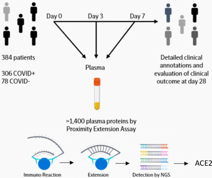
Data were downloaded from https://www.olink.com/mgh-covid-study/ on 14 Sep 2020. A detailed description is available online (https://www.olink.com/mgh-covid-study/) (Fig 1) and has been published previously [29]. Subjects included patients 18 years or older, who were in acute respiratory distress with a clinical concern for COVID-19 upon arrival to the Emergency Department. Of the 384 patients enrolled, 306 (80%) tested positive and 78 tested negative for SARS-CoV-2. COVID-19-positive patients had blood samples drawn on days 0, 3, and 7, while virus-negative patients only had samples drawn on day 0. Clinical data collected included 28-day outcome classification based on the maximal illness severity (Acuity max, A1-A5) experience during the first 28 days after enrollment: A1 = Death. A2 = Intubated, ventilated, survived. A3 = Hospitalized, supplementary O2 required. A4 = Hospitalized, no supplementary O2 required. A5 = Discharged directly from ED and not subsequently hospitalized within 28 days. We also defined a dichotomous severity outcome with severe as A1-A2, and non-severe as A3-5. Discharged directly from ED and not subsequently hospitalized within 28 days [30]. Patient age, body mass index (BMI) and pre-existing medical conditions were recorded, including heart conditions (coronary artery disease, congestive heart failure, valvular disease), kidney disease (chronic kidney disease, baseline creatinine >1.5, end stage renal disease), lung disease (asthma, chronic obstructive lung disease, requiring home O2, any chronic lung condition), diabetes (pre-diabetes, insulin and non-insulin dependent diabetes), and immunosuppressive conditions (active cancer, chemotherapy, transplant, immunosuppressive agents, asplenia). Laboratory tests used in the analyses of this study were C-reactive protein (CRP), absolute neutrophil count, and D-dimer. Continuous variables were categorized to make the dataset anonymized. Age (years) categories were 1 = 20–34, 2 = 35–49, 3 = 50–64, 4 = 65–79, 5 = 80+. BMI (kg/m2) categories were 0 = <18.5 (underweight), 1 = 18.5–24.9 (normal), 2 = 25.0–29.9 (overweight), 3 = 30.0–39.9 (obese), 4 = 40+ (severely obese), 5 = Unknown (these were excluded from analysis on BMI). CRP (mg/L) categories were 1 = 0–19.9, 2 = 20–59.0, 3 = 60–99.9, 4 = 100–179, 5 = 180+. Absolute neutrophil count (109/L) categories were 1 = 0–0.99, 2 = 1.0–3.99, 3 = 4.0–7.99, 4 = 8.0–11.99, 5 = 12+. D-dimer (fibrinogen-equivalent units) categories were 1 = 0–499, 2 = 500–999, 3 = 1000–1999, 4 = 2000–3999, 5 = 4000+.

Fig 1. Diagram of study work flow.

Fig 1

Link https://www.olink.com/mgh-covid-study/. Modified and used with permission.

COVID-19-negative subjects enrolled were older than COVID-19-positive patients, less Hispanic, and with greater baseline burden of chronic illnesses. Of the 78 COVID-19-negative subjects, 37 (47%) were diagnosed with non-COVID-19 pneumonia or acute lung injury (e.g., aspiration), 12 (15%) with congestive heart failure exacerbation, 6 (7.7%) with COPD exacerbation, 3 (3.8%) with acute pulmonary embolus, 11 (14%) with non-pulmonary sepsis or infection, and 8 (10%) with other illnesses. COVID-19-negative patients were significantly less inflamed than COVID-19-positive patients, median CRP 22 [IQR 9–67] versus 105 [IQR 48–161], p-value < 0.05, but illness acuity and outcomes were very similar between the two groups [29].

Ethics

Sample collection and analysis was approved by Partners Human Research Committee (PHRC). The need for informed consent was waived by this committee.

Quantification of ACE2 and Olink data analysis

Detailed description is available online (https://www.olink.com/mgh-covid-study/) (Fig 1) and has been published previously [29]. Briefly, the samples were analyzed by the Olink® Explore 1536 platform which includes measurement of the ACE2 protein. The Olink platform is based on Proximity Extension Assay (PEA) technology and has been validated previously [31]. Data generation consists of three main steps: normalization to known standard (extension control), log2-transformation, and level adjustment using the plate control. The generated data represent relative protein values, Normalized Protein eXpression (NPX), on a log2 scale where a larger number represents a higher protein level in the sample. For more information about Olink® Explore 1536, PEA and NPX, please visit www.olink.com.

Statistical analyses

Group comparisons were made using non-parametric tests. The diagnostic value of plasma ACE2 at baseline was further tested with a receiver operating characteristic (ROC) curve for severe (A1-A2) vs non-severe (A3-A5). The comparison of outcome group differences between COVID-19 positive and negative patients was done using a two-way ANOVA. The association between clinical outcome (response variable) and plasma ACE2 (explanatory variable) was also investigated using ordered logistic regression and results are presented as odds ratios (OR) for worse outcome scores per unit increase in plasma ACE2. The specific statistical method tests used for each analysis is described in the table and figure legends. For all tests, the level of significance was a two-sided P value of less than 0.05. Figures were made using GraphPad Prism 8 for Mac (GraphPad Software).

Results

Association between day 0 plasma ACE2 and worst clinical outcome group during the 28-day period in COVID-19 patients

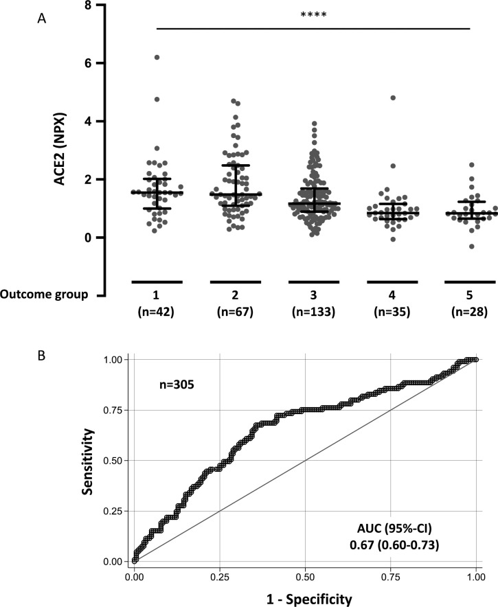
First, we investigated the association between circulating ACE2 at day 0 and maximal acuity outcome group during the 28-day study period (Acuity max, A1-A5). Elevated baseline plasma ACE2 from COVID-19 patients was significantly associated with Acuity max with OR = 1.8, 95%-CI: 1.4–2.3 (P < 0.0001) (Table 1 and Fig 2A). The receiver operating characteristic (ROC) curve in Fig 2B depicts ability for ACE2 level at day 0 to discriminate severe (A1-A2) versus non-severe (A3-A5) outcome during the 28-day study period; the area under the curve (AUC) was 0.67, 95%-CI: 0.60–0.73. The association between circulating ACE2 at day 0 and Acuity max was also tested in regression models with correction for baseline characteristics, pre-existing medical conditions, and laboratory test results. In models correcting for age, body mass index, hypertension, and pre-existing heart conditions, kidney disease, lung disease, diabetes, and immunosuppressive conditions, significant associations were still observed between plasma ACE2 at day 0 and Acuity max (Table 1). Furthermore, we analyzed the association between plasma ACE2 at day 0 and Acuity max after correcting for CRP, absolute neutrophil count, and D-dimer to evaluate whether plasma ACE2 adds to the information already achieved by these laboratory test results. The association between elevated plasma ACE2 and Acuity max remained significant (Table 1).

Table 1. Associations between day 0 plasma ACE2 and maximal acuity within 28 days in COVID-19 positive patients in ordered logistic regression models.

Model n OR 95%-CI p
1 285 1.8 1.4–2.3 <0.0001
2 285 1.9 1.4–2.4 <0.0001
3 285 1.9 1.4–2.4 <0.0001
4 265 1.4 1.1–1.9 0.007

The outcome is the maximum acuity score during the 28-day period with death (A1) being the highest acuity possible, and discharge without requiring admission within 28 days (A5) the least acuity possible.

Models (predictors): 1) Plasma ACE2. 2) Plasma ACE2, age, body mass index (BMI). 3) Plasma ACE2, age, body mass index (BMI), pre-existing hypertension, pre-existing heart disease, pre-existing lung disease, pre-existing kidney disease, pre-existing diabetes, pre-existing immunosuppressive condition. 4) Plasma ACE2, C-reactive protein (CRP), absolute neutrophile count, and D-dimer.

n = number of patients included in statistical analysis. OR = Odds ratio for a higher clinical outcome category per unit increase in plasma ACE2, 95%-CI = 95% confidence interval, p = p-value.

Fig 2. Day 0 plasma ACE2 in COVID-19 positive patients by clinical outcome group and ROC curve.

Fig 2

A. Acuity max group is the maximum acuity score attained within the first 28 days with death being the maximum possible. A1 = Death within 28 days. A2 = Intubated, ventilated, survived to 28 days. A3 = Hospitalized, supplementary O2 required. A4 = Hospitalized, no supplementary O2 required. A5 = Discharged directly from ED and not subsequently hospitalized within 28 days. Data were analyzed using the Kruskal-Wallis test. Bars indicate median and interquartile range. **** P < 0.0001. B. Receiver operating characteristic (ROC) curve comparing severe outcome groups A1-A2 vs non-severe outcome groups A3-A5 during the 28-day period.

Association between plasma ACE2 at day 0, day 3, and day 7 and acuity group A2 (intubated) versus A3-A4 (not intubated) in hospitalized COVID-19 patients

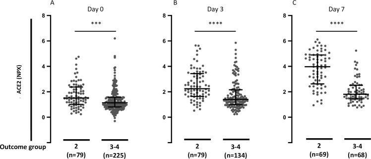
Next, we tested whether circulating ACE2 at day 0, day 3, and day 7 was associated with clinical status at the time of blood sampling. Hospitalized patients were grouped according to outcome categories A2 (intubated at the time of sample collection) or A3-A4 (not intubated at the time of sample collection). Elevated plasma ACE2 in COVID-19 patients was significantly associated with higher acuity category at the time of blood sampling at day 0, day 3, and day 7 (P = 0.0004, P < 0.0001, and P < 0.0001, respectively) (Fig 3). We performed the same analysis in the patients with a plasma ACE2 measurement for all three time points (n = 130). In this analysis as well, plasma ACE2 in COVID-19 patients was significantly elevated in patients with outcome category A2 (intubated and survived) compared with outcome categories A3-A4 (not intubated) at the time of blood sampling at day 0, day 3, and day 7 (P = 0.0016, P < 0.0001, and P < 0.0001, respectively) (S1 Fig).

Fig 3. Plasma ACE2 in hospitalized COVID-19 positive patients segregated by acuity groups A2 (intubated) versus A3-A4 (not intubated).

Fig 3

A. Day 0 plasma ACE2 in hospitalized COVID-19 positive patients by acuity groups A2 (intubated) or A3-A4 (not intubated) for day 0 study window (enrollment plus 24 hours). B. Day 3 plasma ACE2 in hospitalized COVID-19 positive patients by acuity groups A2 versus A3-A4 for day 3 study window. C. Day 7 plasma ACE2 in hospitalized COVID-19 positive patients by acuity groups A2 or A3-A4 for day 7 study window. Acuity categories: A2 = Intubated, ventilated. A3 = Hospitalized, supplementary O2 required. A4 = Hospitalized, no supplementary O2 required. Bars indicate median and interquartile range. Data were analyzed using the Mann Whitney test. *** P< 0.001. **** P < 0.0001.

Association between plasma ACE2 and comorbidities in COVID-19 patients

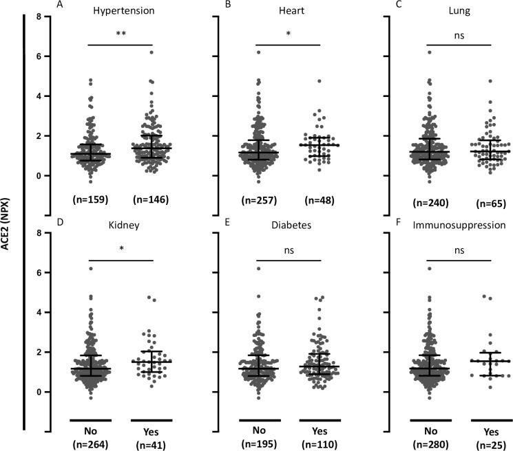
We then analyzed the relationship between ACE2 and comorbidities. Circulating ACE2 in COVID-19-positive patients with hypertension was significantly elevated compared with patients without hypertension (P = 0.0045) (Fig 4). Circulating ACE2 was also significantly elevated in patients with pre-existing heart conditions and in patients with pre-existing kidney disease compared with patients without these pre-existing conditions (P = 0.0363 and P = 0.0303, respectively) (Fig 4). There was no significant difference in plasma ACE2 comparing patients with or without pre-existing lung disease, diabetes, or immunosuppressive conditions (P = 0.953, P = 0.291, and P = 0.237, respectively) (Fig 4).

Fig 4. Plasma ACE2 in COVID-19-positive patients with comorbidities.

Fig 4

A. Plasma ACE2 in COVID-19-positive patients with or without pre-existing hypertension. B. Plasma ACE2 in COVID-19-positive patients with or without pre-existing heart disease (coronary artery disease, congestive heart failure, valvular disease). C. Plasma ACE2 in COVID-19-positive patients with or without pre-existing lung disease (asthma, COPD, requiring home O2, any chronic lung condition). D. Plasma ACE2 in COVID-19-positive patients with or without pre-existing kidney disease (chronic kidney disease, baseline creatinine >1.5, ESRD). E. Plasma ACE2 in COVID-19-positive patients with or without pre-existing diabetes (pre-diabetes, insulin and non-insulin dependent diabetes). F. Plasma ACE2 in COVID-19-positive patients with or without pre-existing immunocompromised condition (active cancer, chemotherapy, transplant, immunosuppressant agents, asplenic). Data were analyzed using the Mann Whitney test. Bars indicate median and interquartile range. * P< 0.05. ** P< 0.01. ns = not significant.

Association of plasma ACE2 with age and BMI in COVID-19 patients

We further tested associations of circulating ACE2 with age and body mass index (BMI). Elevated plasma ACE2 in COVID-19-positive patients was significantly associated with increasing age (P = 0.0001) (Fig 5). There was no significant association between plasma ACE2 and BMI (P = 0.497) (Fig 5).

Fig 5. Plasma ACE2 in COVID-19-positive patients in relation to age and BMI.

Fig 5

A. Plasma ACE2 in COVID-19-positive patients by age groups. B. Plasma ACE2 in COVID-19 positive patients by body mass index (BMI) groups. Data were analyzed using the Kruskal-Wallis test. *** P< 0.001. ns = not significant.

Association of day 0 plasma ACE2 with severe (A1-A2) versus non-severe (A3-A5) in COVID-19 positive and negative patients

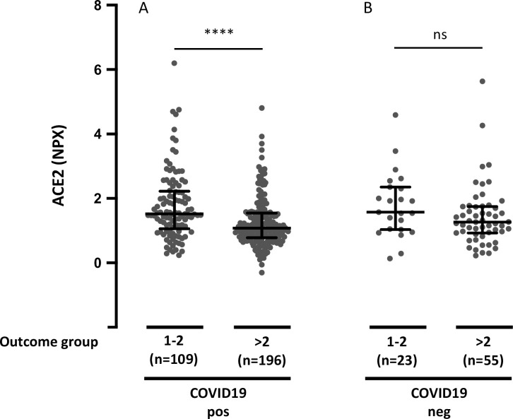
Next, we analyzed whether circulating ACE2 differed between COVID-19 patients and non-COVID-19 patients with respiratory symptoms. Plasma ACE2 showed a clear overlap between the two groups. There was no significant difference between plasma ACE2 in COVID-19-positive versus negative patients (P = 0.13). Finally, we tested whether the association between plasma ACE2 and the maximal acuity score during the 28-day period was found in both COVID-19 positive and negative patients. The patients were grouped according to severe (A1-A2) or non-severe (A3-A5). Elevated plasma ACE2 in COVID-19-positive patients was significantly associated with maximal severity during the 28-day period (P < 0.0001), whereas there was no significant association between plasma ACE2 and outcome category in COVID-19 negative patients (P = 0.085) (Fig 6). To examine whether plasma ACE2 was differentially distributed among the clinical outcome groups in COVID-19-positive versus negative patients, we also conducted a two-way analysis of variance (ANOVA). Elevated plasma ACE2 was significantly associated with higher Acuity max during the 28-day period in COVID-19-positive patients and not in COVID-19-negative patients. However, differences were not statistically different (Table 2).

Fig 6. Plasma ACE2 and maximal acuity group during the 28-day period in COVID-19 positive and negative patients.

Fig 6

A. Plasma ACE2 and outcome group in COVID-19-positive patients. B. Plasma ACE2 and outcome group in COVID-19 negative patients. The acuity group is the maximum acuity group during the 28-day period with death being the maximum possible. A1 = Death within 28 days. A2 = Intubated, ventilated, survived to 28 days. A3 = Hospitalized, supplementary O2 required. A4 = Hospitalized, no supplementary O2 required. A5 = Discharged directly from ED and not subsequently hospitalized within 28 days. Data were analyzed using the Mann Whitney test. Bars indicate median and interquartile range. **** P < 0.0001. ns = not significant.

Table 2. Associations between day 0 plasma ACE2 and maximal acuity group during the 28-day period (Acuity max) in COVID-19-positive and negative patients in two-way analysis of variance (ANOVA).

Outcome group A1-2 vs. A3-5 p 95%-CI
COVID-19-negative 0.33 0.15 -0.12–0.77
COVID-19-positive 0.49 <0.0001 0.27–0.71
Difference 0.16 0.52 -0.34–0.66

Maximal acuity category at 28 days is the maximum score within first 28 days with death (A1) being the highest acuity possible, and discharge without requiring admission within 28 days (A5) the least acuity possible.

p = p-value, 95%-CI = 95% confidence interval.

Discussion

SARS-CoV-2 uses ACE2 as a functional receptor for entry into cells [32]. Circulating ACE2 is increased in patients with active COVID-19 infection compared with healthy controls [2123]. Our analysis is the first description of circulating ACE2 in association with clinical outcomes in patients with COVID-19 disease. Data were obtained from a large longitudinal COVID-19 study with proteomics and clinical data, including disease severity evaluated through 28 days [29].

Elevated baseline plasma ACE2 in COVID-19 patients was significantly associated with increased disease severity during the 28-day study period. This indicates that abundant ACE2 production could be involved in increased viral spread and disease burden, as previously shown in experimental models of SARS-CoV infection [32, 33], with similar mechanisms postulated to be relevant for SARS-CoV-2 infection [34]. The association of elevated plasma ACE2 with higher clinical acuity and worse outcomes would in theory support blockade of ACE2 as a therapeutic strategy. ACE2 is expressed in the type II alveolar pneumocytes [12], which produce surfactant and act as progenitors for the type I alveolar pneumocytes [35]. The binding of the virus to ACE2 on the type II alveolar pneumocytes and subsequent infection leads to depletion of these cells, resulting in a decrease in the production and secretion of surfactant, as well as a lack of ability to regenerate and repair injured lung tissue, leading to the exacerbation of lung injury in severe COVID-19 [35, 36]. This interpretation is however not supported by findings in animal models, showing how ACE2 protects from severe acute lung injury induced by acid aspiration or sepsis [37]. After induction of severe acute lung injury through acid aspiration, ACE2-knockout mice had worsened oxygenation, massive pulmonary edema, and increased inflammatory cell infiltration compared to the wild type mice. In wild type mice, acid aspiration provoked a marked downregulation of ACE2 and an increase in Ang II levels without affecting ACE levels. In ACE2-knockout mice, Ang II levels increased to a greater extent, promoting further lung damage. Moreover, Ang II has been found to activate several cells within the immune system, such as macrophages, leading to a higher production of proinflammatory cytokines like IL-6 and TNFα [35, 36, 38]. Therefore, the direct blockade of ACE2, by removing the physiological brakes on the angiotensin system, could have detrimental effects on COVID-19 disease. For these reasons, an alternative strategy to inhibit the interaction of SARS-CoV-2 with ACE2 could be the administration of recombinant soluble ACE2 as a decoy receptor, which has been previously tested in small clinical trials in acute respiratory distress syndrome [39] and is now being explored in COVID-19 disease [4042]. However, the level of circulating ACE2 does not necessarily reflect the expression of ACE2 in the plasma membrane of host cells. Thus, high levels of ACE2 in the plasma might result from increased lysis of ACE2-expressing cells as a consequence of a more severe infection.

It is desirable to predict disease outcomes in order to specialize treatment, since the drugs used in the treatment of COVID-19 can have deleterious side effects [43]. The ROC curve showed moderate value of baseline ACE2 levels for discriminating patients with severe (death or intubation) vs non-severe disease within 28 days. Further, difference in median plasma ACE2 levels between groups A2 (intubated, survived) versus A3-A4 (hospitalized but not intubated) increased over time on day 3 and day 7 of admission. Therefore, repeated analysis of plasma ACE2 may provide increased predictive value. This is in line with a recent study showing increasing SARS-CoV-2 viral load during hospitalization and prolonged viral shedding in more severe COVID-19 disease [44]. Further, it is interesting that the association between plasma ACE2 and maximal acuity within 28 days in COVID-19 patients was statistically significant also after correction for age, BMI, pre-existing medical conditions, and the laboratory tests CRP, absolute neutrophil count and D-dimer. This suggests that measuring plasma ACE2 adds to the value of clinically available data to help predict disease outcome in COVID-19. ACE2 is a membrane-bound enzyme, and therefore measuring the circulating and urine levels of ACE2 is complex [38]. Soluble ACE2 is the result of the cleavage of the membrane-bound ACE2 by disintegrin and metalloprotease 17. Interestingly, there is a correlation between plasma ACE1 activity and ACE2 activity in healthy individuals [45]. If this same correlation is seen in patients with COVID-19, plasma ACE1 levels (serum ACE) could be used to approximate the activity of ACE2 in COVID-19 positive patients. This is noteworthy because serum ACE analysis is a standardized test in most international hospitals.

Circulating ACE2 in COVID-19-positive patients with hypertension was significantly increased compared with plasma from patients without hypertension. Patients with hypertension are often treated with ACE-inhibitors and AT1R-blockers. During this pandemic, it has been highly debated whether or not the use of these antihypertensive medications should be discontinued in patients with COVID-19. A study performed on rats showed that the use of ACE inhibitors and/or use of AT1R-blocker led to an increased expression of ACE2 in cardiac tissue [46]. Others found a decrease of kidney ACE2 expression and no effect on lung ACE2 expression with ACE-inhibitors and AT1R-blockers [47]. These findings have raised the concern that patients with hypertension, who are treated with ACE2-modulating drugs like ACE-inhibitors or AT1R-blockers might be at a higher risk for severe COVID-19 infection, since it could alter the entry-way for the virus [48]. A recent study showed that plasma ACE2 activity is increased in COVID-19 patients treated with ACE inhibitors [49]. However, more severe COVID-19 disease in patients treated with ACE-inhibitors or AT1R-blockers has not been supported by recent population based data [50]. Circulating ACE2 was also significantly increased in patients with pre-existing heart conditions and in patients with pre-existing kidney disease. This is in line with the central role of RAAS in these conditions. Importantly, the changes in circulating ACE2 in COVID-19 disease observed here are small and are likely not to be of any significance regarding the metabolism of the substrates of ACE2.

In contrast, differences in plasma ACE2 does not seem to explain the risk of severe COVID-19 disease associated with pre-existing lung disease, diabetes, or immunosuppression. Plasma ACE2 was associated with age, which is in line with recent observations showing higher serum levels of ACE2 in adults compared to a pediatric cohort [51]. There were no associations between plasma ACE2 and BMI.

Baseline plasma ACE2 was not significantly different between COVID-19-positive and negative patients, which is in line with the results of a recent study with a much smaller sample size [52]. The association between elevated plasma ACE2 and maximal acuity within 28 days were more pronounced in COVID-19-positive patients compared with COVID-19-negative patients, but the difference was not significant in a two-way ANOVA analysis. It is therefore not possible with this dataset to conclude that the observed association between circulating ACE2 and disease outcome is specific to patients with COVID-19-positive respiratory disease.

There are some overall limitations of this study. First, plasma ACE2 was measured as relative protein concentrations using NPX (Normalized Protein eXpression) values. Therefore, it is not possible to determine a plasma ACE2 cut-off value to predict severe outcome or to compare findings in this study with results in studies measuring protein concentration or enzymatic activity. Second, several continuous variables were categorized in the publicly available data set. This decreases the statistical power of some of the analysis. Third, some potentially important variables such as gender and treatment were not available. Fourth, severity driven recruitment criteria (respiratory distress at emergency department) might have introduced a bias. Thus, patients with more severe COVID-19 at admission have both higher ACE2 at time of inclusion and a poorer prognosis. However, our study reflects clinical practice where laboratory tests are performed when patients are admitted to the hospital. Finally, causality of associations between plasma ACE2 and severity of COVID-19 disease cannot be drawn from this study.

Conclusions

Overall, this study suggests a potential utility of measuring ACE2 in COVID-19 to predict disease outcome. Further, circulating ACE2 could be a link between severe COVID-19 disease and its risk factors, namely hypertension, pre-existing heart disease and pre-existing kidney disease. The design of the data analysis using the Olink platform does not allow assessment of quantitative differences. However, previous studies have described a positive correlation between plasma ACE2 and ACE1 activity. This is interesting because ACE1 (serum ACE) analysis is a standardized test in most hospital laboratories. Therefore, our study encourages quantitative investigations of both plasma ACE 1 and 2 in COVID-19.

Supporting information

S1 Fig. Plasma ACE2 in hospitalized COVID-19 positive patients with samples analyzed at all time points divided in clinical outcome groups 2 (intubated) or 3–4 (not intubated).

A. Day 0 plasma ACE2 in hospitalized COVID-19 positive patients divided in outcome groups 2 (intubated) or 3–4 (not intubated) for day 0 study window (enrollment plus 24 hours). B. Day 3 plasma abundance of ACE2 in hospitalized COVID-19 positive patients divided in outcome groups 2 (intubated) or 3–4 (not intubated) for day 3 study window. C. Day 7 plasma ACE2 in hospitalized COVID-19 positive patients divided in outcome groups 2 (intubated) or 3–4 (not intubated) for day 7 study window. The acuity groups: 2 = Intubated, ventilated. 3 = Hospitalized, supplementary O2 required. 4 = Hospitalized, no supplementary O2 required. Bars indicate median and interquartile range. Data were analyzed using the Mann Whitney test. ** P< 0.01. **** P < 0.0001.

(DOCX)

Acknowledgments

Data provided by the MGH Emergency Department COVID-19 Cohort (Filbin, Goldberg, Hacohen) with Olink Proteomics. We want to thank Aparna Udupi, Department of Biostatistics, Aarhus University for technical assistance running the statistical analyses.

Abbreviations

ACE

Angiotensin converting enzyme

Ang I

Angiotensin I

Ang II

Angiotensin II

ANOVA

Analysis of variance

AT1R

Type 1 angiotensin II receptor

AUC

Area under the curve

BMI

Body mass index

CoV

Coronavirus

COVID-19

Corona virus disease 2019

CRP

C-reactive protein

IL

Interleukin

NPX

Normalized Protein eXpression

PEA

Proximity extension assay

RAAS

Renin-angiotensin-aldosterone-system

ROC

receiver operating characteristic

SARS-CoV-2

Severe acute respiratory syndrome coronavirus 2

TNF

Tumor necrosis factor

Data Availability

Data is available from https://www.olink.com/mgh-covid-study/.

Funding Statement

American Lung Association COVID-19 Action Initiative award (MBG). Independent Research Fund Denmark clinician scientist award (9039-00015B, TWK). Olink Proteomics financed and performed the proteomics assays presented in this work as part of the collaboration with Massachusetts General Hospital (MGH and the Broad Institute on the MGH Emergency Department COVID-19 Cohort. Olink Proteomics provided support in the form of salaries for IG, but did not have any additional role in the study design, data collection and analysis, decision to publish, or preparation of the manuscript. The specific roles of all authors are articulated in the ‘author contributions’ section.

References

  • 1.Cummings MJ, Baldwin MR, Abrams D, Jacobson SD, Meyer BJ, Balough EM, et al. Epidemiology, clinical course, and outcomes of critically ill adults with COVID-19 in New York City: a prospective cohort study. Lancet. 2020;395(10239):1763–70. doi: 10.1016/S0140-6736(20)31189-2 [DOI] [PMC free article] [PubMed] [Google Scholar]
  • 2.Banerjee A, Pasea L, Harris S, Gonzalez-Izquierdo A, Torralbo A, Shallcross L, et al. Estimating excess 1-year mortality associated with the COVID-19 pandemic according to underlying conditions and age: a population-based cohort study. Lancet. 2020;395(10238):1715–25. doi: 10.1016/S0140-6736(20)30854-0 [DOI] [PMC free article] [PubMed] [Google Scholar]
  • 3.Zhou F, Yu T, Du R, Fan G, Liu Y, Liu Z, et al. Clinical course and risk factors for mortality of adult inpatients with COVID-19 in Wuhan, China: a retrospective cohort study. Lancet. 2020;395(10229):1054–62. doi: 10.1016/S0140-6736(20)30566-3 [DOI] [PMC free article] [PubMed] [Google Scholar]
  • 4.Zhou P, Yang XL, Wang XG, Hu B, Zhang L, Zhang W, et al. A pneumonia outbreak associated with a new coronavirus of probable bat origin. Nature. 2020;579(7798):270–3. doi: 10.1038/s41586-020-2012-7 [DOI] [PMC free article] [PubMed] [Google Scholar]
  • 5.Wan Y, Shang J, Graham R, Baric RS, Li F. Receptor Recognition by the Novel Coronavirus from Wuhan: an Analysis Based on Decade-Long Structural Studies of SARS Coronavirus. J Virol. 2020;94(7). doi: 10.1128/JVI.00127-20 [DOI] [PMC free article] [PubMed] [Google Scholar]
  • 6.Ou X, Liu Y, Lei X, Li P, Mi D, Ren L, et al. Characterization of spike glycoprotein of SARS-CoV-2 on virus entry and its immune cross-reactivity with SARS-CoV. Nat Commun. 2020;11(1):1620. doi: 10.1038/s41467-020-15562-9 [DOI] [PMC free article] [PubMed] [Google Scholar]
  • 7.Hoffmann M, Kleine-Weber H, Schroeder S, Krüger N, Herrler T, Erichsen S, et al. SARS-CoV-2 Cell Entry Depends on ACE2 and TMPRSS2 and Is Blocked by a Clinically Proven Protease Inhibitor. Cell. 2020;181(2):271–80.e8. doi: 10.1016/j.cell.2020.02.052 [DOI] [PMC free article] [PubMed] [Google Scholar]
  • 8.Lores E, Wysocki J, Batlle D. ACE2, the kidney and the emergence of COVID-19 two decades after ACE2 discovery. Clin Sci (Lond). 2020;134(21):2791–805. doi: 10.1042/CS20200484 [DOI] [PubMed] [Google Scholar]
  • 9.Lee IT, Nakayama T, Wu CT, Goltsev Y, Jiang S, Gall PA, et al. ACE2 localizes to the respiratory cilia and is not increased by ACE inhibitors or ARBs. Nat Commun. 2020;11(1):5453. doi: 10.1038/s41467-020-19145-6 [DOI] [PMC free article] [PubMed] [Google Scholar]
  • 10.Serfozo P, Wysocki J, Gulua G, Schulze A, Ye M, Liu P, et al. Ang II (Angiotensin II) Conversion to Angiotensin-(1–7) in the Circulation Is POP (Prolyloligopeptidase)-Dependent and ACE2 (Angiotensin-Converting Enzyme 2)-Independent. Hypertension. 2020;75(1):173–82. doi: 10.1161/HYPERTENSIONAHA.119.14071 [DOI] [PMC free article] [PubMed] [Google Scholar]
  • 11.Hamming I, Timens W, Bulthuis ML, Lely AT, Navis G, van Goor H. Tissue distribution of ACE2 protein, the functional receptor for SARS coronavirus. A first step in understanding SARS pathogenesis. J Pathol. 2004;203(2):631–7. doi: 10.1002/path.1570 [DOI] [PMC free article] [PubMed] [Google Scholar]
  • 12.Zou X, Chen K, Zou J, Han P, Hao J, Han Z. Single-cell RNA-seq data analysis on the receptor ACE2 expression reveals the potential risk of different human organs vulnerable to 2019-nCoV infection. Front Med. 2020;14(2):185–92. doi: 10.1007/s11684-020-0754-0 [DOI] [PMC free article] [PubMed] [Google Scholar]
  • 13.Boulpaep WBE. Medical Physiology. 3 ed: Elsevier; 2017. [Google Scholar]
  • 14.Atlas SA. The renin-angiotensin aldosterone system: pathophysiological role and pharmacologic inhibition. J Manag Care Pharm. 2007;13(8 Suppl B):9–20. doi: 10.18553/jmcp.2007.13.s8-b.9 [DOI] [PMC free article] [PubMed] [Google Scholar]
  • 15.Paz Ocaranza M, Riquelme JA, García L, Jalil JE, Chiong M, Santos RAS, et al. Counter-regulatory renin-angiotensin system in cardiovascular disease. Nat Rev Cardiol. 2020;17(2):116–29. doi: 10.1038/s41569-019-0244-8 [DOI] [PMC free article] [PubMed] [Google Scholar]
  • 16.Forrester SJ, Booz GW, Sigmund CD, Coffman TM, Kawai T, Rizzo V, et al. Angiotensin II Signal Transduction: An Update on Mechanisms of Physiology and Pathophysiology. Physiol Rev. 2018;98(3):1627–738. doi: 10.1152/physrev.00038.2017 [DOI] [PMC free article] [PubMed] [Google Scholar]
  • 17.Epelman S, Shrestha K, Troughton RW, Francis GS, Sen S, Klein AL, et al. Soluble angiotensin-converting enzyme 2 in human heart failure: relation with myocardial function and clinical outcomes. J Card Fail. 2009;15(7):565–71. doi: 10.1016/j.cardfail.2009.01.014 [DOI] [PMC free article] [PubMed] [Google Scholar]
  • 18.Tikellis C, Bialkowski K, Pete J, Sheehy K, Su Q, Johnston C, et al. ACE2 deficiency modifies renoprotection afforded by ACE inhibition in experimental diabetes. Diabetes. 2008;57(4):1018–25. doi: 10.2337/db07-1212 [DOI] [PubMed] [Google Scholar]
  • 19.Yamaleyeva LM, Gilliam-Davis S, Almeida I, Brosnihan KB, Lindsey SH, Chappell MC. Differential regulation of circulating and renal ACE2 and ACE in hypertensive mRen2.Lewis rats with early-onset diabetes. Am J Physiol Renal Physiol. 2012;302(11):F1374–84. doi: 10.1152/ajprenal.00656.2011 [DOI] [PMC free article] [PubMed] [Google Scholar]
  • 20.Patel SK, Juno JA, Lee WS, Wragg KM, Hogarth PM, Kent SJ, et al. Plasma ACE2 activity is persistently elevated following SARS-CoV-2 infection: implications for COVID-19 pathogenesis and consequences. European Respiratory Journal. 2021:2003730. doi: 10.1183/13993003.03730-2020 [DOI] [PMC free article] [PubMed] [Google Scholar]
  • 21.Lundström A, Ziegler L, Havervall S, Rudberg A-S, von Meijenfeldt F, Lisman T, et al. Soluble angiotensin-converting enzyme 2 is transiently elevated in COVID-19 and correlates with specific inflammatory and endothelial markers. medRxiv. 2021:2021.03.03.21252841. [DOI] [PMC free article] [PubMed] [Google Scholar]
  • 22.Nagy B Jr., Fejes Z, Szentkereszty Z, Suto R, Varkonyi I, Ajzner E, et al. A dramatic rise in serum ACE2 activity in a critically ill COVID-19 patient. Int J Infect Dis. 2021;103:412–4. doi: 10.1016/j.ijid.2020.11.184 [DOI] [PMC free article] [PubMed] [Google Scholar]
  • 23.van Lier D, Kox M, Santos K, van der Hoeven H, Pillay J, Pickkers P. Increased blood angiotensin converting enzyme 2 activity in critically ill COVID-19 patients. ERJ Open Res. 2021;7(1). doi: 10.1183/23120541.00848-2020 [DOI] [PMC free article] [PubMed] [Google Scholar]
  • 24.Sama IE, Ravera A, Santema BT, van Goor H, Ter Maaten JM, Cleland JGF, et al. Circulating plasma concentrations of angiotensin-converting enzyme 2 in men and women with heart failure and effects of renin-angiotensin-aldosterone inhibitors. Eur Heart J. 2020;41(19):1810–7. doi: 10.1093/eurheartj/ehaa373 [DOI] [PMC free article] [PubMed] [Google Scholar]
  • 25.Fagyas M, Kertesz A, Siket IM, Banhegyi V, Kracsko B, Szegedi A, et al. Level of the SARS-CoV-2 receptor ACE2 activity is highly elevated in old-aged patients with aortic stenosis: implications for ACE2 as a biomarker for the severity of COVID-19. Geroscience. 2021. doi: 10.1007/s11357-020-00300-2 [DOI] [PMC free article] [PubMed] [Google Scholar]
  • 26.Narula S, Yusuf S, Chong M, Ramasundarahettige C, Rangarajan S, Bangdiwala SI, et al. Plasma ACE2 and risk of death or cardiometabolic diseases: a case-cohort analysis. Lancet. 2020;396(10256):968–76. doi: 10.1016/S0140-6736(20)31964-4 [DOI] [PMC free article] [PubMed] [Google Scholar]
  • 27.Emilsson V, Gudmundsson EF, Aspelund T, Jonsson BG, Gudjonsson A, Launer LJ, et al. ACE2 levels are altered in comorbidities linked to severe outcome in COVID-19. medRxiv. 2020. doi: 10.1101/2020.06.04.20122044 [DOI] [PMC free article] [PubMed] [Google Scholar]
  • 28.Chan KK, Dorosky D, Sharma P, Abbasi SA, Dye JM, Kranz DM, et al. Engineering human ACE2 to optimize binding to the spike protein of SARS coronavirus 2. Science. 2020;369(6508):1261–5. doi: 10.1126/science.abc0870 [DOI] [PMC free article] [PubMed] [Google Scholar]
  • 29.Filbin MR, Mehta A, Schneider AM, Kays KR, Guess JR, Gentili M, et al. Plasma proteomics reveals tissue-specific cell death and mediators of cell-cell interactions in severe COVID-19 patients. bioRxiv. 2020:2020.11.02.365536. doi: 10.1101/2020.11.02.365536 [DOI] [PMC free article] [PubMed] [Google Scholar]
  • 30.Characterisation WHOWGotC, Management of C-i. A minimal common outcome measure set for COVID-19 clinical research. Lancet Infect Dis. 2020;20(8):e192–e7. doi: 10.1016/S1473-3099(20)30483-7 [DOI] [PMC free article] [PubMed] [Google Scholar]
  • 31.Assarsson E, Lundberg M, Holmquist G, Bjorkesten J, Thorsen SB, Ekman D, et al. Homogenous 96-plex PEA immunoassay exhibiting high sensitivity, specificity, and excellent scalability. PLoS One. 2014;9(4):e95192. doi: 10.1371/journal.pone.0095192 [DOI] [PMC free article] [PubMed] [Google Scholar]
  • 32.Li W, Moore MJ, Vasilieva N, Sui J, Wong SK, Berne MA, et al. Angiotensin-converting enzyme 2 is a functional receptor for the SARS coronavirus. Nature. 2003;426(6965):450–4. doi: 10.1038/nature02145 [DOI] [PMC free article] [PubMed] [Google Scholar]
  • 33.Hofmann H, Geier M, Marzi A, Krumbiegel M, Peipp M, Fey GH, et al. Susceptibility to SARS coronavirus S protein-driven infection correlates with expression of angiotensin converting enzyme 2 and infection can be blocked by soluble receptor. Biochem Biophys Res Commun. 2004;319(4):1216–21. doi: 10.1016/j.bbrc.2004.05.114 [DOI] [PMC free article] [PubMed] [Google Scholar]
  • 34.Perrotta F, Matera MG, Cazzola M, Bianco A. Severe respiratory SARS-CoV2 infection: Does ACE2 receptor matter? Respir Med. 2020;168:105996. doi: 10.1016/j.rmed.2020.105996 [DOI] [PMC free article] [PubMed] [Google Scholar]
  • 35.Rivellese F, Prediletto E. ACE2 at the centre of COVID-19 from paucisymptomatic infections to severe pneumonia. Autoimmun Rev. 2020:102536. doi: 10.1016/j.autrev.2020.102536 [DOI] [PMC free article] [PubMed] [Google Scholar]
  • 36.Verdecchia P, Cavallini C, Spanevello A, Angeli F. The pivotal link between ACE2 deficiency and SARS-CoV-2 infection. Eur J Intern Med. 2020. doi: 10.1016/j.ejim.2020.04.037 [DOI] [PMC free article] [PubMed] [Google Scholar]
  • 37.Imai Y, Kuba K, Rao S, Huan Y, Guo F, Guan B, et al. Angiotensin-converting enzyme 2 protects from severe acute lung failure. Nature. 2005;436(7047):112–6. doi: 10.1038/nature03712 [DOI] [PMC free article] [PubMed] [Google Scholar]
  • 38.Kreutz R, Algharably EAE, Azizi M, Dobrowolski P, Guzik T, Januszewicz A, et al. Hypertension, the renin-angiotensin system, and the risk of lower respiratory tract infections and lung injury: implications for COVID-19. Cardiovasc Res. 2020. [DOI] [PMC free article] [PubMed] [Google Scholar]
  • 39.Khan A, Benthin C, Zeno B, Albertson TE, Boyd J, Christie JD, et al. A pilot clinical trial of recombinant human angiotensin-converting enzyme 2 in acute respiratory distress syndrome. Crit Care. 2017;21(1):234. doi: 10.1186/s13054-017-1823-x [DOI] [PMC free article] [PubMed] [Google Scholar]
  • 40.Monteil V, Kwon H, Prado P, Hagelkruys A, Wimmer RA, Stahl M, et al. Inhibition of SARS-CoV-2 Infections in Engineered Human Tissues Using Clinical-Grade Soluble Human ACE2. Cell. 2020;181(4):905–13 e7. doi: 10.1016/j.cell.2020.04.004 [DOI] [PMC free article] [PubMed] [Google Scholar]
  • 41.Batlle D, Wysocki J, Satchell K. Soluble angiotensin-converting enzyme 2: a potential approach for coronavirus infection therapy? Clin Sci (Lond). 2020;134(5):543–5. doi: 10.1042/CS20200163 [DOI] [PubMed] [Google Scholar]
  • 42.Wysocki J, Ye M, Hassler L, Gupta AK, Wang Y, Nicoleascu V, et al. A Novel Soluble ACE2 Variant with Prolonged Duration of Action Neutralizes SARS-CoV-2 Infection in Human Kidney Organoids. J Am Soc Nephrol. 2021. doi: 10.1681/ASN.2020101537 [DOI] [PMC free article] [PubMed] [Google Scholar]
  • 43.Putman M, Chock YPE, Tam H, Kim AHJ, Sattui SE, Berenbaum F, et al. Antirheumatic Disease Therapies for the Treatment of COVID-19: A Systematic Review and Meta-analysis. Arthritis Rheumatol. 2020. doi: 10.1002/art.41469 [DOI] [PMC free article] [PubMed] [Google Scholar]
  • 44.Munker D, Osterman A, Stubbe H, Muenchhoff M, Veit T, Weinberger T, et al. Dynamics of SARS-CoV-2 shedding in the respiratory tract depends on the severity of disease in COVID-19 patients. European Respiratory Journal. 2021:2002724. doi: 10.1183/13993003.02724-2020 [DOI] [PMC free article] [PubMed] [Google Scholar]
  • 45.Yang CW, Lu LC, Chang CC, Cho CC, Hsieh WY, Tsai CH, et al. Imbalanced plasma ACE and ACE2 level in the uremic patients with cardiovascular diseases and its change during a single hemodialysis session. Ren Fail. 2017;39(1):719–28. doi: 10.1080/0886022X.2017.1398665 [DOI] [PMC free article] [PubMed] [Google Scholar]
  • 46.Ferrario CM, Jessup J, Chappell MC, Averill DB, Brosnihan KB, Tallant EA, et al. Effect of angiotensin-converting enzyme inhibition and angiotensin II receptor blockers on cardiac angiotensin-converting enzyme 2. Circulation. 2005;111(20):2605–10. doi: 10.1161/CIRCULATIONAHA.104.510461 [DOI] [PubMed] [Google Scholar]
  • 47.Wysocki J, Lores E, Ye M, Soler MJ, Batlle D. Kidney and Lung ACE2 Expression after an ACE Inhibitor or an Ang II Receptor Blocker: Implications for COVID-19. J Am Soc Nephrol. 2020;31(9):1941–3. doi: 10.1681/ASN.2020050667 [DOI] [PMC free article] [PubMed] [Google Scholar]
  • 48.Fang L, Karakiulakis G, Roth M. Are patients with hypertension and diabetes mellitus at increased risk for COVID-19 infection? Lancet Respir Med. 2020;8(4):e21. doi: 10.1016/S2213-2600(20)30116-8 [DOI] [PMC free article] [PubMed] [Google Scholar]
  • 49.Kintscher U, Slagman A, Domenig O, Rohle R, Konietschke F, Poglitsch M, et al. Plasma Angiotensin Peptide Profiling and ACE (Angiotensin-Converting Enzyme)-2 Activity in COVID-19 Patients Treated With Pharmacological Blockers of the Renin-Angiotensin System. Hypertension. 2020;76(5):e34–e6. doi: 10.1161/HYPERTENSIONAHA.120.15841 [DOI] [PMC free article] [PubMed] [Google Scholar]
  • 50.Reynolds HR, Adhikari S, Pulgarin C, Troxel AB, Iturrate E, Johnson SB, et al. Renin-Angiotensin-Aldosterone System Inhibitors and Risk of Covid-19. N Engl J Med. 2020;382(25):2441–8. doi: 10.1056/NEJMoa2008975 [DOI] [PMC free article] [PubMed] [Google Scholar]
  • 51.Pavel AB, Wu J, Renert-Yuval Y, Del Duca E, Glickman JW, Miller RL, et al. SARS-CoV-2 receptor ACE2 protein expression in serum is significantly associated with age. Allergy. 2020. doi: 10.1111/all.14522 [DOI] [PMC free article] [PubMed] [Google Scholar]
  • 52.Rieder M, Wirth L, Pollmeier L, Jeserich M, Goller I, Baldus N, et al. Serum ACE-2, angiotensin II, and aldosterone levels are unchanged in patients with COVID-19. Am J Hypertens. 2020. [DOI] [PMC free article] [PubMed] [Google Scholar]

Decision Letter 0

Michael Bader

30 Mar 2021

PONE-D-21-06434

Plasma ACE2 levels predict outcome of COVID-19 in hospitalized patients

PLOS ONE

Dear Dr. Kragstrup,

Thank you for submitting your manuscript to PLOS ONE. After careful consideration, we feel that it has merit but does not fully meet PLOS ONE’s publication criteria as it currently stands. Therefore, we invite you to submit a revised version of the manuscript that addresses the points raised during the review process.

Please submit your revised manuscript by May 14 2021 11:59PM. If you will need more time than this to complete your revisions, please reply to this message or contact the journal office at plosone@plos.org. When you're ready to submit your revision, log on to https://www.editorialmanager.com/pone/ and select the 'Submissions Needing Revision' folder to locate your manuscript file.

Please include the following items when submitting your revised manuscript:

  • A rebuttal letter that responds to each point raised by the academic editor and reviewer(s). You should upload this letter as a separate file labeled 'Response to Reviewers'.

  • A marked-up copy of your manuscript that highlights changes made to the original version. You should upload this as a separate file labeled 'Revised Manuscript with Track Changes'.

  • An unmarked version of your revised paper without tracked changes. You should upload this as a separate file labeled 'Manuscript'.

If you would like to make changes to your financial disclosure, please include your updated statement in your cover letter. Guidelines for resubmitting your figure files are available below the reviewer comments at the end of this letter.

If applicable, we recommend that you deposit your laboratory protocols in protocols.io to enhance the reproducibility of your results. Protocols.io assigns your protocol its own identifier (DOI) so that it can be cited independently in the future. For instructions see: http://journals.plos.org/plosone/s/submission-guidelines#loc-laboratory-protocols. Additionally, PLOS ONE offers an option for publishing peer-reviewed Lab Protocol articles, which describe protocols hosted on protocols.io. Read more information on sharing protocols at https://plos.org/protocols?utm_medium=editorial-email&utm_source=authorletters&utm_campaign=protocols.

We look forward to receiving your revised manuscript.

Kind regards,

Michael Bader

Academic Editor

PLOS ONE

Journal Requirements:

When submitting your revision, we need you to address these additional requirements.

1. Please ensure that your manuscript meets PLOS ONE's style requirements, including those for file naming. The PLOS ONE style templates can be found at

https://journals.plos.org/plosone/s/file?id=wjVg/PLOSOne_formatting_sample_main_body.pdf and

https://journals.plos.org/plosone/s/file?id=ba62/PLOSOne_formatting_sample_title_authors_affiliations.pdf

2. Thank you for including your ethics statement:

"Patients were enrolled in the Emergency Department of a large, urban, academic hospital from 3/24/2020 to 4/30/2020 in Boston during the peak of the COVID-19 surge, with an institutional IRB-approved waiver of informed consent."

a. Please amend your current ethics statement to include the full name of the ethics committee/institutional review board(s) that approved your specific study.

b. Once you have amended this/these statement(s) in the Methods section of the manuscript, please add the same text to the “Ethics Statement” field of the submission form (via “Edit Submission”).

For additional information about PLOS ONE ethical requirements for human subjects research, please refer to http://journals.plos.org/plosone/s/submission-guidelines#loc-human-subjects-research.

3. We suggest you thoroughly copyedit your manuscript for language usage, spelling, and grammar. If you do not know anyone who can help you do this, you may wish to consider employing a professional scientific editing service. 

Whilst you may use any professional scientific editing service of your choice, PLOS has partnered with both American Journal Experts (AJE) and Editage to provide discounted services to PLOS authors. Both organizations have experience helping authors meet PLOS guidelines and can provide language editing, translation, manuscript formatting, and figure formatting to ensure your manuscript meets our submission guidelines. To take advantage of our partnership with AJE, visit the AJE website (http://learn.aje.com/plos/) for a 15% discount off AJE services. To take advantage of our partnership with Editage, visit the Editage website (www.editage.com) and enter referral code PLOSEDIT for a 15% discount off Editage services.  If the PLOS editorial team finds any language issues in text that either AJE or Editage has edited, the service provider will re-edit the text for free.

Upon resubmission, please provide the following:

●             The name of the colleague or the details of the professional service that edited your manuscript

●             A copy of your manuscript showing your changes by either highlighting them or using track changes (uploaded as a *supporting information* file)

●             A clean copy of the edited manuscript (uploaded as the new *manuscript* file)

4. Thank you for stating the following in the Competing Interests section:

'IG is an employee of Olink Proteomics. The authors declare no other potential conflicts of interest.'

We note that one or more of the authors have an affiliation to the commercial funders of this research study : Olink Proteomics.

a. Please provide an amended Funding Statement declaring this commercial affiliation, as well as a statement regarding the Role of Funders in your study. If the funding organization did not play a role in the study design, data collection and analysis, decision to publish, or preparation of the manuscript and only provided financial support in the form of authors' salaries and/or research materials, please review your statements relating to the author contributions, and ensure you have specifically and accurately indicated the role(s) that these authors had in your study. You can update author roles in the Author Contributions section of the online submission form.

Please also include the following statement within your amended Funding Statement.

“The funder provided support in the form of salaries for authors [insert relevant initials], but did not have any additional role in the study design, data collection and analysis, decision to publish, or preparation of the manuscript. The specific roles of these authors are articulated in the ‘author contributions’ section.”

If your commercial affiliation did play a role in your study, please state and explain this role within your updated Funding Statement.

b. Please also provide an updated Competing Interests Statement declaring this commercial affiliation along with any other relevant declarations relating to employment, consultancy, patents, products in development, or marketed products, etc. 

Within your Competing Interests Statement, please confirm that this commercial affiliation does not alter your adherence to all PLOS ONE policies on sharing data and materials by including the following statement: "This does not alter our adherence to  PLOS ONE policies on sharing data and materials.” (as detailed online in our guide for authors http://journals.plos.org/plosone/s/competing-interests). If this adherence statement is not accurate and  there are restrictions on sharing of data and/or materials, please state these. Please note that we cannot proceed with consideration of your article until this information has been declared.

c. Please include both an updated Funding Statement and Competing Interests Statement in your cover letter. We will change the online submission form on your behalf.

Please know it is PLOS ONE policy for corresponding authors to declare, on behalf of all authors, all potential competing interests for the purposes of transparency. PLOS defines a competing interest as anything that interferes with, or could reasonably be perceived as interfering with, the full and objective presentation, peer review, editorial decision-making, or publication of research or non-research articles submitted to one of the journals. Competing interests can be financial or non-financial, professional, or personal. Competing interests can arise in relationship to an organization or another person. Please follow this link to our website for more details on competing interests: http://journals.plos.org/plosone/s/competing-interests

5. We note that Figure 1 in your submission contains copyrighted images.

All PLOS content is published under the Creative Commons Attribution License (CC BY 4.0), which means that the manuscript, images, and Supporting Information files will be freely available online, and any third party is permitted to access, download, copy, distribute, and use these materials in any way, even commercially, with proper attribution. For more information, see our copyright guidelines: http://journals.plos.org/plosone/s/licenses-and-copyright.

We require you to either (a) present written permission from the copyright holder to publish this figure specifically under the CC BY 4.0 license, or (b) remove the figure from your submission:

a. You may seek permission from the original copyright holder of Figure 1 to publish the content specifically under the CC BY 4.0 license.

We recommend that you contact the original copyright holder with the Content Permission Form (http://journals.plos.org/plosone/s/file?id=7c09/content-permission-form.pdf) and the following text:

“I request permission for the open-access journal PLOS ONE to publish XXX under the Creative Commons Attribution License (CCAL) CC BY 4.0 (http://creativecommons.org/licenses/by/4.0/). Please be aware that this license allows unrestricted use and distribution, even commercially, by third parties. Please reply and provide explicit written permission to publish XXX under a CC BY license and complete the attached form.”

Please upload the completed Content Permission Form or other proof of granted permissions as an "Other" file with your submission. 

In the figure caption of the copyrighted figure, please include the following text: “Reprinted from [ref] under a CC BY license, with permission from [name of publisher], original copyright [original copyright year].”

b.  If you are unable to obtain permission from the original copyright holder to publish this figure under the CC BY 4.0 license or if the copyright holder’s requirements are incompatible with the CC BY 4.0 license, please either i) remove the figure or ii) supply a replacement figure that complies with the CC BY 4.0 license. Please check copyright information on all replacement figures and update the figure caption with source information. If applicable, please specify in the figure caption text when a figure is similar but not identical to the original image and is therefore for illustrative purposes only.

6. Please amend your list of authors on the manuscript to ensure that each author is linked to an affiliation. Authors’ affiliations should reflect the institution where the work was done (if authors moved subsequently, you can also list the new affiliation stating “current affiliation:….” as necessary).

[Note: HTML markup is below. Please do not edit.]

Reviewers' comments:

Reviewer's Responses to Questions

Comments to the Author

1. Is the manuscript technically sound, and do the data support the conclusions?

The manuscript must describe a technically sound piece of scientific research with data that supports the conclusions. Experiments must have been conducted rigorously, with appropriate controls, replication, and sample sizes. The conclusions must be drawn appropriately based on the data presented.

Reviewer #1: No

Reviewer #2: Yes

**********

2. Has the statistical analysis been performed appropriately and rigorously?

Reviewer #1: I Don't Know

Reviewer #2: Yes

**********

3. Have the authors made all data underlying the findings in their manuscript fully available?

The PLOS Data policy requires authors to make all data underlying the findings described in their manuscript fully available without restriction, with rare exception (please refer to the Data Availability Statement in the manuscript PDF file). The data should be provided as part of the manuscript or its supporting information, or deposited to a public repository. For example, in addition to summary statistics, the data points behind means, medians and variance measures should be available. If there are restrictions on publicly sharing data—e.g. participant privacy or use of data from a third party—those must be specified.

Reviewer #1: No

Reviewer #2: Yes

**********

4. Is the manuscript presented in an intelligible fashion and written in standard English?

PLOS ONE does not copyedit accepted manuscripts, so the language in submitted articles must be clear, correct, and unambiguous. Any typographical or grammatical errors should be corrected at revision, so please note any specific errors here.

Reviewer #1: Yes

Reviewer #2: Yes

**********

5. Review Comments to the Author

Please use the space provided to explain your answers to the questions above. You may also include additional comments for the author, including concerns about dual publication, research ethics, or publication ethics. (Please upload your review as an attachment if it exceeds 20,000 characters)

Reviewer #1: In the current paper, the authors investigate the link of ACE2 signals obtained from a proximal extension assay multiplex panel in a cohort of 306 COVID-19 patients and 78 SARS-CoV-2 negative controls. The authors suggest a link between baseline signals for ACE2 obtained from a commercially available proximal extension assay and the outcome of COVID-19 disease. The findings are interesting and in-line with previous observations regarding ACE2 regulation during COVID-19. The authors should also check for more recent literature on the topic, describing a link between soluble ACE2 activity and COVID-19 severity.

The aspect of a prognostic value of baseline ACE2 for COVID-19 outcome appears to be interesting at the first glance, but considering the design of the clinical readout, it might be introduced by the fact that "Day 0" in the current study was actually the admission to the emergency department respiratory distress, which could happen at very variable time points related to disease onset. Knowing that COVID-19 severity is linked to plasma ACE2 activity with a peak in concentrations between 1 and 2 weeks after disease onset, severity driven recruitment criteria (respiratory distress at emergency department) might have introduces a certain bias, as more severe cases have higher ACE2 at time of inclusion, with obviously having poorer outcomes.

In other words, patients being included with a more severe disease manifestation ion day 0 (e.g. higher ACE2), may just be at another time-point in the course of COVID-19 disease, with an already pre-determined worse outcome. It would add a lot of valuer to the study, if the measurement time points would be related to the time of symptom onset, first positive test result or any other earlier time point that may would assure the direct comparability of disease severity groups. The authors should further describe the control group of 78 Covid-negative patients described more clearly. Why did these patients at emergency units with respiratory distress?

Finally, the use of terminology within the whole paper is misleading, as the authors keep using the terms "ACE2 levels" and "ACE2 concentrations". Of note, the result of the proximal extension assay the authors used as a basis for their interpretation is given in the form of an artificial unit (NPX), that has been calculated by the manufacturer by arithmetically linking a series of Ct value based correction algorithms. Even the manufacturers point out on their webpage that the given readout cannot be compared to actual protein levels, which disables comparability to other studies.

From a technical perspective, the big open question is why no calibration of the readout is performed to be able to provide actual protein levels. With a highly reproducible and standardized method as described by the manufacturers on their webpage, it should be easy to retrospectively include a valid calibration allowing for providing actual ACE2 concentrations instead of manufacturer invented units that prevent comparability with other studies. Moreover, a certain analytical validation vor ACE2 in the used PEA panel should be shown that compares the used "NPX" values with real concentration units or standard activity units in a defined set of clinical samples. It should also be noted that reading the method section needs some revision as it currently reads like an advertisement for the used (commercially available) technology rather than an objective method description to be published in a research paper.

Reviewer #2: .This study investigated the association between plasma ACE2 levels and outcomes of COVID-19 patients, using clinical data and plasma samples from 306 COVID-19 positive and 76 COVID-19 negative patients. High baseline plasma ACE2 levels are reported to be associated with worse COVID-19 outcomes and patients with hypertension, pre-existing heart conditions or kidney disease had higher plasma ACE2 levels than those without.

Even as a marker I doubt very much that it will be useful for COVID-19 because the changes found in figure 2 are so small.

Main criticisms and suggestions for improvement:

- Overall, the paper is an excellent contribution but the authors should acknowledge the limitations of measurements of ACE2 in plasma where the levels are usually very low and even when mildly elevated in pathological conditions the significance remains uncertain. Specifically, it must be stated that ACE2 is a tissue enzyme and that the levels in the circulation are low in all species studied, including humans. Appropriate references should be given.

Please acknowledge that the changes in COVID-19 are so small that they are not likely to be of any significance regarding the metabolism of the substrates of ACE2

- In the introduction the part on ACE2 receptor distribution must be modified to include the kidney as a main site of ACE2 . The authors do not seem aware of a critical important observation, namely that the expression of ACE2 in the lung is very low as shown by Serfozo et al using western blot and confirmed by others

See https://doi.org/10.1161/HYPERTENSIONAHA.119.14071 and https://doi.org/10.1038/s41467-020-19145-6

These references must be cited as well as others showing that the kidney is next to the intestine the organ that has the highest abundance of ACE2.

- Perhaps the authors are not aware that ACE2 RNA levels do not necessarily imply protein levels. Actually, ACE2 can be post translationally regulated and be increased when the levels of mRNA are not. The authors may consult and cite a review on this by Lores et al. https://doi.org/10.1042/CS20200484

- In the discussion it is stated that “ACE2 as a decoy receptor… is now being explored in COVID-19 disease.” Appropriate references should be given to be faithful to the literature. Perhaps they are not aware of these papers because they are recent but they should be cited. For instance, https://doi.org/10.1042/CS20200163 and https://doi.org/10.1681/ASN.2020101537

- “A study performed on rats showed that the use of ACE inhibitors and/or use of AT1R-blocker led to an increased expression of ACE2”. It should be specified that in ref 34 ACE2 was only examined in cardiac tissue and not kidney or lung. Others found a decrease of kidney ACE2 after ACEi and ARBs and no effect on lungs. For instance, see https://doi.org/10.1681/ASN.2020050667

- “The population sampled on day 3 and day 7 therefore consists of patients with more severe disease compared with day 0.” Maybe ACE2 for day 3 and day 7 should be shown in a way that they are only compared to their matching samples of day 0?

**********

6. PLOS authors have the option to publish the peer review history of their article (what does this mean?). If published, this will include your full peer review and any attached files.

If you choose “no”, your identity will remain anonymous but your review may still be made public.

Do you want your identity to be public for this peer review? For information about this choice, including consent withdrawal, please see our Privacy Policy.

Reviewer #1: No

Reviewer #2: No

[NOTE: If reviewer comments were submitted as an attachment file, they will be attached to this email and accessible via the submission site. Please log into your account, locate the manuscript record, and check for the action link "View Attachments". If this link does not appear, there are no attachment files.]

While revising your submission, please upload your figure files to the Preflight Analysis and Conversion Engine (PACE) digital diagnostic tool, https://pacev2.apexcovantage.com/. PACE helps ensure that figures meet PLOS requirements. To use PACE, you must first register as a user. Registration is free. Then, login and navigate to the UPLOAD tab, where you will find detailed instructions on how to use the tool. If you encounter any issues or have any questions when using PACE, please email PLOS at figures@plos.org. Please note that Supporting Information files do not need this step.

PLoS One. 2021 Jun 4;16(6):e0252799. doi: 10.1371/journal.pone.0252799.r002

Author response to Decision Letter 0


20 May 2021

Response letter

Dear editor and reviewers,

We are very pleased with a constructive review of our paper. We readily acknowledge that reviewers have in depth knowledge on ACE2 physiology and the role of this protein in COVID-19 disease. We are therefore very happy to accommodate all suggestions made by the reviewers.

Three native speaking authors have carefully proof read the manuscript. This included a few changes worth mentioning.

1) The Odds ratios have been inverted to report odds ratios for a smaller category instead of a larger category. Seems like it would make more intuitive sense to have higher severity as the primary outcome in analyses. The text as written supports this approach.

2) Fig 2B has been changed to better align with the other figures (no colors).

We have made two general corrections throughout the manuscript based on reviewer suggestions and co-author comments after seeing the review. These changes are not highlighted in the manuscript because the corrections are found abundantly throughout.

1) As suggested by reviewer #1 we have replaced all mentions of ”levels” and ”concentrations” with ”plasma ACE2” or ”circulating ACE2”.

2) We have also systematically changed the terminology of the disease groups from “WHO groups” to “clinical outcome groups/Acuity groups”. “WHO groups” was part of the publicly available information downloaded from the source link at the Olink website. However, the scores were not based on the WHO Outcome scale. This is because COVID19 is not treated with non-invasive ventilation or high-flow nasal cannula (therefore, no patients were categorized in previous group 3). The group definitions are the same but have been renamed accordingly. 1 = Death. 2 = Intubated, ventilated, survived. 3 = Hospitalized, supplementary O2 required. 4 = Hospitalized, no supplementary O2 required. 5 = Not hospitalized. These changes are not highlighted in the manuscript.

All other changes have been highlighted in the revised manuscript.

We believe the paper has been greatly improved. Thank you.

On behalf of the authors,

Tue Wenzel Kragstrup

Tue Wenzel Kragstrup

Associate Professor, MD, PhD

E-mail: kragstrup@biomed.au.dk

Biomedicine, Health

Aarhus University

Wilhelm Meyers Alle 4, 4fl.

DK-8000 Aarhus C

1. Please ensure that your manuscript meets PLOS ONE's style requirements, including those for file naming. The PLOS ONE style templates can be found at

https://journals.plos.org/plosone/s/file?id=wjVg/PLOSOne_formatting_sample_main_body.pdf and

https://journals.plos.org/plosone/s/file?id=ba62/PLOSOne_formatting_sample_title_authors_affiliations.pdf

Response: We have made all formatting according to PLOS ONE's style requirements.

2. Thank you for including your ethics statement:

"Patients were enrolled in the Emergency Department of a large, urban, academic hospital from 3/24/2020 to 4/30/2020 in Boston during the peak of the COVID-19 surge, with an institutional IRB-approved waiver of informed consent."

a. Please amend your current ethics statement to include the full name of the ethics committee/institutional review board(s) that approved your specific study.

Response: We have included full name of ethics committee in manuscript (Partners Human Research Committee).

b. Once you have amended this/these statement(s) in the Methods section of the manuscript, please add the same text to the “Ethics Statement” field of the submission form (via “Edit Submission”).

Response: We have included full name of ethics committee in submission form (Partners Human Research Committee).

For additional information about PLOS ONE ethical requirements for human subjects research, please refer to http://journals.plos.org/plosone/s/submission-guidelines#loc-human-subjects-research.

3. We suggest you thoroughly copyedit your manuscript for language usage, spelling, and grammar. If you do not know anyone who can help you do this, you may wish to consider employing a professional scientific editing service.

Whilst you may use any professional scientific editing service of your choice, PLOS has partnered with both American Journal Experts (AJE) and Editage to provide discounted services to PLOS authors. Both organizations have experience helping authors meet PLOS guidelines and can provide language editing, translation, manuscript formatting, and figure formatting to ensure your manuscript meets our submission guidelines. To take advantage of our partnership with AJE, visit the AJE website (http://learn.aje.com/plos/) for a 15% discount off AJE services. To take advantage of our partnership with Editage, visit the Editage website (www.editage.com) and enter referral code PLOSEDIT for a 15% discount off Editage services. If the PLOS editorial team finds any language issues in text that either AJE or Editage has edited, the service provider will re-edit the text for free.

Upon resubmission, please provide the following:

● The name of the colleague or the details of the professional service that edited your manuscript

Response: Three native speaking authors have carefully proof read the manuscript (AM, MBG, and MF). Both reviewers find the manuscript presented in an intelligible fashion and written in standard English.

● A copy of your manuscript showing your changes by either highlighting them or using track changes (uploaded as a *supporting information* file)

Response: We uploaded version with changes highlighted.

● A clean copy of the edited manuscript (uploaded as the new *manuscript* file)

Response: We uploaded clean version.

4. Thank you for stating the following in the Competing Interests section:

'IG is an employee of Olink Proteomics. The authors declare no other potential conflicts of interest.'

We note that one or more of the authors have an affiliation to the commercial funders of this research study : Olink Proteomics.

a. Please provide an amended Funding Statement declaring this commercial affiliation, as well as a statement regarding the Role of Funders in your study. If the funding organization did not play a role in the study design, data collection and analysis, decision to publish, or preparation of the manuscript and only provided financial support in the form of authors' salaries and/or research materials, please review your statements relating to the author contributions, and ensure you have specifically and accurately indicated the role(s) that these authors had in your study. You can update author roles in the Author Contributions section of the online submission form.

Please also include the following statement within your amended Funding Statement.

“The funder provided support in the form of salaries for authors [insert relevant initials], but did not have any additional role in the study design, data collection and analysis, decision to publish, or preparation of the manuscript. The specific roles of these authors are articulated in the ‘author contributions’ section.”

If your commercial affiliation did play a role in your study, please state and explain this role within your updated Funding Statement.

Response: We have included the below statement about the role of Olink Proteomics.

”Olink Proteomics financed and performed the proteomics assays presented in this work as part of the collaboration with Massachusetts General Hospital (MGH and the Broad Institute on the MGH Emergency Department COVID-19 Cohort.”

We already stated that IG (Ida Grundberg) is an employee of Olink. Now, as per your request we also included the statement below.

”Olink Proteomics provided support in the form of salaries for IG, but did not have any additional role in the study design, data collection and analysis, decision to publish, or preparation of the manuscript. The specific roles of all authors are articulated in the ‘author contributions’ section”

b. Please also provide an updated Competing Interests Statement declaring this commercial affiliation along with any other relevant declarations relating to employment, consultancy, patents, products in development, or marketed products, etc.

Within your Competing Interests Statement, please confirm that this commercial affiliation does not alter your adherence to all PLOS ONE policies on sharing data and materials by including the following statement: "This does not alter our adherence to PLOS ONE policies on sharing data and materials.” (as detailed online in our guide for authors http://journals.plos.org/plosone/s/competing-interests). If this adherence statement is not accurate and there are restrictions on sharing of data and/or materials, please state these. Please note that we cannot proceed with consideration of your article until this information has been declared.

Response: We already stated that IG (Ida Grundberg) is an employee of Olink. Now, as per your request we also included the statement below.

”This does not alter our adherence to PLOS ONE policies on sharing data and materials”.

c. Please include both an updated Funding Statement and Competing Interests Statement in your cover letter. We will change the online submission form on your behalf.

Response: We included Funding Statement and Competing Interests Statement in the updated Cover letter.

Please know it is PLOS ONE policy for corresponding authors to declare, on behalf of all authors, all potential competing interests for the purposes of transparency. PLOS defines a competing interest as anything that interferes with, or could reasonably be perceived as interfering with, the full and objective presentation, peer review, editorial decision-making, or publication of research or non-research articles submitted to one of the journals. Competing interests can be financial or non-financial, professional, or personal. Competing interests can arise in relationship to an organization or another person. Please follow this link to our website for more details on competing interests: http://journals.plos.org/plosone/s/competing-interests

5. We note that Figure 1 in your submission contains copyrighted images.

All PLOS content is published under the Creative Commons Attribution License (CC BY 4.0), which means that the manuscript, images, and Supporting Information files will be freely available online, and any third party is permitted to access, download, copy, distribute, and use these materials in any way, even commercially, with proper attribution. For more information, see our copyright guidelines: http://journals.plos.org/plosone/s/licenses-and-copyright.

We require you to either (a) present written permission from the copyright holder to publish this figure specifically under the CC BY 4.0 license, or (b) remove the figure from your submission:

Response: We have uploaded written permission to publish this figure specifically under the CC BY 4.0 license.

a. You may seek permission from the original copyright holder of Figure 1 to publish the content specifically under the CC BY 4.0 license.

We recommend that you contact the original copyright holder with the Content Permission Form (http://journals.plos.org/plosone/s/file?id=7c09/content-permission-form.pdf) and the following text:

“I request permission for the open-access journal PLOS ONE to publish XXX under the Creative Commons Attribution License (CCAL) CC BY 4.0 (http://creativecommons.org/licenses/by/4.0/). Please be aware that this license allows unrestricted use and distribution, even commercially, by third parties. Please reply and provide explicit written permission to publish XXX under a CC BY license and complete the attached form.”

Please upload the completed Content Permission Form or other proof of granted permissions as an "Other" file with your submission.

In the figure caption of the copyrighted figure, please include the following text: “Reprinted from [ref] under a CC BY license, with permission from [name of publisher], original copyright [original copyright year].”

b. If you are unable to obtain permission from the original copyright holder to publish this figure under the CC BY 4.0 license or if the copyright holder’s requirements are incompatible with the CC BY 4.0 license, please either i) remove the figure or ii) supply a replacement figure that complies with the CC BY 4.0 license. Please check copyright information on all replacement figures and update the figure caption with source information. If applicable, please specify in the figure caption text when a figure is similar but not identical to the original image and is therefore for illustrative purposes only.

6. Please amend your list of authors on the manuscript to ensure that each author is linked to an affiliation. Authors’ affiliations should reflect the institution where the work was done (if authors moved subsequently, you can also list the new affiliation stating “current affiliation:….” as necessary).

Response: The author affiliations are correct.

Reviewers' comments:

Reviewer's Responses to Questions

Comments to the Author

1. Is the manuscript technically sound, and do the data support the conclusions?

The manuscript must describe a technically sound piece of scientific research with data that supports the conclusions. Experiments must have been conducted rigorously, with appropriate controls, replication, and sample sizes. The conclusions must be drawn appropriately based on the data presented.

Reviewer #1: No

Reviewer #2: Yes

2. Has the statistical analysis been performed appropriately and rigorously?

Reviewer #1: I Don't Know

Reviewer #2: Yes

3. Have the authors made all data underlying the findings in their manuscript fully available?

The PLOS Data policy requires authors to make all data underlying the findings described in their manuscript fully available without restriction, with rare exception (please refer to the Data Availability Statement in the manuscript PDF file). The data should be provided as part of the manuscript or its supporting information, or deposited to a public repository. For example, in addition to summary statistics, the data points behind means, medians and variance measures should be available. If there are restrictions on publicly sharing data—e.g. participant privacy or use of data from a third party—those must be specified.

Response: The data is fully available without restriction and can be downloaded from https://www.olink.com/mgh-covid-study/. This has been highlighted in the Methods section.

Reviewer #1: No

Reviewer #2: Yes

4. Is the manuscript presented in an intelligible fashion and written in standard English?

PLOS ONE does not copyedit accepted manuscripts, so the language in submitted articles must be clear, correct, and unambiguous. Any typographical or grammatical errors should be corrected at revision, so please note any specific errors here.

Reviewer #1: Yes

Reviewer #2: Yes

5. Review Comments to the Author

Please use the space provided to explain your answers to the questions above. You may also include additional comments for the author, including concerns about dual publication, research ethics, or publication ethics. (Please upload your review as an attachment if it exceeds 20,000 characters)

Reviewer #1: In the current paper, the authors investigate the link of ACE2 signals obtained from a proximal extension assay multiplex panel in a cohort of 306 COVID-19 patients and 78 SARS-CoV-2 negative controls. The authors suggest a link between baseline signals for ACE2 obtained from a commercially available proximal extension assay and the outcome of COVID-19 disease. The findings are interesting and in-line with previous observations regarding ACE2 regulation during COVID-19. The authors should also check for more recent literature on the topic, describing a link between soluble ACE2 activity and COVID-19 severity.

Response: We would like to thank the Reviewer for taking time to carefully read our paper. We agree with all his/her comments. We believe that we have addressed all comments and this is a great improvement to the manucript.

We reviewed the literature again and found the very recent publications below evaluating circulating ACE2 in COVID-19. They have been cited in the manuscript.

Lundström A, Ziegler L, Havervall S, Rudberg A-S, von Meijenfeldt F, Lisman T, et al. Soluble angiotensin-converting enzyme 2 is transiently elevated in COVID-19 and correlates with specific inflammatory and endothelial markers. medRxiv. 2021:2021.03.03.21252841. doi: 10.1101/2021.03.03.21252841.

Nagy B, Jr., Fejes Z, Szentkereszty Z, Suto R, Varkonyi I, Ajzner E, et al. A dramatic rise in serum ACE2 activity in a critically ill COVID-19 patient. Int J Infect Dis. 2021;103:412-4. Epub 2020/11/30. doi: 10.1016/j.ijid.2020.11.184. PubMed PMID: 33249290;

van Lier D, Kox M, Santos K, van der Hoeven H, Pillay J, Pickkers P. Increased blood angiotensin converting enzyme 2 activity in critically ill COVID-19 patients. ERJ Open Res. 2021;7(1). Epub 2021/03/20. doi: 10.1183/23120541.00848-2020.

Fagyas M, Kertesz A, Siket IM, Banhegyi V, Kracsko B, Szegedi A, et al. Level of the SARS-CoV-2 receptor ACE2 activity is highly elevated in old-aged patients with aortic stenosis: implications for ACE2 as a biomarker for the severity of COVID-19. Geroscience. 2021. Epub 2021/01/21. doi: 10.1007/s11357-020-00300-2.

And per request from reviewer #2 we added these publications better refelcting the current litterature:

Lores E, Wysocki J, Batlle D. ACE2, the kidney and the emergence of COVID-19 two decades after ACE2 discovery. Clin Sci (Lond). 2020;134(21):2791-2805.

Lee IT, Nakayama T, Wu CT, et al. ACE2 localizes to the respiratory cilia and is not increased by ACE inhibitors or ARBs. Nat Commun. 2020;11(1):5453.

Serfozo P, Wysocki J, Gulua G, et al. Ang II (Angiotensin II) Conversion to Angiotensin-(1-7) in the Circulation Is POP (Prolyloligopeptidase)-Dependent and ACE2 (Angiotensin-Converting Enzyme 2)-Independent. Hypertension. 2020;75(1):173-182.

Batlle D, Wysocki J, Satchell K. Soluble angiotensin-converting enzyme 2: a potential approach for coronavirus infection therapy? Clin Sci (Lond). 2020;134(5):543-545.

Wysocki J, Ye M, Hassler L, et al. A Novel Soluble ACE2 Variant with Prolonged Duration of Action Neutralizes SARS-CoV-2 Infection in Human Kidney Organoids. J Am Soc Nephrol. 2021.

Epelman S, Shrestha K, Troughton RW, et al. Soluble angiotensin-converting enzyme 2 in human heart failure: relation with myocardial function and clinical outcomes. J Card Fail. 2009;15(7):565-571.

Tikellis C, Bialkowski K, Pete J, et al. ACE2 deficiency modifies renoprotection afforded by ACE inhibition in experimental diabetes. Diabetes. 2008;57(4):1018-1025.

Yamaleyeva LM, Gilliam-Davis S, Almeida I, Brosnihan KB, Lindsey SH, Chappell MC. Differential regulation of circulating and renal ACE2 and ACE in hypertensive mRen2.Lewis rats with early-onset diabetes. Am J Physiol Renal Physiol. 2012;302(11):F1374-1384.

Wysocki J, Lores E, Ye M, Soler MJ, Batlle D. Kidney and Lung ACE2 Expression after an ACE Inhibitor or an Ang II Receptor Blocker: Implications for COVID-19. J Am Soc Nephrol. 2020;31(9):1941-1943.

The aspect of a prognostic value of baseline ACE2 for COVID-19 outcome appears to be interesting at the first glance, but considering the design of the clinical readout, it might be introduced by the fact that "Day 0" in the current study was actually the admission to the emergency department respiratory distress, which could happen at very variable time points related to disease onset. Knowing that COVID-19 severity is linked to plasma ACE2 activity with a peak in concentrations between 1 and 2 weeks after disease onset, severity driven recruitment criteria (respiratory distress at emergency department) might have introduces a certain bias, as more severe cases have higher ACE2 at time of inclusion, with obviously having poorer outcomes. In other words, patients being included with a more severe disease manifestation ion day 0 (e.g. higher ACE2), may just be at another time-point in the course of COVID-19 disease, with an already pre-determined worse outcome. It would add a lot of valuer to the study, if the measurement time points would be related to the time of symptom onset, first positive test result or any other earlier time point that may would assure the direct comparability of disease severity groups.

Response: We agree with this comment. However, we are not able to get access to a longitudinal study of samples included at symptom onset or at the time of a positive test result. We have included the comment from the reviewer in the discussion because it is obviously a limitation to the study (page 22 line 16). We also mention that our study likely reflects clinical pratice were prognostic tests will not be carried our with no or mild symptoms. We would like to thank the Reviewer for bringing up this relevant point.

”Fourth, severity driven recruitment criteria (respiratory distress at emergency department) might have introduces a bias. Thus, patients with more severe COVID-19 at admission have both higher ACE2 at time of inclusion and a poorer prognosis. However, our study reflects clinical practice where laboratory tests are performed when patients are admitted to the hospital.”

The authors should further describe the control group of 78 Covid-negative patients described more clearly. Why did these patients at emergency units with respiratory distress?

Response: We agree with this comment. We included a description of the control group as below, while we are also referencing the original study where full patient characteristics are provided (page 7 line 5).

”COVID-19-negative subjects enrolled were older than COVID-19-positive patients, less Hispanic, and with greater baseline burden of chronic illnesses. Of the 78 COVID-19-negative subjects, 37 (47%) were diagnosed with non-COVID-19 pneumonia or acute lung injury (e.g., aspiration), 12 (15%) with congestive heart failure exacerbation, 6 (7.7%) with COPD exacerbation, 3 (3.8%) with acute pulmonary embolus, 11 (14%) with non-pulmonary sepsis or infection, and 8 (10%) with other illnesses. COVID-19-negative patients were significantly less inflamed than COVID-19-positive patients, median CRP 22 [IQR 9-67] versus 105 [IQR 48-161], p-value < 0.05, but illness acuity and outcomes were very similar between the two groups.”

Finally, the use of terminology within the whole paper is misleading, as the authors keep using the terms "ACE2 levels" and "ACE2 concentrations". Of note, the result of the proximal extension assay the authors used as a basis for their interpretation is given in the form of an artificial unit (NPX), that has been calculated by the manufacturer by arithmetically linking a series of Ct value based correction algorithms. Even the manufacturers point out on their webpage that the given readout cannot be compared to actual protein levels, which disables comparability to other studies. From a technical perspective, the big open question is why no calibration of the readout is performed to be able to provide actual protein levels. With a highly reproducible and standardized method as described by the manufacturers on their webpage, it should be easy to retrospectively include a valid calibration allowing for providing actual ACE2 concentrations instead of manufacturer invented units that prevent comparability with other studies. Moreover, a certain analytical validation vor ACE2 in the used PEA panel should be shown that compares the used "NPX" values with real concentration units or standard activity units in a defined set of clinical samples.

Response: We of course agree with these limitations. Unfortunately, many factors hinder us in generating actual protein concentrations. The manufacturer cannot generate protein levels from current data, we are not able to re-analyse the same samples in a different assay, and we are not in a position to collect samples from a validation cohort. We highlighted this in the limitations as described below (page 22 line 10). We have also reworded all mentions of ”levels” and ”concentrations” with ”plasma ACE2” or ”circulating ACE”.

“The plasma ACE2 levels were measured as relative protein concentrations using NPX (Normalized Protein eXpression) values. Therefore, it is not possible to determine a plasma ACE2 cut-off value to predict severe outcome or to compare findings in this study with results in studies measuring protein concentration or enzymatic activity.”

It should also be noted that reading the method section needs some revision as it currently reads like an advertisement for the used (commercially available) technology rather than an objective method description to be published in a research paper.

Response: We specifically referred to the website where the data is publicly available, although we agree that the methods should be tailored for scientists and we have therefore removed the unnecessary text about the technology. Thanks for this comment.

”Detailed description is available online (https://www.olink.com/mgh-covid-study/) (Fig 1) and has been published previously.[29] Briefly, the samples were analyzed by the Olink® Explore 1536 platform which includes measurement of the ACE2 protein. The Olink platform is based on Proximity Extension Assay (PEA) technology and has been validated previously.[31] Data generation consists of three main steps: normalization to known standard (extension control), log2-transformation, and level adjustment using the plate control. The generated data represent relative protein values, Normalized Protein eXpression (NPX), on a log2 scale where a larger number represents a higher protein level in the sample. For more information about Olink® Explore 1536, PEA and NPX, please visit www.olink.com.”

Reviewer #2: .This study investigated the association between plasma ACE2 levels and outcomes of COVID-19 patients, using clinical data and plasma samples from 306 COVID-19 positive and 76 COVID-19 negative patients. High baseline plasma ACE2 levels are reported to be associated with worse COVID-19 outcomes and patients with hypertension, pre-existing heart conditions or kidney disease had higher plasma ACE2 levels than those without.

Even as a marker I doubt very much that it will be useful for COVID-19 because the changes found in figure 2 are so small.

Response: We would like to thank the Reviewer for taking the time to help improving our manuscript. We really appreciate it. We agree with the comment that changes in ACE2 are small. Our study will require validation by others in assays designed to measure protein concentration or as mentioned further investigation with measuring ACE(1) as mentioned in the paper. We have reviewed our discussion and believe that the conclusions are balanced and in line with the reviewers comments, particularly as we have now included additional limitations of the study.

Main criticisms and suggestions for improvement:

- Overall, the paper is an excellent contribution but the authors should acknowledge the limitations of measurements of ACE2 in plasma where the levels are usually very low and even when mildly elevated in pathological conditions the significance remains uncertain. Specifically, it must be stated that ACE2 is a tissue enzyme and that the levels in the circulation are low in all species studied, including humans. Appropriate references should be given.

Response: Thank you for the comment. We completely agree that citing previous studies on measuring circulating ACE2 levels is a valuable adition. We have now added the following referneces and included a comment to highlight the limitation that ACE2 levels in circulations are low (page 4 line 22).

Epelman S, Shrestha K, Troughton RW, et al. Soluble angiotensin-converting enzyme 2 in human heart failure: relation with myocardial function and clinical outcomes. J Card Fail. 2009;15(7):565-571.

Tikellis C, Bialkowski K, Pete J, et al. ACE2 deficiency modifies renoprotection afforded by ACE inhibition in experimental diabetes. Diabetes. 2008;57(4):1018-1025.

Yamaleyeva LM, Gilliam-Davis S, Almeida I, Brosnihan KB, Lindsey SH, Chappell MC. Differential regulation of circulating and renal ACE2 and ACE in hypertensive mRen2.Lewis rats with early-onset diabetes. Am J Physiol Renal Physiol. 2012;302(11):F1374-1384.

”ACE2 is a tissue enzyme and circulation levels are low and the significance of measuring circulating ACE2 in pathologic conditions remains uncertain.”

Please acknowledge that the changes in COVID-19 are so small that they are not likely to be of any significance regarding the metabolism of the substrates of ACE2

Response: Thank you for bringing this to our attention. We did not consider this previously. It is an important perspective. We have mentioned this in the Discussion with the below wording (page 21 line 19):

”Importantly, changes in ACE2 levels in COVID-19 disease observed here are small and are likely not to be of any significance regarding the metabolism of the substrates of ACE2.”

- In the introduction the part on ACE2 receptor distribution must be modified to include the kidney as a main site of ACE2 . The authors do not seem aware of a critical important observation, namely that the expression of ACE2 in the lung is very low as shown by Serfozo et al using western blot and confirmed by others

See https://doi.org/10.1161/HYPERTENSIONAHA.119.14071 and https://doi.org/10.1038/s41467-020-19145-6

These references must be cited as well as others showing that the kidney is next to the intestine the organ that has the highest abundance of ACE2.

Response: We agree and have added the below sentence including the mentioned references (page 4 line 12).

- Perhaps the authors are not aware that ACE2 RNA levels do not necessarily imply protein levels. Actually, ACE2 can be post translationally regulated and be increased when the levels of mRNA are not. The authors may consult and cite a review on this by Lores et al. https://doi.org/10.1042/CS20200484

Response: We would like to thank the reviewer for underlining this, we have now included this important reference. However, the Olink assay measures protein, albeit with all the limitations already discussed above, therefore we did not measure RNA levels of ACE2.

- In the discussion it is stated that “ACE2 as a decoy receptor… is now being explored in COVID-19 disease.” Appropriate references should be given to be faithful to the literature. Perhaps they are not aware of these papers because they are recent but they should be cited. For instance, https://doi.org/10.1042/CS20200163 and https://doi.org/10.1681/ASN.2020101537

Response: Thank you very much for bringing these references to our attention. Indeed, we were not aware of this progress. We have included both citations.

- “A study performed on rats showed that the use of ACE inhibitors and/or use of AT1R-blocker led to an increased expression of ACE2”. It should be specified that in ref 34 ACE2 was only examined in cardiac tissue and not kidney or lung. Others found a decrease of kidney ACE2 after ACEi and ARBs and no effect on lungs. For instance, see https://doi.org/10.1681/ASN.2020050667

Response: Again we can only thank the reviewer for updating us and improving the manuscript. We have included this in the paper.

”Others found a decrease of kidney ACE2 expression and no effect on lung ACE2 expression with ACE-inhibitors and AT1R-blockers.”

- “The population sampled on day 3 and day 7 therefore consists of patients with more severe disease compared with day 0.” Maybe ACE2 for day 3 and day 7 should be shown in a way that they are only compared to their matching samples of day 0?

Response: Thank you for this suggestion. We agree that this gives a nice representation of the data. We have included this in the Results section and shown data as Supplementary figure S1. The results are the same. We have therefore removed the above mentioned sentance in the Discussion.

6. PLOS authors have the option to publish the peer review history of their article (what does this mean?). If published, this will include your full peer review and any attached files.

If you choose “no”, your identity will remain anonymous but your review may still be made public.

Do you want your identity to be public for this peer review? For information about this choice, including consent withdrawal, please see our Privacy Policy.

Reviewer #1: No

Reviewer #2: No

[NOTE: If reviewer comments were submitted as an attachment file, they will be attached to this email and accessible via the submission site. Please log into your account, locate the manuscript record, and check for the action link "View Attachments". If this link does not appear, there are no attachment files.]

While revising your submission, please upload your figure files to the Preflight Analysis and Conversion Engine (PACE) digital diagnostic tool, https://pacev2.apexcovantage.com/. PACE helps ensure that figures meet PLOS requirements. To use PACE, you must first register as a user. Registration is free. Then, login and navigate to the UPLOAD tab, where you will find detailed instructions on how to use the tool. If you encounter any issues or have any questions when using PACE, please email PLOS at figures@plos.org. Please note that Supporting Information files do not need this step.

Attachment

Submitted filename: Response letter.docx

Decision Letter 1

Michael Bader

24 May 2021

Plasma ACE2 predicts outcome of COVID-19 in hospitalized patients

PONE-D-21-06434R1

Dear Dr. Kragstrup,

We’re pleased to inform you that your manuscript has been judged scientifically suitable for publication and will be formally accepted for publication once it meets all outstanding technical requirements.

Within one week, you’ll receive an e-mail detailing the required amendments. When these have been addressed, you’ll receive a formal acceptance letter and your manuscript will be scheduled for publication.

An invoice for payment will follow shortly after the formal acceptance. To ensure an efficient process, please log into Editorial Manager at http://www.editorialmanager.com/pone/, click the 'Update My Information' link at the top of the page, and double check that your user information is up-to-date. If you have any billing related questions, please contact our Author Billing department directly at authorbilling@plos.org.

If your institution or institutions have a press office, please notify them about your upcoming paper to help maximize its impact. If they’ll be preparing press materials, please inform our press team as soon as possible -- no later than 48 hours after receiving the formal acceptance. Your manuscript will remain under strict press embargo until 2 pm Eastern Time on the date of publication. For more information, please contact onepress@plos.org.

Kind regards,

Michael Bader

Academic Editor

PLOS ONE

Additional Editor Comments (optional):

Reviewers' comments:

Acceptance letter

Michael Bader

26 May 2021

PONE-D-21-06434R1

Plasma ACE2 predicts outcome of COVID-19 in hospitalized patients

Dear Dr. Kragstrup:

I'm pleased to inform you that your manuscript has been deemed suitable for publication in PLOS ONE. Congratulations! Your manuscript is now with our production department.

If your institution or institutions have a press office, please let them know about your upcoming paper now to help maximize its impact. If they'll be preparing press materials, please inform our press team within the next 48 hours. Your manuscript will remain under strict press embargo until 2 pm Eastern Time on the date of publication. For more information please contact onepress@plos.org.

If we can help with anything else, please email us at plosone@plos.org.

Thank you for submitting your work to PLOS ONE and supporting open access.

Kind regards,

PLOS ONE Editorial Office Staff

on behalf of

Prof. Michael Bader

Academic Editor

PLOS ONE

Associated Data

    This section collects any data citations, data availability statements, or supplementary materials included in this article.

    Supplementary Materials

    S1 Fig. Plasma ACE2 in hospitalized COVID-19 positive patients with samples analyzed at all time points divided in clinical outcome groups 2 (intubated) or 3–4 (not intubated).

    A. Day 0 plasma ACE2 in hospitalized COVID-19 positive patients divided in outcome groups 2 (intubated) or 3–4 (not intubated) for day 0 study window (enrollment plus 24 hours). B. Day 3 plasma abundance of ACE2 in hospitalized COVID-19 positive patients divided in outcome groups 2 (intubated) or 3–4 (not intubated) for day 3 study window. C. Day 7 plasma ACE2 in hospitalized COVID-19 positive patients divided in outcome groups 2 (intubated) or 3–4 (not intubated) for day 7 study window. The acuity groups: 2 = Intubated, ventilated. 3 = Hospitalized, supplementary O2 required. 4 = Hospitalized, no supplementary O2 required. Bars indicate median and interquartile range. Data were analyzed using the Mann Whitney test. ** P< 0.01. **** P < 0.0001.

    (DOCX)

    Attachment

    Submitted filename: Response letter.docx

    Data Availability Statement

    Data is available from https://www.olink.com/mgh-covid-study/.


    Articles from PLoS ONE are provided here courtesy of PLOS

    RESOURCES