Abstract
HIV-1 evolved from SIV during cross-species transmission events, though viral genetic changes are not well understood. Here we studied the evolution of SIVcpzLB715 into HIV-1 Group M using humanized mice. High viral loads, rapid CD4+ T-cell decline, and non-synonymous substitutions were identified throughout the viral genome suggesting viral adaptation.
Keywords: HIV-1 viral evolution from SIV, SIV chimpanzee evolution towards HIV-1, Cross-species SIV transmission, SIVcpz infection in humanized mice, hu-HSC mice for cross-species viral transmission
INTRODUCTION
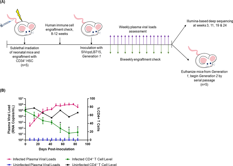
Many genetic changes across the viral genome were necessary for the successful transformation of SIVs into pathogenic HIVs due to differences in host T-cell receptor structure, restriction factors, and immune system barriers.1–7 Here, we experimentally simulated the human exposures of SIVcpzLB715 and serial propagation in multiple human immune environments to determine the mutations that led to the generation of the pathogenic HIV-1 Group M virus. We used humanized hematopoietic stem cell (hu-HSC) mice harboring a complete repertoire of human immune cells susceptible to SIV and HIV infection to model these genetic adaptions.8–15 SIVcpzLB715 was used to infect a cohort of hu-HSC mice and passaged into a second generation of mice (Figure 1A). Plasma viral loads (PVL), CD4+ T-cell decline and Illumina-based deep sequencing at multiple time points were used to determine the adaptation of this virus over time.
Figure 1. SIVcpzLB715 infection kinetics during the second generation in hu-HSC mice.
(A) Experimental scheme for SIVcpzLB715 infection of hu-HSC mice. Neonatal mice are sublethally irradiated and inoculated intrahepatically with CD34+ hematopoietic stem cells. Following SIVcpzLB715 inoculation, PVL were assessed weekly and CD4+ T-cell decline bimonthly. (B) Second generation PVL and CD4+ T-cell decline seen in SIVcpzLB715 infected hu-HSC mice. Statistically significant CD4+ T-cell depletion occurred by the end of the second generation in infected mice relative to the uninfected mice (two-tailed Student’s t-test, *p<0.001).
MATERIALS & METHODS
Generation of humanized mice and ethics
Humanized mice were generated according to methods previously described (Figure 1A).16–20 All mice were maintained at the Colorado State University Painter Animal Center. The studies conducted here have all been reviewed and approved by the CSU Institutional Animal Care and Use Committee.
LB715 viral propagation, in vivo infection, and serial passage
The SIVcpzLB715 plasmid was obtained from Dr. Preston Marx and used in cell transfections to generate stock virus as described previously.10 For the initial infection, 200 μl (TCID50 2.0×10⁵) virus was injected intraperitoneally into five (>75% CD45+, >60% CD4+) hu-HSC mice. SIVcpzLB715 infected mice with the highest viral titer after 6 months were euthanized and infected tissues were cultured to harvest the first passage stock virus as described previously.13,14 For the next generation, a new cohort of five hu-HSC mice were injected with 200 μl of first passage virus.
Determination of PVL and CD4+ T-cell decline
To evaluate PVL and CD4+ T-cell decline, peripheral blood was obtained on a weekly and bimonthly basis, respectively. Plasma RNA was extracted utilizing the E.Z.N.A. Viral RNA kit (Omega bio-tek, Norcross, CA) and the manufacturer’s instructions. Viral load was quantified using the iScript One-Step RT-PCR kit with SYBR Green (BioRad, Hercules, CA) per the manufacturer’s instructions as described previously.10,14 CD4+ T-cell decline was determined by staining whole blood with fluorophore conjugated anti-human CD45-FitC (eBioscience), CD3-PE (eBioscience), and CD4-PE/Cy5 (BD Pharmigen, San Jose, CA) antibodies. BD Accuri C6 FACS Analyzer was used to determine cell counts as described previously.13,14 The CD4+ T-cell decline between the infected and uninfected mice was assessed using a two-tailed Student’s t-test (p<0.001).
Illumina-based deep sequencing and analysis
Viral RNA samples from timepoints 3, 11, 19- and 24-weeks post-inoculation were used to generate amplicons for sequencing. Whole-genome primer pools were made with Primal Scheme.21 Amplicons were prepared using the TruSeq Nano DNA HT Library Preparation Kit and sequenced utilizing the MiSeq Illumina sequencer (Invitrogen, Carlsbad, CA). Reads were mapped to the SIVcpzLB715 stock virus (GenBank accession number: KP861923.1) using bowtie2 software v2.2.5.22 This output was used as input for lofreq software v2.1.2 to call variants.23 Each variant required >20 reads depth of coverage, >20% last timepoint frequency, a change of at least 30% frequency over time and were present in at least 5 datasets.
RESULTS
Virus from the first hu-mice passage was used to infect a second cohort of five hu-mice to evaluate the potential evolution of SIVcpzLB715 into HIV-1 Group M (Figure 1A). Viral loads showed an approximate 4.5-log increase throughout the duration of infection (Figure 1B). CD4+ T-cell depletion occurred within 14 days and significantly declined (p<0.001) throughout the duration of the second-generation infection (Figure 1B). These data show SIVcpzLB715 can establish chronic viremia upon serial passage that produces significant CD4+ T-cell decline by the end of the second generation.
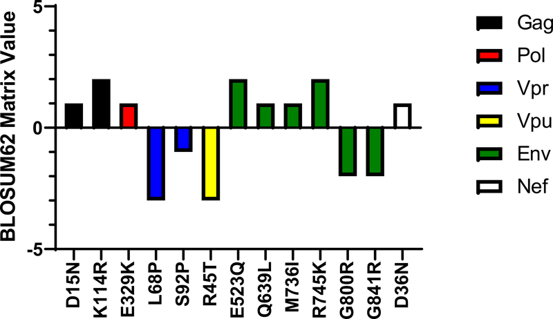
Viral RNA was extracted from plasma of viremic mice at weeks 3, 11, 19 and 24 and subjected to deep sequencing. Detected amino acid substitutions were scored using a BLOSUM62 matrix to determine the likelihood of any given residue substitution based on known protein alignments (Figure 2). Mutations in gag, pol, nef and env were identified as more favorable for viral protein structure.
Figure 2. BLOSUM62 matrix scores of identified residue changes.
Variant residues were identified as described in the results and methods. Some variants in Vpr, Vpu and Env appear to be disfavored, yet still became more frequent in the viral population.
DISCUSSION
We assessed the phenotypic and genetic changes that facilitated the evolution of SIVcpzLB715 towards HIV-1 Group M utilizing a humanized mouse model to recapitulate cross-species transmission. Hu-mice became infected with SIVcpzLB715 within 2 weeks after initial viral challenge. Enhanced CD4+ T-cell loss was observed during the second serial passage suggesting increased pathogenicity and viral fitness. Additionally, one of the mutations in Env gene (E523Q) located in the CD4 binding site appears to have a favorable BLOSUM62 score (Figure 2).24,25 This implies that the CD4 binding site is adapting to improve virus-receptor binding. With increased knowledge of how SIVcpzLB715 first adapted to the human host, we may be able to gain better insight on how SIVs cross over into the human population.
ACKNOWLEDGEMENTS
This work was supported by NIH, USA grant RO1 AI123234 to R.A, and was also funded in part by the NIH, USA grant P51OD011106 at the Wisconsin National Primate Research Center National Center for Research Resources and the Office of Research Infrastructure Programs (ORIP) of the NIH through grant OD011104 at the Tulane National Primate Research Center. Computational resources were supported by NIH-NCATS Colorado CTSA Grant Number UL1 TR002535.
Footnotes
CONFLICT OF INTEREST STATEMENT
The authors confirm there are no conflicts of interest with these studies.
REFERENCES
- 1.Wain LV, Bailes E, Bibollet-Ruche F, et al. Adaptation of HIV-1 to its human host. Mol Biol Evol. 2007;24(8):1853–1860. [DOI] [PMC free article] [PubMed] [Google Scholar]
- 2.Sauter D, Kirchhoff F. Key Viral Adaptations Preceding the AIDS Pandemic. Cell Host Microbe. 2019;25(1):27–38. [DOI] [PubMed] [Google Scholar]
- 3.Hvilsom C, Carlsen F, Siegismund HR, Corbet S, Nerrienet E, Fomsgaard A. Genetic subspecies diversity of the chimpanzee CD4 virus-receptor gene. Genomics. 2008;92(5):322–328. [DOI] [PubMed] [Google Scholar]
- 4.Locatelli S, Peeters M. Cross-species transmission of simian retroviruses: how and why they could lead to the emergence of new diseases in the human population. AIDS. 2012;26(6):659–673. [DOI] [PubMed] [Google Scholar]
- 5.Keele BF, Van Heuverswyn F, Li Y, et al. Chimpanzee reservoirs of pandemic and nonpandemic HIV-1. Science. 2006;313(5786):523–526. [DOI] [PMC free article] [PubMed] [Google Scholar]
- 6.Sharp PM, Hahn BH. Origins of HIV and the AIDS pandemic. Cold Spring Harb Perspect Med. 2011;1(1):a006841. [DOI] [PMC free article] [PubMed] [Google Scholar]
- 7.Hemelaar J The origin and diversity of the HIV-1 pandemic. Trends Mol Med. 2012;18(3):182–192. [DOI] [PubMed] [Google Scholar]
- 8.Akkina R, Allam A, Balazs AB, et al. Improvements and Limitations of Humanized Mouse Models for HIV Research: NIH/NIAID “Meet the Experts” 2015 Workshop Summary. AIDS Res Hum Retroviruses. 2016;32(2):109–119. [DOI] [PMC free article] [PubMed] [Google Scholar]
- 9.Charlins P, Schmitt K, Remling-Mulder L, et al. A humanized mouse-based HIV-1 viral outgrowth assay with higher sensitivity than in vitro qVOA in detecting latently infected cells from individuals on ART with undetectable viral loads. Virology. 2017;507:135–139. [DOI] [PMC free article] [PubMed] [Google Scholar]
- 10.Curlin J, Schmitt K, Remling-Mulder L, et al. SIVcpz cross-species transmission and viral evolution toward HIV-1 in a humanized mouse model. J Med Primatol. 2020;49(1):40–43. [DOI] [PMC free article] [PubMed] [Google Scholar]
- 11.Denton PW, Garcia JV. Humanized mouse models of HIV infection. AIDS Rev. 2011;13(3):135–148. [PMC free article] [PubMed] [Google Scholar]
- 12.Sato K, Misawa N, Takeuchi JS, et al. Experimental Adaptive Evolution of Simian Immunodeficiency Virus SIVcpz to Pandemic Human Immunodeficiency Virus Type 1 by Using a Humanized Mouse Model. J Virol. 2018;92(4). [DOI] [PMC free article] [PubMed] [Google Scholar]
- 13.Schmitt K, Curlin J, Kumar DM, et al. SIV progenitor evolution toward HIV: A humanized mouse surrogate model for SIVsm adaptation toward HIV-2. J Med Primatol. 2018;47(5):298–301. [DOI] [PMC free article] [PubMed] [Google Scholar]
- 14.Schmitt K, Mohan Kumar D, Curlin J, et al. Modeling the evolution of SIV sooty mangabey progenitor virus towards HIV-2 using humanized mice. Virology. 2017;510:175–184. [DOI] [PMC free article] [PubMed] [Google Scholar]
- 15.Yuan Z, Kang G, Ma F, et al. Recapitulating Cross-Species Transmission of Simian Immunodeficiency Virus SIVcpz to Humans by Using Humanized BLT Mice. J Virol. 2016;90(17):7728–7739. [DOI] [PMC free article] [PubMed] [Google Scholar]
- 16.Garcia S, Freitas AA. Humanized mice: current states and perspectives. Immunol Lett. 2012;146(1–2):1–7. [DOI] [PubMed] [Google Scholar]
- 17.Akkina RK, Rosenblatt JD, Campbell AG, Chen IS, Zack JA. Modeling human lymphoid precursor cell gene therapy in the SCID-hu mouse. Blood. 1994;84(5):1393–1398. [PubMed] [Google Scholar]
- 18.Bai J, Gorantla S, Banda N, Cagnon L, Rossi J, Akkina R. Characterization of anti-CCR5 ribozyme-transduced CD34+ hematopoietic progenitor cells in vitro and in a SCID-hu mouse model in vivo. Mol Ther. 2000;1(3):244–254. [DOI] [PubMed] [Google Scholar]
- 19.Berges BK, Akkina SR, Folkvord JM, Connick E, Akkina R. Mucosal transmission of R5 and X4 tropic HIV-1 via vaginal and rectal routes in humanized Rag2−/− gammac −/− (RAG-hu) mice. Virology. 2008;373(2):342–351. [DOI] [PMC free article] [PubMed] [Google Scholar]
- 20.Shultz LD, Brehm MA, Garcia-Martinez JV, Greiner DL. Humanized mice for immune system investigation: progress, promise and challenges. Nat Rev Immunol. 2012;12(11):786–798. [DOI] [PMC free article] [PubMed] [Google Scholar]
- 21.Quick J, Grubaugh ND, Pullan ST, et al. Multiplex PCR method for MinION and Illumina sequencing of Zika and other virus genomes directly from clinical samples. Nat Protoc. 2017;12(6):1261–1276. [DOI] [PMC free article] [PubMed] [Google Scholar]
- 22.Langmead B, Salzberg SL. Fast gapped-read alignment with Bowtie 2. Nat Methods. 2012;9(4):357–359. [DOI] [PMC free article] [PubMed] [Google Scholar]
- 23.Wilm A, Aw PP, Bertrand D, et al. LoFreq: a sequence-quality aware, ultra-sensitive variant caller for uncovering cell-population heterogeneity from high-throughput sequencing datasets. Nucleic Acids Res. 2012;40(22):11189–11201. [DOI] [PMC free article] [PubMed] [Google Scholar]
- 24.Chen B, Vogan EM, Gong H, Skehel JJ, Wiley DC, Harrison SC. Structure of an unliganded simian immunodeficiency virus gp120 core. Nature. 2005;433(7028):834–841. [DOI] [PubMed] [Google Scholar]
- 25.Yu B, Fonseca DP, O’Rourke SM, Berman PW. Protease cleavage sites in HIV-1 gp120 recognized by antigen processing enzymes are conserved and located at receptor binding sites. J Virol. 2010;84(3):1513–1526. [DOI] [PMC free article] [PubMed] [Google Scholar]


