. 2021 Jun 4;10:e65745. doi: 10.7554/eLife.65745

Figure 5. CO2-dependent pathway from TD neurons to RPNs.

(A) Comparison of underlying connectivity of TD (CO2) neurons via interneurons to the RPNs, with the predicted outcome of mean activity (with an activation factor of 2; when more than 5% of presynaptic neurons are active, interneurons become activated up to an activity of 50%) of RPNs, and the outcome of CaMPARI-2 CO2 experiments. FFN diffusion model reliably shows modulation of the RPNs. Please note that the circled numbers in the path analysis refer to the total number of neurons, not neuron identification number. (B) Using the combination of connectivity, prediction, and functional imaging experiments, a new sensory to endocrine neural circuit can be derived. TD (CO2) neurons at the trachea respond to CO2 levels and communicate predominantly via a core set of thoracic interneurons to DH44 and CRZ, which show release sites in the CC and AO. (C) Connectivity of the single thoracic interneurons (hemilateral pairs) to presynaptic sensory origins and to the distinct postsynaptic RPN groups. Thoracic interneurons receive additionally other sensory modalities apart from TD (CO2) neurons and target different combinations of RPNs. (D) CRZ interneurons: hive plot showing the polysynaptic pathways from all sensory origins to all RPN target groups using the interneurons (synaptic threshold = 3) that target CRZ. Main sensory origins are enteric, pharyngeal, and CO2. DH44 interneurons: TD (CO2) represent the most dominant polysynaptic path from sensory origins to DH44. Note that monosynaptic connections from sensory neurons to RPNs are shown in gray. AO: aorta; CA-LP: corpus allatum innervating neurosecretory neurons of the lateral protocerebrum; CaMPARI-2: calcium-modulated photoactivatable ratiometric integrator 2; CAPA: capability neurons; CC: corpora cardiaca; CO2: carbon dioxide; CRZ: corazonin neurons; DH44: diuretic hormone 44 neurons; DMS: Drosophila myosuppressin neurons; EH: eclosion hormone neurons; FFN: feed forward network; HugRG: hugin neurons innervating ring gland; IPCs: insulin-producing cells; ITP: ion transport peptide neurons; PTTH: prothoracicotropic hormone neurons; RPNs: ring gland projection neurons; ssTEM: serial section transmission electron microscope; TD: tracheal dendritic neurons.

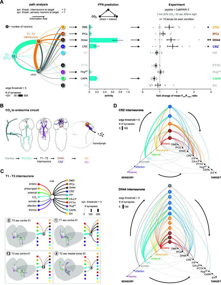
Figure 5.

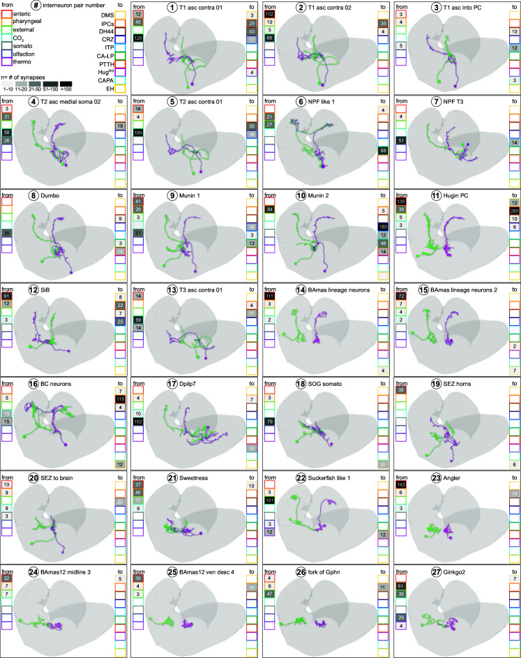
Figure 5—figure supplement 1. Neuron catalogue of interneurons receiving sensory input.

Figure 5—figure supplement 1.

Absolute synapse numbers from sensory neurons to interneurons, and from interneurons to RPN target groups, are displayed in color-coded boxes. Strength of synaptic contacts is heatmap coded in grayscale. For abbreviations, see Figure 3—figure supplement 1.
Figure 5—figure supplement 2. Neuron catalogue of interneurons receiving sensory input.

Figure 5—figure supplement 2.

Absolute synapse numbers from sensory neurons to interneurons, and from interneurons to RPN target groups, are displayed in color-coded boxes. Strength of synaptic contacts is heatmap coded in grayscale. For abbreviations, see Figure 3—figure supplement 1.
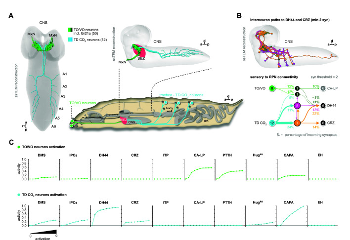
Figure 5—figure supplement 3. Impact of Gr21a candidate neurons on modulation of RPN activity.

Figure 5—figure supplement 3.

(A) Ventral and lateral 3D representations of reconstructed chemosensory neurons of the TO, VO (Miroschnikow et al., 2018), and TD (CO2) neurons in the CNS (top left). Note that Gr21a is expressed in TO ganglion cell 6 and projects via the MxN to the SEZ in the brain (Kwon et al., 2011). Schematic side view of a Drosophila larva. Colored dots and lines represent the location and projection of TOG and TDs (CO2). (B) Shortest path (2-hops) analysis reveals 17 interneurons relaying TO, VO, and TD (CO2) sensory information to DH44 and CRZ neuroendocrine RPNs. Path threshold = minimum two synapses. Neuron colors correspond to respective nodes in connectivity graph. Note that only 8 of the 50 TO and VO neurons are weakly connected to DH44 and CRZ via two interneurons. Percentage represents the fraction of synapses from upstream neurons (arrows). Numbers in circles represent number of cells. (C) Activation of all neurons of the TOG and VO in the FFN, using all reconstructed interneurons to the RPNs, did not alter modulation of the strong CO2-responsive RPNs (e.g., DH44 and CRZ). Instead, TOG/VO neurons are capable of modulating the activity of a different set of RPNs, namely PTTH and CA-LP. Slight activity modulation was observed for DMS and CAPA. Thus it seems unlikely, given the connectome-based modulation of RPNs via the FFN, that Gr21a neurons have an effect on RPN activity based on the CO2 pathway originating from TDs (CO2). A potential implication in terms of valence has to be determined in future experiments. a: anterior; CA-LP: corpus allatum innervating neurosecretory neurons of the lateral protocerebrum; CAPA: capability neurons; CO2: carbon dioxide; CRZ: corazonin neurons; d: dorsal; DH44: diuretic hormone 44 neurons; DMS: Drosophila myosuppressin neurons; EH: eclosion hormone neurons; FFN: feed forward network; HugRG: hugin neurons innervating ring gland; IPCs: insulin-producing cells; ITP: ion transport peptide neurons; MxN: maxillary nerve; p: posterior; PTTH: prothoracicotropic hormone neurons; r: right; RPNs: ring gland projection neurons; SEZ: subesophageal zone; ssTEM: serial section transmission electron microscope; TD: tracheal dendritic neurons; TO: terminal organ; TOG: terminal organ ganglion; VO: ventral organ.