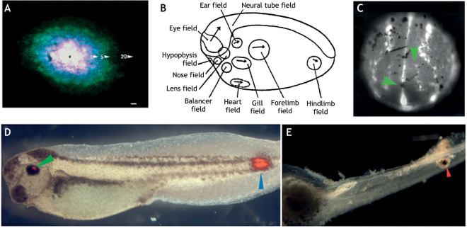
Fig. 3.
Bioelectric association with and modulation of regional developmental fields. (A) Regional transmission of electrically charged lucifer yellow within the feather tract placodal epidermis of chick embryos. An overlay of images taken at 1, 5 and 20 min after injection of charged dye into a single placodal cell (marked with an asterisk) reveals bounded coupling within the feather placode during development. Image reproduced from Serras et al. (1993). (B) Fate map of regional morphogenic fields in a developing Xenopus embryo. Image reproduced, with permission, from Huxley and De Beer (1936). (C) Staining of a developing Xenopus embryo showing regionalization of voltage-sensitive dyes; brighter areas represent relatively hyperpolarized cells (green arrowheads), whereas dimmer staining indicates relatively depolarized cells. Image reproduced, with permission, from Adams et al. (2016). (D,E) Ectopic eye formation in Xenopus non-neurogenic tissue after ectopically altering potassium channel activity during early development. Red staining marks tissue positive for b-crystallin, which is found in normal eye tissue (green arrowhead) and in ectopic tissue formed in caudal regions of the embryo (blue arrowhead); E indicates a further example of pigmented eye tissue forming in caudal domains. Images reproduced from Pai et al. (2012).

