Abstract
Objectives:
To investigate the effects of light and heavy forces with corticotomy on tooth movement rate, alveolar bone response, and root resorption in a rat model.
Materials and Methods:
The right and left sides of 40 male Wistar rats were randomly assigned using the split-mouth design to two groups: light force with corticotomy (LF) and heavy force with corticotomy (HF). Tooth movement was performed on the maxillary first molars using a nickel-titanium closed-coil spring delivering either 10 g (light force) or 50 g (heavy force). Tooth movement and alveolar bone response were assessed by micro–computed tomography (micro-CT) at day 0 as the baseline and on days 7, 14, 21, and 28. Root resorption was examined by histomorphometric analysis at day 28.
Results:
Micro-CT analysis showed a significantly greater tooth movement in the HF group at days 7 and 14 but no difference in bone volume fraction at any of the observed periods. Histomorphometric analysis found no significant difference in root resorption between the LF and HF groups at day 28.
Conclusions:
Heavy force with corticotomy increased tooth movement at days 7 and 14 but did not show any difference in alveolar bone change or root resorption.
Keywords: Corticotomy, Root resorption, Micro-CT
INTRODUCTION
Orthodontic force can be categorized as light or heavy. Previous studies showed that light forces generated favorable tooth movement and minimal root damage. On the other hand, heavy forces produced hyalinization of the periodontal ligament (PDL) and greater root resorption.1,2
A previous study reported that higher bone metabolism rates could accelerate tooth movement without increasing root resorption.3 There are many methods to increase the rate of tooth movement and bone remodeling. One method is corticotomy, which is a surgical intervention that partially cuts the cortical plate with minimal penetration into the bone marrow. This technique activates bone metabolism and accelerates tooth movement.4,5 Based on the regional acceleratory phenomenon (RAP) concept,6 physiologic healing of injured tissues is activated and causes reduction of the bone density and increased bone remodeling.7 Previous studies showed that tooth movement after corticotomy was faster than conventional techniques.7,8 Moreover, less hyalinization of the PDL and decreased root resorption were observed.9
Many studies in corticotomy-assisted orthodontics have been published. However, the optimum force for efficient tooth movement in corticotomy-assisted orthodontics is still controversial. Chung et al.10 recommended a heavy force combined with corticotomy to expand the envelope of orthodontic tooth movement, whereas Iino et al.9 suggested that conventional orthodontic force could increase tooth movement velocity by the RAP concept. The aim of this study was to compare the rate of tooth movement, alveolar bone response, and root resorption after the application of light or heavy forces with corticotomy.
MATERIALS AND METHODS
Study Design
This study followed a randomized split-mouth experimental design. Forty adult male Wistar rats, aged 10 to 12 weeks and weighing 250 to 300 g, were used in this study. Approval was received from The Animal Ethics Committee at Prince of Songkla University (MOE 0521.11/602). The rats were acclimatized for at least 1 week before the experiment started. A total of 8 rats were used as control (day 0) without any intervention. The rats received a corticotomy combined with randomly assigned orthodontic force of 10 g (light force [LF]) or 50 g (heavy force [HF]) on each side of the maxilla. The rats were analyzed for the amount of tooth movement and bone volume fraction (BV/TV) at days 0, 7, 14, 21, and 28. Root resorption was compared between the LF and HF groups at day 28.
Surgical Procedure
The rats were accurately weighed for anesthetic injection. Intraperitoneal injection was done using 90 mg/kg of ketamine hydrochloride (Ketamin-hameln, Hameln Pharmaceuticals Gmbh, Germany) and 10 mg/kg of xylazine hydrochloride (X-lazine, L.B.S. Laboratory Ltd, Thailand).
Corticotomy was performed by making a sulcular incision from the distal line angle of the maxillary first molar that extended 5 mm in a mesial direction from the mesial aspect of the maxillary first molar to the edentulous ridge. Mucoperiosteal flaps were elevated on the buccal and palatal sides, and two-point decortications were made on both the buccal and palatal sides using a slow-speed handpiece with a 0.5-mm round carbide bur under sterile water irrigation. Each decortication mark was drilled to a depth that was half the diameter of the bur. The flaps were sutured with 3-0 absorbable material (Vicryl, Ethicon, Georgia, USA), The operations were done by the same operator.
Tooth Movement Procedure
Ultra-light and light-type nickel-titanium closed-coil springs (Dentos, Daegu, Korea), 8 mm in length and 1.5 mm in diameter, were stretched between the maxillary first molars and incisors. The springs were activated to generate 10 g and 50 g of force, depending on the type of spring, and then fixed to the teeth with ligature wires (Figure 1). The forces generated by the nickel-titanium closed-coil springs were measured with a force gauge (ATG, Shanghai Sunlight Electronic Technology, China). Composite resin was applied to the incisors to prevent slipping and gingival irritation. The appliances were monitored each week after force application. At each endpoint, the rats were sacrificed with a high dose of anesthetic drug.
Figure 1.
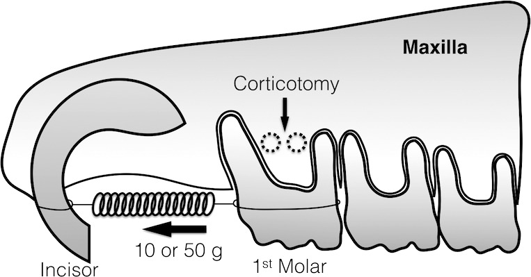
Schematic figure of tooth movement and corticotomy area.
Micro–computed tomography Analysis
The maxillae were removed after euthanasia and placed immediately in 10% formalin for 7 days. Micro–computed tomography (micro-CT) analysis (Scanco μCT 35, Scanco Medical, Bassersdorf, Switzerland) was performed at 70 kVp and 114 μA with an exposure time of 256 ms and a voxel size of 10 μm. After calibration, 40 maxillae were scanned parallel to the occlusal surface of the maxillary second molar. Reconstruction of the scanned data was done with built-in software. The measured parameters were the amount of tooth movement and BV/TV (the ratio of the mineralized tissue considered to be bone [BV] to the total volume that is surrounded by the contours [TV]). The amount of tooth movement was measured from a two-dimensional transverse section. Measurement was made from the most distal point of the first molar crown to the most mesial point of the second molar crown (Figure 2A). The rate of tooth movement was obtained by dividing the total amount of movement by the number of weeks at the time of evaluation.
Figure 2.
(A) Tooth movement measurement using micro-CT. (B) ROI for alveolar bone analysis. M indicates mesial root; Mid-B, mid-buccal; MP, mesiopalatal; DB, distobuccal; and DP, distopalatal. (C) ROI in sagittal view.
A standard hydroxyapatite was used for measurement of the same parameters at baseline. A region of interest (ROI) was contoured in a transverse plane surrounding the interradicular area of the maxillary first molar (Figure 2B). Vertically, it was defined as the most occlusal point of the furcation to the apex of the shortest root (most often, the mid-buccal root). The serial images in the ROI were used for analysis of the BV/TV.
Histological Procedure
After micro-CT scanning, the eight maxillae of the 28-day group were decalcified with 10% EDTA solution (Fluka, Sigma-Aldrich Inc, Singapore) at pH 7 and room temperature for 28 days. The maxillae were then dehydrated with graded alcohol and embedded in paraffin blocks. The histological sections were horizontally parallel to the occlusal plane of the maxillary first molar. Each section was 3-μm thick at 100-μm intervals. The sections were stained with hematoxylin and eosin to evaluate the root resorption area.
Root Resorption Analysis
The distobuccal and distopalatal roots of the maxillary first molar were chosen for histomorphometric analysis. To calculate the resorption area, the roots were divided into mesial and distal sides by using the most buccal and most lingual points on the root surface as the border. The root resorption area was defined as the enclosed area of resorptive root surface and the line that connected the margins of each discontinuity of root surface. The root resorption areas on the mesial aspect were compared with the sum of root resorption areas and dentin areas on the mesial side by percentage by using the captured picture at 4× magnification (Figure 3). To measure the root resorption area and dentin area, the program NIS Elements D version 4.20 (Nikon Instruments, Inc, New York, USA) was used. The mean and standard deviation of percentage root resorption were calculated from five random sections. The percentages of root resorption were compared between the LF and HF groups at day 28.
Figure 3.
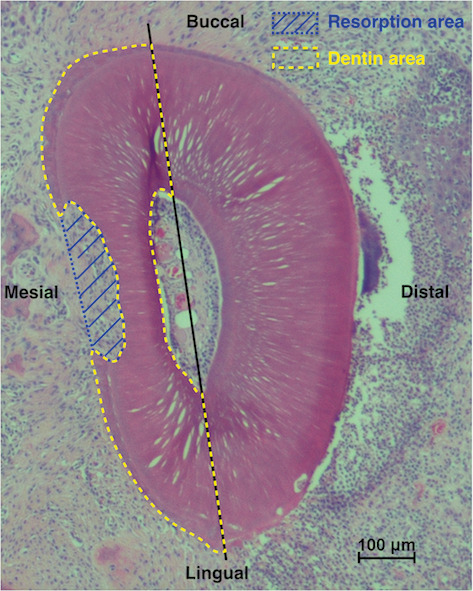
Histomorphometric analysis on distobuccal root.
Statistical Analysis
All sample measurements were repeated twice at a 1-month interval, and intraobserver reliability was assessed using intraclass correlation coefficients. All data were analyzed by SPSS version 17 (SPSS, Chicago, Ill). The results are shown as mean and standard deviation. The Shapiro-Wilk test showed normal distribution of data. The t-test was used to compare the difference between the LF and HF groups. One-way analysis of variance (ANOVA) was used to compare intragroup changes. A difference of probabilities of less than 5% (P < .05) was considered statistically significant.
RESULTS
During the experiment, all springs remained intact and all maxillary first molars moved in the proper direction. The intraclass correlation coefficients were .99 for the amount of tooth movement, .93 for bone volumetric analysis, and .96 for histomorphometric analysis, which indicated excellent reliability.
Amount and Rate of Tooth Movement
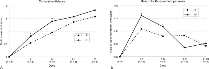
The amount of tooth movement in the HF group was higher than in the LF group from the beginning to 28 days. The HF group showed a significantly greater amount of tooth movement than the LF group, which was approximately 1.5 times greater at day 7 (0.81 ± 0.24 mm vs 0.55 ± 0.1 mm) and at day 14 (1.39 ± 0.1 mm vs 0.95 ± 0.14 mm). However, there were no significant differences in the amount of tooth movement between the two groups at days 21 and 28 (Figure 4A).
Figure 4.
(A) Cumulative tooth movement of LF and HF groups. (B) Rate of tooth movement of LF and HF groups. *P < .05.
The rate of tooth movement in both groups showed a sharp increase in the first 7 days followed by a continuous decrease from day 14 to day 28. There was a significantly higher tooth movement velocity in the HF group at 0–7 days (0.81 ± 0.24 mm/wk) and at 7–14 days (0.59 ± 0.1 mm/wk) than the LF group at 0–7 days (0.55 ± 0.1 mm/wk) and at 7–14 days (0.4 ± 0.14 mm/wk). On day 21, the HF group showed a dramatic tooth movement deceleration that resulted in slower tooth movement than the LF group, which demonstrated a constant velocity. However, no significant difference was detected (Figure 4B).
Alveolar Bone Change
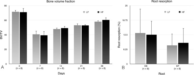
The initial BV/TV values between the LF group and HF group were not significantly different (71.65% ± 1.99% vs 71.07% ± 5.14%, respectively). The BV/TV values in the LF and HF groups showed similar patterns at all time points. There was a significantly lower BV/TV after corticotomy and force applications at days 7, 14, 21, and 28 than at baseline (day 0) in both groups. The results showed that the BV/TV quickly and significantly decreased from baseline to the lowest point at 7 days after treatment in both the LF and HF groups (40.68% ± 4.48% vs 39.35% ± 5.14%, respectively). The BV/TV then gradually increased from day 14 and was restored to near the baseline level at day 28. There were no significant differences between the LF and the HF groups at all observed time periods (Figure 5A).
Figure 5.
Comparison between LF and HF groups. (A) BV/TV. (B) Root resorption.
Root Resorption
Histomorphometric analysis was used to evaluate the mesial half of the distobuccal and distopalatal roots of the maxillary first molar. The percentage of resorbed area of the distobuccal root in the LF group was higher than the HF group (13.25% ± 10.3% vs 12.54% ± 5.82%), while the distopalatal root had a greater percentage of root resorption in the HF group than in the LF group (9.05% ± 6.19% vs 7.95% ± 4.93%). However, no significant differences were detected (Figures 5B and 6).
Figure 6.
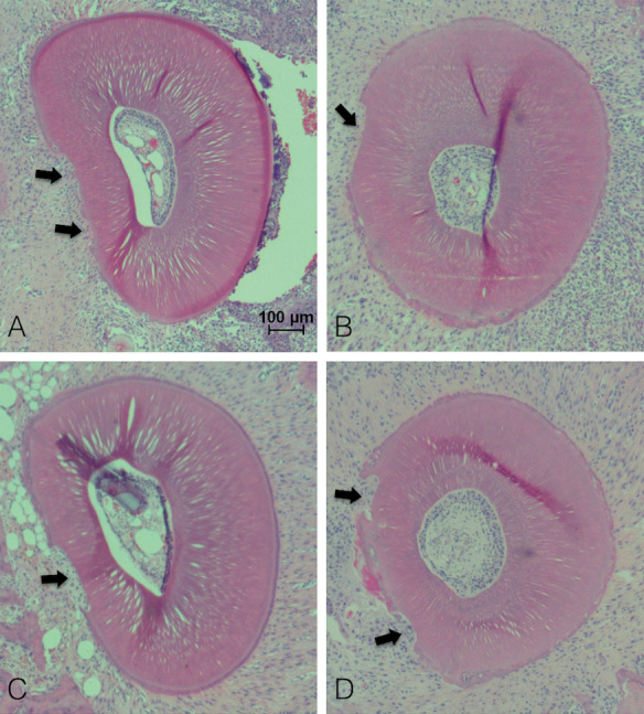
Root resorption evaluation. (A) Distobuccal root of LF. (B) Distopalatal root of LF. (C) Distobuccal root of HF. (D) Distopalatal root of HF. Arrows indicate the resorptions present on the mesial side.
DISCUSSION
The results showed that orthodontic force application with corticotomy promoted early rapid tooth movement. These findings are in agreement with the results of Baloul et al.11 and Tsai et al.,12 who reported that the tooth movement rate in the surgically assisted group increased to the highest point in the first week. The reason for accelerated tooth movement is explained by the RAP concept, which is a transient process in which localized increased alveolar bone remodeling results in decreased bone density.4,11 In addition, the results illustrated earlier and faster tooth movement in HF (50 g) compared with LF (10 g) combined with corticotomy. Murphy et al.13 also found earlier and faster tooth movement at day 7 in HF compared with LF when corticision was performed. However, the current results differed from conventional tooth movement, in which heavier forces did not increase the initial tooth movement and delayed initial tooth movement due to hyalinized tissue.2,14 It was assumed that the increase of alveolar bone reaction from corticotomy might lead to rapid elimination of PDL hyalinization,9 resulting in an increased amount of tooth movement in the early stage in the HF group. However, a slower rate of tooth movement in the HF group was found on day 21. This finding possibly occurred from diminished RAP, and consequently, the hyalinized tissue was possibly removed slowly, which induced a lag phase. On the other hand, LF produced less or no hyalinization. Therefore, tooth movement velocity was not dramatically reduced despite a decrease in the RAP. However, the amount of tooth movement measured by micro-CT may be overestimated because corticotomy moves the center of rotation of tooth displacement more apically, and consequently, the tooth movement observed was mainly due to tipping movement.15
Alveolar bone change at the maxillary first molar showed that the lowest bone volume fraction was found 7 days after force application, which corresponded to the early accelerated tooth movement in the results. A previous study also found that the most catabolic activity occurred during the first week following the operation.11 A decreased BV/TV explains the transient osteopenia, which occurs following tissue injury such as flap elevation,16 bone fracture, and osteotomy.17 Murphy et al.13 evaluated BV/TV after corticision and the application of light and heavy forces at 14 days. They showed no significant difference among the corticision groups. This study agreed with Murphy et al., in that both groups exhibited the same trends of alveolar bone change at all time periods. It is assumed that the injury from either an LF or HF is low compared with alveolar bone decortication. Thus, it may be suggested that the increased force magnitude in corticotomy-assisted tooth movement does not increase alveolar bone resorption. However, differences in bone density do not always indicate faster tooth movement. The present study found a higher rate of tooth movement in the HF group during the first 2 weeks, but no differences were found in the BV/TV.
Root resorption from orthodontic treatment is a part of the hyaline tissue elimination process.18 During hyaline tissue removal, the nearby layer of cementum tissue can be damaged. In conventional tooth movement, several studies reported that higher force magnitudes were related to greater hyalinization19 and root resorption.2 They found that HF (50–100 g) applications increased the amount of root resorption compared with LF (10 g). The current results were in contrast with previous findings, in which a force of a higher magnitude combined with corticotomy did not increase the amount of root resorption. A recent finding by Murphy et al.20 also showed similar results. They found no differences in the resorption areas between a 10 g and a 100 g force with corticision at 14 days. After the corticotomy, tissues were activated by increased osteoclastogenesis that resulted in decreased bone density.21 Deguchi et al.22 found that increased root resorption was related to decreased bone turnover. It has been assumed that increased bone turnover leads to less PDL hyalinization and root resorption in both LF and HF groups.3
Histomorphometric measurements provided a high resolution for root resorption identification. However, this method may not disclose all of the areas of resorption around the root since it may occur in three dimensions. A three-dimensional evaluation may be needed to be fully accurate. In addition, histologic examinations at early time points are needed to provide information on the process of hyalinization.
From these results, the HF magnitude combined with corticotomy might accelerate tooth movement at early time points when bone density is dramatically reduced, and without the risk of increased root resorption. After that, an LF magnitude provides the benefit of steady tooth movement.
CONCLUSIONS
The amount of tooth movement using 50 g of force with corticotomy was higher than 10 g of force during the first 2 weeks, but there was no difference by the end of the study period (4 weeks).
The application of 10 g or 50 g with corticotomy did not show significant differences in bone volume fraction or root resorption.
REFERENCES
- 1.Nakano T, Hotokezaka H, Hashimoto M, et al. Effects of different types of tooth movement and force magnitudes on the amount of tooth movement and root resorption in rats. Angle Orthod. 2014;84:1079–1085. doi: 10.2319/121913-929.1. [DOI] [PMC free article] [PubMed] [Google Scholar]
- 2.Gonzales C, Hotokezaka H, Yoshimatsu M, Yozgatian JH, Darendeliler MA, Yoshida N. Force magnitude and duration effects on amount of tooth movement and root resorption in the rat molar. Angle Orthod. 2008;78:502–509. doi: 10.2319/052007-240.1. [DOI] [PubMed] [Google Scholar]
- 3.Verna C, Dalstra M, Melsen B. Bone turnover rate in rats does not influence root resorption induced by orthodontic treatment. Eur J Orthod. 2003;25:359–363. doi: 10.1093/ejo/25.4.359. [DOI] [PubMed] [Google Scholar]
- 4.Sebaoun JD, Kantarci A, Turner JW, Carvalho RS, Van Dyke TE, Ferguson DJ. Modeling of trabecular bone and lamina dura following selective alveolar decortication in rats. J Periodontol. 2008;79:1679–1688. doi: 10.1902/jop.2008.080024. [DOI] [PMC free article] [PubMed] [Google Scholar]
- 5.Nimeri G, Kau CH, Abou-Kheir NS, Corona R. Acceleration of tooth movement during orthodontic treatment-a frontier in orthodontics. Prog Orthod. 2013;14:42. doi: 10.1186/2196-1042-14-42. [DOI] [PMC free article] [PubMed] [Google Scholar]
- 6.Frost HM. The regional acceleratory phenomenon: a review. Henry Ford Hosp Med J. 1983;31(1):3–9. [PubMed] [Google Scholar]
- 7.Wilcko MT, Wilcko WM, Pulver JJ, Bissada NF, Bouquot JE. Accelerated osteogenic orthodontics technique: a 1-stage surgically facilitated rapid orthodontic technique with alveolar augmentation. J Oral Maxillofac Surg. 2009;67:2149–2159. doi: 10.1016/j.joms.2009.04.095. [DOI] [PubMed] [Google Scholar]
- 8.Mostafa YA, Mohamed Salah Fayed M, Mehanni S, ElBokle NN, Heider AM. Comparison of corticotomy-facilitated vs standard tooth-movement techniques in dogs with miniscrews as anchor units. Am J Orthod Dentofacial Orthop. 2009;136:570–577. doi: 10.1016/j.ajodo.2007.10.052. [DOI] [PubMed] [Google Scholar]
- 9.Iino S, Sakoda S, Ito G, Nishimori T, Ikeda T, Miyawaki S. Acceleration of orthodontic tooth movement by alveolar corticotomy in the dog. Am J Orthod Dentofacial Orthop. 2007;131(448):e441–e448. doi: 10.1016/j.ajodo.2006.08.014. [DOI] [PubMed] [Google Scholar]
- 10.Chung K-R, Mitsugi M, Lee B-S, Kanno T, Lee W, Kim S-H. Speedy surgical orthodontic treatment with skeletal anchorage in adults-sagittal correction and open bite correction. J Oral Maxillofac Surg. 2009;67:2130–2148. doi: 10.1016/j.joms.2009.07.002. [DOI] [PubMed] [Google Scholar]
- 11.Baloul SS, Gerstenfeld LC, Morgan EF, Carvalho RS, Van Dyke TE, Kantarci A. Mechanism of action and morphologic changes in the alveolar bone in response to selective alveolar decortication-facilitated tooth movement. Am J Orthod Dentofacial Orthop. 2011;139(4 suppl):S83–S101. doi: 10.1016/j.ajodo.2010.09.026. [DOI] [PubMed] [Google Scholar]
- 12.Tsai CY, Yang TK, Hsieh HY, Yang LY. Comparison of the effects of micro-osteoperforation and corticision on the rate of orthodontic tooth movement in rats. Angle Orthod. 2016;86:558–564. doi: 10.2319/052015-343.1. [DOI] [PMC free article] [PubMed] [Google Scholar]
- 13.Murphy CA, Chandhoke T, Kalajzic Z, et al. Effect of corticision and different force magnitudes on orthodontic tooth movement in a rat model. Am J Orthod Dentofacial Orthop. 2014;146:55–66. doi: 10.1016/j.ajodo.2014.03.024. [DOI] [PubMed] [Google Scholar]
- 14.Yee JA, Türk T, Elekdağ-Türk S, Cheng LL, Darendeliler MA. Rate of tooth movement under heavy and light continuous orthodontic forces. Am J Orthod Dentofacial Orthop. 2009;136:150.e1–9. doi: 10.1016/j.ajodo.2009.03.026. [DOI] [PubMed] [Google Scholar]
- 15.Verna C, Cattaneo PM, Dalstra M. Corticotomy affects both the modus and magnitude of orthodontic tooth movement. Eur J Orthod. 2018;40:107–112. doi: 10.1093/ejo/cjx041. [DOI] [PubMed] [Google Scholar]
- 16.Owen KM, Campbell PM, Feng JQ, Dechow PC, Buschang PH. Elevation of a full-thickness mucoperiosteal flap alone accelerates orthodontic tooth movement. Am J Orthod Dentofacial Orthop. 2017;152:49–57. doi: 10.1016/j.ajodo.2016.11.026. [DOI] [PubMed] [Google Scholar]
- 17.Teng GYY, Liou EJW. Interdental osteotomies induce regional acceleratory phenomenon and accelerate orthodontic tooth movement. J Oral Maxillofac Surg. 2014;72:19–29. doi: 10.1016/j.joms.2013.09.012. [DOI] [PubMed] [Google Scholar]
- 18.Brudvik P, Rygh P. Non-clast cells start orthodontic root resorption in the periphery of hyalinized zones. Eur J Orthod. 1993;15:467–480. doi: 10.1093/ejo/15.6.467. [DOI] [PubMed] [Google Scholar]
- 19.Tomizuka R, Shimizu Y, Kanetaka H, et al. Histological evaluation of the effects of initially light and gradually increasing force on orthodontic tooth movement. Angle Orthod. 2007;77:410–416. doi: 10.2319/0003-3219(2007)077[0410:HEOTEO]2.0.CO;2. [DOI] [PubMed] [Google Scholar]
- 20.Murphy C, Kalajzic Z, Chandhoke T, Utreja A, Nanda R, Uribe F. The effect of corticision on root resorption with heavy and light forces. Angle Orthod. 2016;86:17–23. doi: 10.2319/112514-843.1. [DOI] [PMC free article] [PubMed] [Google Scholar]
- 21.Dutra EH, Ahmida A, Lima A, Schneider S, Nanda R, Yadav S. The effects of alveolar decortications on orthodontic tooth movement and bone remodelling in rats. Eur J Orthod. 2017] doi: 10.1093/ejo/cjx080. [published online October 28. [DOI] [PMC free article] [PubMed]
- 22.Deguchi T, Seiryu M, Daimaruya T, Garetto LP, Takano-Yamamoto T, Roberts WE. Decreased alveolar bone turnover is related to the occurrence of root resorption during experimental tooth movement in dogs. Angle Orthod. 2015;85:386–393. doi: 10.2319/021714-117.1. [DOI] [PMC free article] [PubMed] [Google Scholar]


