Abstract
Congenital cataract can be caused by several systemic diseases and differential diagnosis should be done between infections, genetic or metabolic diseases. We present a case of a 12-month-old girl with bilateral nuclear cataracts that was referred for investigation. Since she did not present a family history of congenital cataracts or metabolic diseases, and her physical examination was normal, a systemic evaluation was performed. Biochemical studies disclosed abnormal galactose metabolism signs. The diagnosis of galactokinase (GALK1) deficiency was considered and the study of the GALK1 gene allowed identifying a pathogenic genetic variant and a predictably pathogenic missense mutation, previously not described. Dietary measures were imposed with a good evolution.
Keywords: congenital disorders, ophthalmology, diet
Background
Bilateral congenital cataracts can be inherited as an autosomal dominant form or caused by systemic diseases such as infections (TORCH (Toxoplasmosis, Other (syphilis, varicella-zoster, parvovirus B19), Rubella, Cytomegalovirus and Herpes infections) group), genetic diseases (Down syndrome, Lowe syndrome, trisomy 13 and trisomy 15) or metabolic diseases, including galactokinase (GALK1) deficiency.
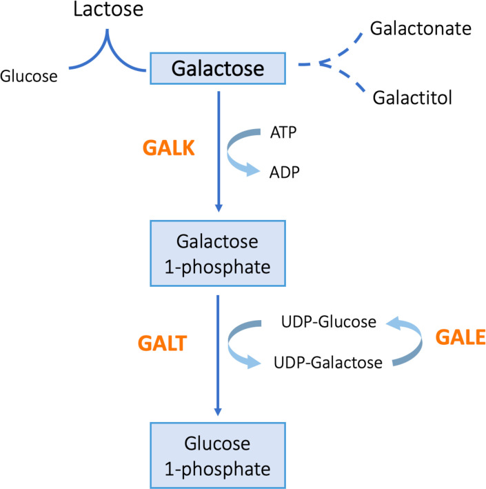
The GALK1 gene, located on chromosome 17q24, encodes GALK1 and is involved in galactose metabolism. GALK1 deficiency is an autosomal recessive disease originally described by Gitzelmann.1 It results from an error in the first committed step of the Leloir pathway—GALK1 catalyses the phosphorylation of galactose with ATP into galactose 1-phosphate (figure 1). This results in the activation of alternate pathways of galactose metabolism, which leads to the accumulation of galactose, galactitol and galactonate. Consequently, high galactitol causes osmotic swelling of lens fibres and protein denaturation, leading to early onset cataracts.2 3
Figure 1.
The Leloir pathway of galactose metabolism. GALK, galactokinase; GALT, galactose 1-phosphate uridyltransferase; GALE, UDP-galactose 4′-epimerase.
The worldwide incidence of GALK1 deficiency is low (1:150.000 to 1:1.000.000), and the highest incidence is found in Romani population (1:40.000), due to a founder mutation.2 4
The most common symptom of patients with GALK1 deficiency is bilateral cataracts with neonatal or juvenile onset. However, neonatal complications have also been described, such as hepatosplenomegaly, hypoglycaemic and hyperbilirubinemia. Additionally, cognitive and/or motor developmental delay, bleeding diathesis and encephalopathy can also occur, yet rarely.5
Galactose and galactitol levels on blood or urine are elevated but, unlike classical galactosemia, galactose-1-phosphate is usually normal. After implementing a dietary treatment, galactose and galactitol concentrations decrease and, therefore, become reliable parameters to understand dietary compliance. Nevertheless, even in patients with good compliance, galactitol concentrations may remain above normal.2 GALK1 deficiency has a relatively benign course even if untreated (except for diet-dependent cataracts and in rare cases of pseudotumor cerebri).
We present a case of a 12 months old girl with bilateral congenital cataracts and referred for investigation, which was diagnosed with GALK1 deficiency.
The aim of this report is to alert medical doctors to this condition, since the diagnosis is infrequent and, therefore, requires a high index of suspicion.
Case presentation
A 12-month-old girl was referred to a general paediatric outpatient clinic of a Tertiary Hospital to investigate bilateral congenital cataracts. A white dot was detected on her pupils since the second month, yet ophthalmologic observation was only performed at 11 months. She was diagnosed with bilateral nuclear cataracts and submitted to surgery, where both her eyes had the lens removed.
During pregnancy, her mother had gestational diabetes controlled with diet, negative serologies and normal ultrasounds. She was born by a caesarean section at a 40 weeks’ gestation with an Apgar score of 10/10/10 and birth weight of 3990 g (P90). The expanded newborn screening for inherited metabolic diseases was normal. Growth was around 50–85th percentile and psychomotor development was age appropriate. Parents are healthy nonconsanguineous Moldovan immigrants and she has a healthy 6-year-old sister. There had no family history of congenital cataracts, early deaths or metabolic diseases. Physical examination was normal, including no dysmorphisms and no palpable abdominal organomegaly.
Investigations
As the child appeared healthy, with no other signs or symptoms except for bilateral cataracts, a disorder in galactose metabolism was considered.
The laboratory results showed high galactose levels (45.2 mg/dL for a reference range <5 mg/dL), with normal galactose-1-P levels (traces, for a reference range <0.9 mmol/L). The remaining analytical study had no abnormalities (including blood count, blood glucose and transaminases). Galactitol measurements were not performed because, at the time, there were no laboratories available in Portugal to do so. The same happened for the measurement of GALK1 enzyme activity.
The GALK1 gene study allowed identifying two heterozygote variants: c.919_921delATG p.(Met307del), a known pathogenic genetic variant, and c.500C>A p.(Ala167Asp), a predictably pathogenic missense mutation, not previously described.
By this time, grounded on symptomatology and genetic results, GALK1 deficiency was the most probable diagnosis.
Differential diagnosis
Since the child had no other clinical manifestations, a normal neurodevelopment and clinical examination, conditions such as genetic syndromes or infections were not feasible.
The main differential diagnosis was classic galactosemia. Classic galactosemia results from galactose-1-phosphate uridyltransferase deficiency, which is more common and severe than GALK1 deficiency. Besides cataracts, clinic manifestations include vomiting, poor feeding, jaundice, hepatic and renal failure and sepsis. GALK1 deficiency differs from classic galactosemia in the absence of severe systemic involvement.
Contrary to medical practices in other countries, in Portugal, newborn screening does not include galactose metabolism disorders, explaining why, in our case, GALK1 deficiency was not detected. Nonetheless, when the acylcarnitine profile reveals high levels of phenylalanine and tyrosine, the laboratory can perform, on medical request, a total galactose measurement.
Treatment
Once considered the diagnosis of GALK1 deficiency, the patient started a galactose restricted diet. Parents received guidance on dietary measures, including about the several origins of the main sources of galactose, how to detect it on labels and a list of permitted and prohibited foods.
Outcome and follow-up
After a 2-year follow-up, the girl is well adapted to the diet, maintains a regular growth (85th percentile) and a normal neurodevelopmental milestone achievement.
Total galactose determination was performed every 6 months, with mean values of 4.95 mg/dL (reference range <5 mg/dL). As expected, she also had normal galactose-1-P levels (0.07 mmol/L, for a reference range <0.9 mmol/L). Routine ophthalmologic evaluations have been normal. As our patient is submitted to a restrictive diet, especially considering calcium intake, she is also regularly monitored by a nutritionist who checks for age-specific nutritional needs. Because her calcium and vitamin D calculated ingestion and plasma levels were always in normal ranges, no vitamin or mineral supplementation has been added so far.
Discussion
In the face of nonsyndromic and nondominant autosomal congenital cataracts, systemic diseases should always be in mind, especially the treatable ones. Bilaterality of cataracts, in the absence of family history, might help evoking the diagnosis of galactose metabolism disorders, namely, GALK1 deficiency.6
The main manifestation of GALK1 deficiency is bilateral cataracts and, while rarely, pseudotumor cerebri can also be found (reported in two cases).3 More recently, it was described that the phenotypic spectrum of GALK1 deficiency can include neonatal elevation of transaminases, bleeding diatheses, encephalopathy and neurodevelopment delay, in addition to cataract. However, their relationship to GALK1 deficiency is still unclear.2 3 5
On suspicion, GALK1 deficiency is easily confirmed by galactose, galactose-1-P and galactitol levels’ determination. Galactitol accumulation, not investigated in our patient, is implicated as the direct cause of cataracts in GALK1 deficiency. Its diagnosis would have been made earlier if it was included in Portugal’s newborn screening.7 8
More than 30 pathogenic variants have been described, but the two most common variants associated with GALK1 deficiency are the founder variant c.82C>A p.(Pro28Thr) and the Osaka variant c.593C>T p.(Ala198Val).5 The founder variant was identified as responsible for GALK1 deficiency in Romani and German immigrants from Bosnia.9 10 Although her Moldovan origin, our patient did not carry the most frequent pathogenic variation reported in that population.
An early start of a galactose-restricted diet results in a significant decrease in galactose and its metabolites, as seen in our patient. It should be maintained beyond infancy in order to prevent secondary cataract recurrence or any other complication.11
Learning points.
The presence of bilateral congenital cataracts requires an aetiologic study.
If cataracts are bilateral, do not miss the diagnosis of inherited galactose metabolism diseases, since they are treatable.
Galactokinase deficiency is a rare, yet important, cause of congenital or infantile bilateral cataracts.
The rarity and variety of clinical presentation (from rare symptoms to neonatal illness) makes this diagnosis easily unthinkable, thus, a high index of suspicion is necessary.
Starting a galactose-restricted diet is the only way to reach normal blood galactose levels, stop disease progression and prevent cataracts development or recurrence.
Acknowledgments
The authors would like to thank Dr Laura Vilarinho (Newborn Screening Unit, Metabolism and Genetics, Department of Human Genetics, National Institute of Health Dr Ricardo Jorge, Oporto) for her help in genetic diagnosis.
Footnotes
Contributors: CC managed the patient, developed the idea for the article, collected clinical data, performed the literature search and wrote the article. PG and DC managed the patient and were involved in critical revision of the manuscript. MO developed the idea for the article and was involved in critical revision of the manuscript.
Funding: The authors have not declared a specific grant for this research from any funding agency in the public, commercial or not-for-profit sectors.
Competing interests: None declared.
Provenance and peer review: Not commissioned; externally peer reviewed.
References
- 1.Gitzelmann R. Hereditary galactokinase deficiency, a newly recognized cause of juvenile Cataracts31. Pediatr Res 1967;1:14–23. 10.1203/00006450-196701000-00002 [DOI] [Google Scholar]
- 2.Hennermann JB, Schadewaldt P, Vetter B, et al. Features and outcome of galactokinase deficiency in children diagnosed by newborn screening. J Inherit Metab Dis 2011;34:399–407. 10.1007/s10545-010-9270-8 [DOI] [PubMed] [Google Scholar]
- 3.Bosch AM, Bakker HD, van Gennip AH, et al. Clinical features of galactokinase deficiency: a review of the literature. J Inherit Metab Dis 2002;25:629–34. 10.1023/A:1022875629436 [DOI] [PubMed] [Google Scholar]
- 4.Kalaydjieva L, Perez-Lezaun A, Angelicheva D, et al. A founder mutation in the GK1 gene is responsible for galactokinase deficiency in Roma (gypsies). J Hum Genet 1999;65:1299–307. 10.1086/302611 [DOI] [PMC free article] [PubMed] [Google Scholar]
- 5.Rubio-Gozalbo ME, Derks B, Das AM, et al. Galactokinase deficiency: lessons from the GalNet registry. Genet Med 2021;23:202–10. 10.1038/s41436-020-00942-9 [DOI] [PMC free article] [PubMed] [Google Scholar]
- 6.Beutler E, Matsumoto F, Kuhl W, et al. Galactokinase deficiency as a cause of cataracts. N Engl J Med 1973;288:1203–6. 10.1056/NEJM197306072882303 [DOI] [PubMed] [Google Scholar]
- 7.Stroek K, Bouva MJ, Schielen PCJI, et al. Recommendations for newborn screening for galactokinase deficiency: a systematic review and evaluation of Dutch newborn screening data. Mol Genet Metab 2018;124:50–6. 10.1016/j.ymgme.2018.03.008 [DOI] [PubMed] [Google Scholar]
- 8.Janzen N, Illsinger S, Meyer U, et al. Early cataract formation due to galactokinase deficiency: impact of newborn screening. Arch Med Res 2011;42:608–12. 10.1016/j.arcmed.2011.11.004 [DOI] [PubMed] [Google Scholar]
- 9.Hunter M, Heyer E, Austerlitz F, et al. The P28T mutation in the GALK1 gene accounts for galactokinase deficiency in Roma (gypsy) patients across Europe. Pediatr Res 2002;51:602–6. 10.1203/00006450-200205000-00010 [DOI] [PubMed] [Google Scholar]
- 10.Reich S, Hennermann J, Vetter B, et al. An unexpectedly high frequency of hypergalactosemia in an immigrant Bosnian population revealed by newborn screening. Pediatr Res 2002;51:598–601. 10.1203/00006450-200205000-00009 [DOI] [PubMed] [Google Scholar]
- 11.Stambolian D, Scarpino-Myers V, Eagle RC, et al. Cataracts in patients heterozygous for galactokinase deficiency. Invest Ophthalmol Vis Sci 1986;27:429–33. [PubMed] [Google Scholar]


