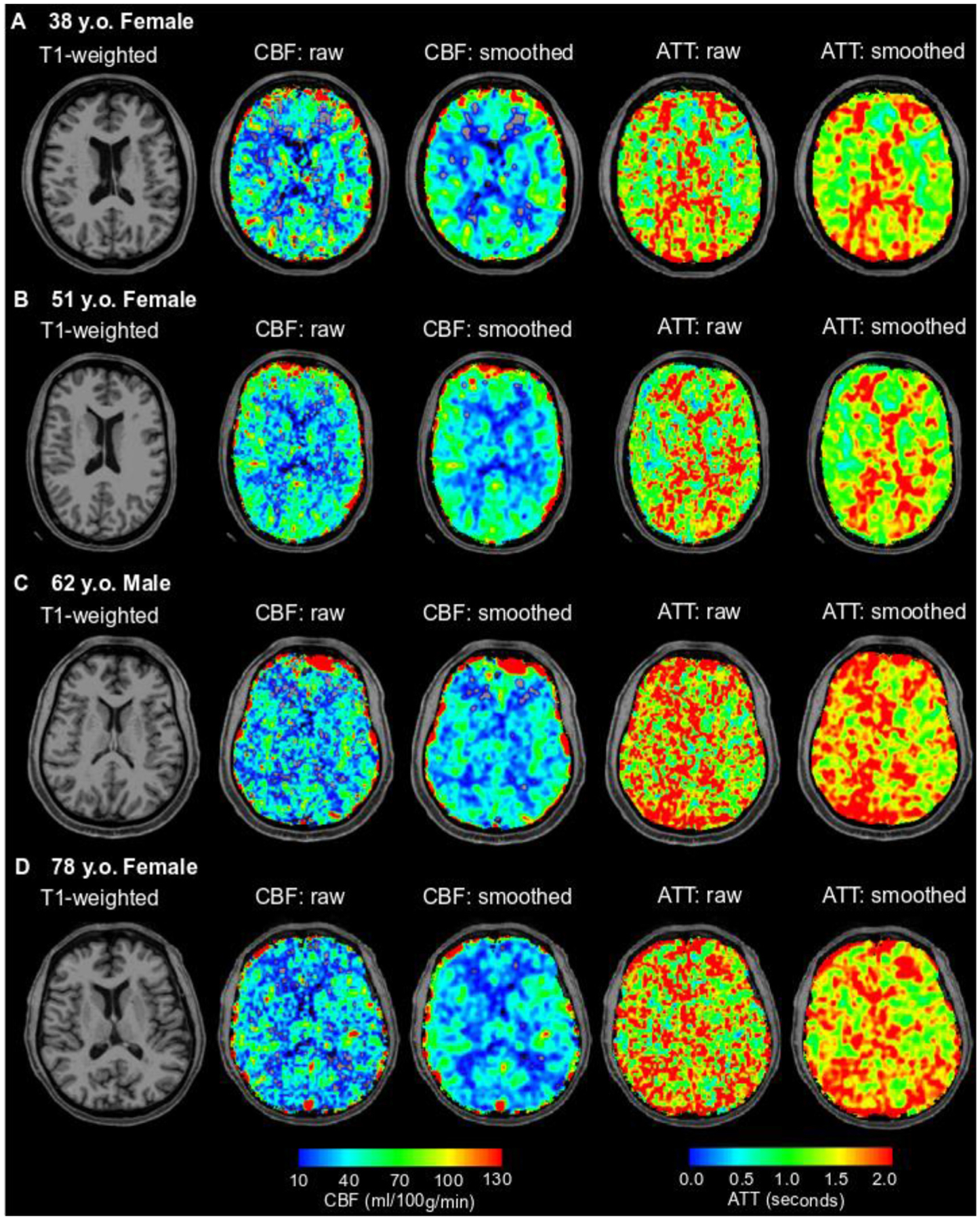
Fig. 8. Representative examples.

T1-weighted (column 1) cerebral blood flow (CBF; columns 2 and 3), and arterial transit time (ATT; columns 4 and 5) are shown here for study participants across each quartile of age: 38 y.o. female (Q1; A), 51 y.o. female (Q2; B), 62 y.o. male (Q3; C), and 78 y.o. female (Q4; D). In addition to CBF and ATT images without any smoothing (raw), which are the basis for the quantifications reported in this manuscript, images smoothed with a 3.75 mm kernel are also shown for visualization of the gray-to-white matter contrast expected from conventional ASL CBF images. The overall group-level findings of this study of decreasing CBF and increasing ATT with age in the gray and white matter can be visualized on a subject-level as observed in these representative cases.
